國
立
交
通
大
學
生物科技學系
博 士 論 文
「固定化酵素動力參數量測平台」之模型建構與計算
A
S
ystematic
a
nd
S
tandardized
A
pproach
t
o
M
odeling
a
nd
M
easuring
I
mmobilized
E
nzyme
K
inetics
研 究 生: 李政哲
指導教授: 楊裕雄 博士
「固定化酵素動力參數量測平台」之模型建構與計算
A Systematic and Standardized Approach to Modeling and
Measuring Immobilized Enzyme Kinetics
研 究 生: 李政哲 Student: Cheng-Che Lee
指導教授: 楊裕雄 Advisor: Yuh-Shyong Yang
國 立 交 通 大 學
生 物 科 技 學 系
博
士
論
文
A Thesis
Submitted to Department of Biological Science and Technology
College of Biological Science and Technology
National Chiao Tung University
in Partial Fulfillment of the Requirements
for the Degree of
Doctor of Philosophy
in
Biological Science and Technology
June 2009
Hsinchu, Taiwan, Republic of China
i
「固定化酵素動力參數量測平台」之模型建構與計算
研究生:李政哲
指導教授:楊裕雄 博士
國 立 交 通 大 學 生 物 科 技 學 院 生 物 科 技 學 系 博 士 班
摘 要
有效整合 top-down 與 bottom up 製程技術於前瞻跨領域「奈米生醫電子」至為關 鍵;其中如何評估局部固定化在元件無機介面上生物分子的工作性能就顯得十分重 要,這些生物分子可以是 DNA、RNA、蛋白質(特別是具有辨識與催化的酵素分子)。 在先期測試中施以掃描電壓於固定化酵素,雖然其活性調控的現象可以被清楚地觀測 到,然而卻無法量測固定化酵素動力參數的即時改變趨勢。因此,本研究團隊在執行 94 年度奈米國家型科技計畫學術卓越計畫─「生化感測與仿生調控功能的奈米結構與 生物分子混成系統之研究」時,也規劃了生物調控器的研究子題;因應評估將酵素嵌 入標準積體電路和微機電系統的平面化技術,我們著手開發了一套可靠合理的即時偵 測與分析固定化酵素動力的量測平台。 在本研究中固定化於二氧化矽基材表面的酵素共有三株,它們分別是老鼠酚亞硫酸 基轉移酵素(E.C.2.8.2.1)、醯亞胺水解酵素(E.C.3.5.2.2)、假絲酵母菌脂肪分解酵素 (E.C.3.1.1.3)。兩項主要的貢獻與理論特徵是:(1)針對符合 Michaelis-Menten 動力學模 式的酵素催化反應並考量質量傳送效應,以系統化、標準化建立表面反應限制的模型 用以量測固定化於平面基材的酵素視動力參數值 K*m 和 V*max;(2)根據上述數學模型建 構標準流程化的實驗操作方式,並對Michaelis-Menten 參數估算提出新的線性圖解法, 其斜率即為視參數 K*m值,且縱座標與橫座標分別具有直觀的物理意義─縱軸表示在 兩個極端基質濃度下反應產率的差值,橫軸在低基質濃度下則近似於基質濃度;此圖 解法亦有利於數值分析求解。 我們以流道高度 167 微米的微流體反應器為實驗平台,成功地量測了老鼠酚亞硫酸 基轉移酵素與假絲酵母菌脂肪分解酵素固定化在二氧化矽基材的及時視動力參數值 K*m 和 V*max;同時本研究也針對固定化酵素失活與基質溶解度限制的實務問題,提出 對應的解決方法。整套量測系統將使我們有能力觀察到電訊號調控固定化酵素活性的 動力參數改變的量值,進而研究相應的調控機制。ii
A Systematic and Standardized Approach to Modeling and Measuring
Immobilized Enzyme Kinetics
Student: Cheng-Che Lee Advisor: Dr. Yuh-Shyong Yang
Department of Biological Science and Technology National Chiao Tung University
Hsinchu, Taiwan, Republic of China
Abstract
How to efficiently combine top-down and bottom-up approaches has become essential in the interdisciplinary field of nano-bio-electronics. It is also important to be able to evaluate the performance of working bio-molecules, like DNA, RNA, proteins (especially for enzymes), immobilized and localized onto the surface of inorganic devices. Although a response of modulated activity was clearly observed via applying a voltage scan onto immobilized enzymes in previous pilot testing, there was no available scheme used to characterize the intrinsic properties of the immobilized enzymes and the corresponding effect of in-situ stresses for further analyzing the modulated mechanism. For developing “ An Artificial-Bio Hybrid Nano-System Capable of Sensing and Regulation,” a reliable and reasonable analysis of immobilized enzyme in situ became a crucial step to embed enzyme onto the planar technology of standard IC and MEMS for the bioregulator subprogram, which belonged to National Research Program for Nanoscience and Technology in the period 2005-2008.
In this study, we have successfully immobilized three enzymes, rat-phenol-sulfotransferase (rat-PST, E.C. 2.8.2.1), D-hydantoinase (E.C. 3.5.2.2), and Candida rugosa lipase (CRL, E.C. 3.1.1.3) onto the silicon dioxide surface. The main contributions and theoretical characteristics should be: (1) A surface reaction limited model, based on systematic and standardized approach, mathematically derived from mass transfer dynamics and Michaelis-Menten equation for measuring apparent K*m and V*max of immobilized enzyme on planar
surface was developed. (2) A new linear plot proposed with a slope K*m, of which axes
containing straightforward, meaningful parameter groups - the difference in reaction yield between two extreme substrate levels (y-axis) versus the reaction conversion fraction (x-axis), is simple to apply either graphically or numerically.
The K*m and V*max of rat-PST and CRL immobilized on silicon oxide surface were
successfully determined in situ. The issues of enzyme inactivation during activity assays and limit of substrate solubility were both concerned in developing measurement approach in this study. Based on this platform, we will be able to make quantitative analysis of electric signal regulation on enzyme activity, and to study its related fundamental mechanisms.
iii
Acknowledgment
還記得陳之藩先生在〈謝天〉這篇文章裡說道:「因為需要感謝的人太多了,就 感謝天吧!」對我而言,生命的成長卻更像演化的縮影,任何「成就」都是一連串阻 力、助力、有意、無意地在時空中交錯醞釀而成,匆匆六年的博士班時光使我學會了 「敬畏天」。 在這段訓練過程中,首先要感謝楊裕雄教授創造一個資源豐富、前瞻與基礎研究 兼顧的跨領域學習環境,營造一個可以相互腦力激盪,又可以動手操作的實驗家天 堂;在這裡我曾和學弟漢平、坤霖一起體會─即使實驗屢次失敗也可以很興奮,很開 心!他是我的指導教授,一個雖不會對學生施以強壓,其領導的研究生也常能不眠地 工作的成功實驗室負責人。 又在研究的結尾也是出發點的此刻,能獲得台灣「半導體之父」張俊彥院士的親 自鼓勵與賞識,彷彿在研究路上打了一劑強心針,由衷感謝這位令我尊敬的長者。其 次也要感謝黃調元教授、林鴻志教授、柯富祥教授,有了他們的愛才惜才與殷殷指 導,才能在混成元件與仿生感控系統的理念建構上,使初生之犢的我能持續保持旺盛 的戰鬥力,不知天高地厚地一路過關斬將,帶著豐碩的戰利品衝刺到終點。又呂平江 教授、徐琅教授、盧向成教授及高健薫組長,您們對我論文的意見和用心,除了讓我 的論文更紮實、完整,同時也啟發我下一階段可以嘗試的研究方向。 六年也是漫長的,家人是最實在的依靠。謝謝爸爸、媽媽、太太得歆毫無怨言的 支持,還有現在兩歲十個月的女兒澄昀經常帶給我們單純快樂的幸福感。除了做研 究,喜歡教書其實是我重回校園的主因;有一個學期我以類似讀書會的方式強迫向學 弟妹推銷所謂「生物電子」的課程,感謝淵仁、程允、秉鈞、漢平和文穎幾乎風雨無 阻,給足了面子。我也抽空陸續輔導明暄、張辰、見陽、詠嫣、芳璿、羿伶、芳謙與 芳諭的數、理、化,我總嘗試在每次 2~3 小時亦師亦友的互動學習中,擺脫升學這種 低層次的枷鎖,希望年輕學子能體會與欣賞數學與自然科學的優美架構;然而也得感 謝這些聰慧高中生的大哉問,使我能重新以初學者的角度看問題,這對研究工作本身 是正面的,因為這常常能使我回到問題的基本面上去。 這頁 A4 的「致謝」看似人名的堆砌,其實正是一段我不想遺忘的記憶…李奇翰 學長、郁吟、慈安、俊榮、羿蓉、靜玟、偉迪、青辰、佩綺、陸宜、音汝、酵素暨蛋 白質實驗室的所有同學、朋友、學弟妹,還有一群「新事」課輔的原住民小朋友 ─ 謝謝大家!iv
Contents
A
A
b
b
s
s
t
t
r
r
a
a
c
c
t
t
(
(
C
C
h
h
i
i
n
n
e
e
s
s
e
e
)
)
i
i
A
A
b
b
s
s
t
t
r
r
a
a
c
c
t
t
(
(
E
E
n
n
g
g
l
l
i
i
s
s
h
h
)
)
i
i
i
i
A
A
c
c
k
k
n
n
o
o
w
w
l
l
e
e
d
d
g
g
m
m
e
e
n
n
t
t
i
i
i
i
i
i
C
C
o
o
n
n
t
t
e
e
n
n
t
t
s
s
i
i
v
v
C
Ch
ha
ap
pt
te
er
r
1
1
In
I
nt
tr
ro
od
du
uc
ct
ti
io
on
n
1.1 B
ackground 11.2 R
eproducible, Reliable and High-sensitivity Nanowire Devices forBiosensing and Bioregulating 8
1.2.1 Perspective and background 8 1.2.2 Specific aims and research approaches 10
1.3 H
ighly Sensitive Detection for Trace Specific Biomolecules and a Novel Bioregulator Controlled by a Locally-intensified Electric Field 151.3.1 Perspective and background 15 1.3.2 Specific aims and research approaches 17
1.4 I
nterface Study and System Integration between 25Nanoelectronic Devices and Biomolecules
1.4.1 Perspective and background 25 1.4.2 Specific aims and research approaches 26
1.5 B
ottle-neck Technologies on interface for sensor andBio-regulator Based on Silicon-based Material 29
1.5.1 Spatially controlled immobilization of biomolecules on silicon surface 29 1.5.2 An Approach to Analyzing and Modeling Immobilized Enzyme Kinetics
29
1.5.3 A Scheme to Measuring and Calculating Immobilized Enzyme Kinetics 31
v
C
Ch
ha
ap
pt
te
er
r
2
2
En
E
nz
zy
ym
me
e
I
Im
m
mo
m
ob
bi
il
li
iz
za
at
ti
io
on
n
2.1 I
ntroduction 342.2 E
xperiment Procedures 372.2.1 Pattern formation processes 37 2.2.2 Chemicals, apparatus and procedures for immobilization 38
2.3 R
esults and Discussion 412.3.1 Immobilization of fluorescent material onto various pattern substrates 41 2.3.2 Enzyme immobilization and its activity
44 2.3.3 Stability of immobilized enzymes 50 2.3.4 Morphology of immobilized enzymes on silicon dioxide Surface 57
C
Ch
ha
ap
pt
te
er
r
3
3
A
A
S
Su
u
r
r
f
f
ac
a
ce
e
L
Li
im
m
it
i
te
ed
d
M
M
od
o
de
el
l
f
fo
or
r
t
t
h
h
e
e
E
Ev
va
al
lu
ua
at
ti
io
on
n
o
of
f
I
Im
m
m
m
ob
o
bi
il
li
iz
z
e
e
d
d
E
En
nz
zy
ym
me
e
o
on
n
P
P
la
l
an
na
ar
r
S
Su
ur
rf
fa
ac
ce
e
3.1 I
ntroduction 593.2 T
heoretical Considerations 623.2.1 Channel Reactor with a One-sided Planar Catalytic Surface 62 3.2.2 Surface Reaction Limited Model 66
3.3 E
xperimental Section 683.3.1 Surface Modification on Silicon Wafer 68 3.3.2 Reactor System and Enzyme Immobilization 69 3.3.3 Enzyme Assay 69
3.4 R
esults and Discussion 713.4.1 Development of Surface Reaction Limited Model for the Derivation of
K*m and V*max
71
vi
3.4.3 Determination of K*m(PAP) through Regression Analysis 74
3.5 C
onclusions 77C
Ch
ha
ap
pt
te
er
r
4
4
A
A
N
N
ew
e
w
A
A
pp
p
pr
ro
oa
ac
ch
h
t
to
o
M
Me
ea
as
su
ur
ri
in
ng
g
I
Im
mm
m
ob
o
bi
il
li
iz
ze
ed
d
E
En
nz
zy
ym
m
e
e
K
Ki
in
ne
et
ti
ic
cs
s
U
U
si
s
in
n
g
g
C
Co
on
nt
ti
in
n
u
u
ou
o
us
s-
-
f
f
lo
l
ow
w
A
A
ss
s
sa
ay
ys
s
4.1 I
ntroduction 794.2 M
ethods 814.3 E
xperimental Section 85 4.3.1 Cleaning and Silanization of Silicon Wafer 854.3.2 Reactor System and Enzyme Immobilization 85
4.3.3 PST Activity Assay 88
4.3.4 CRL Activity Assay 88
4.4 R
esults and Discussion 91 4.4.1 Enzymatic Activity of Surface-immobilized PST, CRL Based on Corresponding Running Controls 914.4.2 Immobilized PST Kinetics: Saturating Substrate Condition to Determine V*max/H and Corresponding Deactivation Curve; Graphical Scheme to Determine K*m(PAP) 92
4.4.3 Immobilized CRL Kinetics: Iterating Scheme to Determine V*max/H, Corresponding Deactivation Curve, and K*m 95
4.5 C
onclusions 98A
Ap
pp
pe
en
nd
di
ix
x
A.1 S
olution of A Partial Differential Equation 99A.2 V
iscosity Measurement of Liquid 103A.3 P
rediction of Liquid Mass Diffusion Coefficient 105A.4 A
pparent Michaelis Constants K*m and V*max for Immobilized Enzymevii
A.5 P
rediction of Damkohler Nunber, Da 112R
R
e
e
f
f
e
e
r
r
e
e
n
n
c
c
e
e
s
s
a
a
n
n
d
d
B
B
i
i
b
b
l
l
i
i
o
o
g
g
r
r
a
a
p
p
h
h
y
y
119C
Cu
ur
rr
ri
ic
cu
ul
lu
um
m
V
V
it
i
ta
ae
e
C.1 E
ducation 133C.2 E
mployment History 133C.3 P
ublication 134C.4 P
atent 1351
-Chapter 1 Introduction
1.1 Background
Owing to the large surface to volume ratio, the conductance of a semiconductor nanowire (or a nanotube) is significantly affected by the surface charge. Many research groups have exploited in this property to develop highly sensitive biosensors. However, essentially all nanowire technologies reported in literatures so far are still having difficulty to efficiently reproduce the biosensors’ performance with nanowire FET devices. This issue limits its expanded purpose in research, not to mention its potential application, especially for detecting a complex system at bioreaction level.
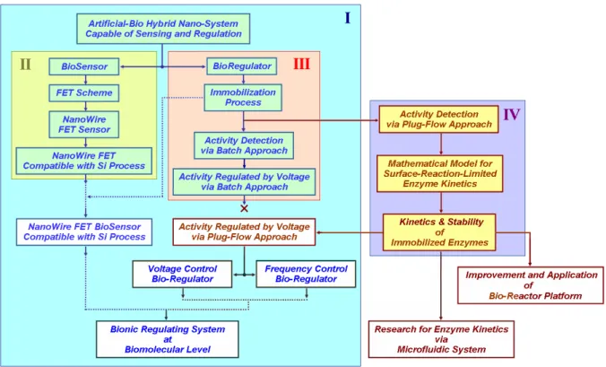
Figure 1. Research flowchart: Block I is the research flowchart for the project “An
Artificial-Bio Hybrid Nano-System Capable of Sensing and Regulation;” Block II shows the stage of research and development for “Nanowire FET Compatible with Si process;” Block III represents the technology development of enzyme immobilization and research of “Activity
2
-Regulated by Voltage via Batch Approach;” Block IV is the progression, “How to systematic and standardized extract the kinetics of immobilized enzyme.”
On the other hand, there are also still few researches on developing bioregulating systems at biocatalytic level directly controlled by electric signals, therefore we try to do it and further combine with biosensor as a bionic system. The three principal goals of this project: 1) to develop a highly sensitive and selective nano-biosensor, of which the scheme must be highly reproducible and compatible with low-cost Si process, 2) to develop a novel bioactivity regulator, which uses a locally-intensified electric field or oscillated electric field to induce the conformational change or to match the turnover frequency of enzymes immobilized on a silicon-based material, and therefore regulates their activities, 3) to construct a bionic system at bioreaction level by integrating the above-mentioned nano-biosensor and bioregulator developed in this project.
In order to meet these goals, it clearly needs three subprograms, which closely support each other in the different researching phases. The strategies, included in these subprograms, are a) to develop a nanowire biosensor with novel and reliable processes, b) to integrate enzyme engineering and SAMs technology for biosensing and bioregulation, c) to construct a bionic system with a cascade of bioreactions on the nano-electronic devices, and d) to integrate and assemble the biosensing, bioregulating, and NEMS technologies into a multi-functioned bionic platform. Figure 1 is the research flowchart, which clearly shows the research strategy and progression. There were preliminary results published in previous studies,1-4 and Block IV in Figure 1 was the major research topic of this thesis.5 The executive summaries of the subprograms are illustrated as follows:
3
-Subprogram 1: Reproducible, reliable and high-sensitivity nanowire
devices for biosensing and bioregulating
Development of highly sensitive nanowire (NW) devices with reproducible and reliable performance is essential for this project. In this subprogram, a novel approach is proposed for the preparation of Si nanowires and the associated device fabrication. In this approach, the Si NWs are formed on the sidewall of a step structure after deposition and etching steps. Accompanied with the patterning of source/drain regions that are formed in a self-aligned manner during the aforementioned etching step, the Si NWs are then employed as the channel portion of a field-effect transistor device. The overall process sequence is very simple and compatible with low-cost Si manufacturing processes and tools, while the feature size of the Si NWs can be well controlled. It is thus very suitable and promising for mass production of future bio-sensing products.
One of the potential issues for the approach is probably the crystallinity of the Si NWs that could be the noise source, and contribute to the leakage current. In line with this, several re-crystallization techniques, such as metal-induced lateral re-crystallization (MILC) and excimer laser annealing (ELA), will be employed to enlarge the grain size. With these advanced methods, quasi-single-crystal Si NWs are expected, which could dramatically improve the sensitivity of the fabricated device. In addition to modifying the material and electrical properties of NWs, we further propose a new concept to enhance the sensitivity of the devices. This is realized by a side-gate design that could modulate the Vth of poly-Si NWs to an appropriate value by applying a voltage to the side-gate. The channel doping step normally used to adjust the Vth of NW required in conventional NW device fabrication could thus be skipped.
The fabricated devices will be used for biosensing purpose by examining the surface bio-reactions on the NW and their effects on the electrical characteristics of the test samples. New
4
-test structure design with a locally-intensified electric filed will also be studied for bioactivity regulation. By combining the biosensors and the bio-regulators, a multi-functioned bionic system could be constructed.
Subprogram 2: Highly sensitive detection for trace specific biomolecules
and a novel bioregulator controlled by a
locally-intensified electric field
Based on the innovative and reliable scheme of silicon nanowire devices, developed in the subprogram 1, this subprogram plans to develop three key applications. The related goals are: (1) a highly sensitive and selective biosensor instrumental for medical diagnosis at detecting range to pM, (2) a revolutionary bioregulator, of which the local conformational states of biomolecules are changed by a locally-intensified electric field, and (3) a multi-functioned bionic system, at bioreaction level, for research and application by integrating the biosensor and the bioregulator technologies mentioned above.
For constructing a highly sensitive biosensor, the spatially controlled monolayer-immobilization of biomolecules on device surfaces is one of the important challenges in this subprogram, and it will strongly affect the sensitivity of biosensors and the commanding of regulators. The nano-tech group, responsible for the subprogram 3, will provide its support to tackle this issue. The bio-research group, in charge of this subprogram, attempts to use this biosensor in medical diagnosis to detect and identify the trace-but-crucial bio-molecules, like beta-endorphin (10~30 pM), neuropeptide Y (29.2+ 3.6 pM), and cholecystokinin (5.2+0.9 pM), etc.
5
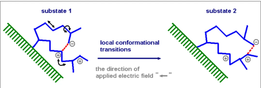
-Figure 2 The local conformation of immobilized biomolecule, under an electric field, is
changed from substate 1 to substate 2. (The conformational transitions among substates are achieved through changing the covalent bond lengths, angles, and dihedral angles.)
Next, based on a similar technology to that of the biosensor, this subprogram will further design and construct a novel bioregulator, through which the local conformation of biomolecules is changed from one substate to another substate by adjusting the intensity or modulating the frequency of electric field. If the biomolecule belongs to a kind of enzyme, then its bio-activity should be regulated. Figure 2 schematically shows the operation principle of this bioregulator. Besides the potential value for biomedical applications, the bio-research group will use this bioregulator scheme as a new skill for studying (a) the allosteric interaction, (b) the disulfide bond formation effected by the enzyme conformation at different substates, and (c) the ion concentration effect of specific metal on the enzymatic catalysis. The locally-intensified electric field will be used to induce the conformational change of the first two cases, and change the local concentration surrounding the enzyme in the last case. Using the bioregulator scheme and by intentionally mutating some selected amino acids away from active site, this subprogram tries to study the phenomena of intra-molecular variations and the effect of local ion concentration on enzymes.
At the third research phase, the goal of this program is to construct a multi-functioned bionic system, which is capable of sensitively detecting and specifically commanding a cascade of bioreactions. For this goal, the bio-research group will be responsible for
6
-designing the related bioreactions and studying the dynamic model in this cascade of bioreactions.
Subprogram 3: Interface study and system integration between
nanoelectronic devices and biomolecules
The subprogram is aimed to study the interface and system integration for the other two subprograms. In the interface study of bionic system, we will immobilize the Si(OR)3(CH2)nR’ molecules (R: CH3, C2H5; R’: -NH2, -SH, -COOH) onto the surface of nanowire to offer the functional groups for further versatile biomolecular reactions of sensing and regulation. The influencing parameters such as the assembling temperature, liquid or gas assembling, vacuum or atmospheric environment, surface cleanliness, and molecule structure will be studied in details. The effect of monolayer/multilayer, packed molecule density and surface clean recipe on the electrical properties of these devices for enzyme sensing and regulation will also be carefully evaluated. Prior to system integration, the suitable immobilization method will be established and standardized for the fabrication of sensor and regulation.
In regard to the integration of electrical devices and fluidic channels, we will focus on investigating the structure fabrication, heterogeneous surface bonding, flowing system design and channel surface treatment. The baby system is achieved after optimization of the above parameters. Then, we will study the biomolecular reaction, immobilization, electrical detection, washing, re-immobilization, detection and so on under 37oC. The system will be applied to control and regulate the biomolecular reaction once the reliability of the system has passed the initial test. The potential barriers are (1) signal interference from micro/nano scale
7
-bubbles, defect particles and memory effect, and (2) poor sensitivity from low immobilization efficiency, assembling molecular instability and contaminated element diffusion into the sensing gate. Figure 3 depicts the bionic system; a normal-regulated biochemical pathway regulated as shown in Figure 3(a), and a pathway of an irregular mechanism regulated by the bionic system as shown in Figure 3(b). In this example, the normal bio-system is able to recognize specific species, A, B, C, D, and α, and catalyze two reactions EA→B and EC→D. If a defective enzyme EC→D leads to a block of certain bio-reaction C→D, then A, B, or α could be accumulated to abounding level. Based on the knowledge obtained from various conditions leads the researchers rather clearly realize this mechanism, the bionic system would be designed and applied to mend the defect, a deficiency of enzyme EC→D, that may be caused by a mutation. As shown in Figure 3(b), the sensing device determines the species α (or A, B), consequentially releases an electronic signal informing the artificial bioregulator to modulate the activity of enzyme EC→D immobilized on the regulating device. Analogous instances occurring in organism are possibly achieved by the bionic system.
Figure 3 Schematic diagram of a bionic system showing a regular mechanism as left Figure
and a EC→D deficient irregular pathway mended by the bionic system, as shown at right. The dark-blue line and orange line represent electric signal and bio-molecular paths, respectively.
8
-1.2 Reproducible, Reliable and High-sensitivity Nanowire
Devices for Biosensing and Bioregulating
1.2.1
Perspective and background
Minimization of silicon electronics is being actively pursued. One-dimensional structures, such as nanowires (NWs), have great potential for testing and understanding fundamental concepts about the roles of dimensionality and size in electrical properties, and could be the ideal building block for nanoelectronics,6 because they can function both as devices and as the interconnect wires that access them. Owing to the inherent high surface-to-volume ratio feature, NWs can suppress short-channel effects encountered in nano-scale MOSFETs7 and provide high surface sensitivity for sensing devices. Many possible applications of Si NWs have been exploited, including nano CMOS,7, 8 memory devices,9 NW TFTs,10 and biosensors.11
Preparation of Si NWs could be categorized into two types, namely, top-down and bottom-up, described as follows:
(I) Top-down: This approach uses advanced lithography techniques, such as deep UV,12 e-beam13 or nanoimprint,14, 15 to generate the NW patterns, followed by an etching step to obtain the NW structures. These techniques are well developed and mature for mass-production purpose. Nevertheless, very expensive equipments and cutting-edge techniques are required. Conventional photolithography processes (e.g., G-line and I-line steppers), though relatively cheap for manufacturing, are not capable of patterning NWs directly. Some special skills such as thermal flow, chemical shrink, and spacer patterning15 have been proposed to help generate the nano-scale patterns using these conventional lithography tools.
(II) Bottom-up: This approach typically utilizes deposition methods to prepare the NWs, and synthesize the NWs on a substrate. Afterwards, these NWs were harvested and dispersed into a solution. Depositing the harvested NWs onto another oxidized substrate and making
9
-electrical contacts to the NWs, will complete the device structure. Many deposition methods have been developed nowadays which include laser ablation catalyst growth,16 chemical deposition catalyst growth17 and oxide-assisted catalyst-free method.18 The first two methods are carried out with metal nanocluster catalyst as the energetically favored sites for absorption of gas-phase reactants, and then the cluster supersaturates and grows a wire-like structure of the materials. The process is based on vapor-liquid-solid (VLS) mechanism. Nanometer-diameter catalyst clusters define the size of wires produced by VLS growth, in which bulk quantities of single crystalline NWs can be obtained. The approach can also be applied to prepare NWs of other materials in addition to Si, such as III-V compounds and II-VI compounds.19 However, metal contamination is a potential concern in this approach. Oxide-assisted catalyst-free method is conducted without metal nanocluster catalyst, and thus is free from metal contamination. Nevertheless, there could be plenty of defects in the wires, and hence it is not applicable to electronic devices. In addition, these aforementioned methods usually suffer from the poor control of structural parameters such as NW’s diameter, length, and orientation.
Methods used to assemble and align the NWs prepared by “bottom-up” approach include electric-field-directed assembly,20 microfluidic channel21 and Langmuir-Blodgett (LB) technique.11 Electric field method is via interaction between electric field of two parallel electrodes and polarity of NWs, and therefore directs the wires. Although electric fields enable more control over assembly, this method is limited by electrostatic interference between nearby electrodes as separations below micrometer level and requirement of extensive lithography to fabricate the electrodes. Fluidic channel method is to align wires by flowing NWs suspension inside a PDMS mold, and could obtain layer-by-layer assembly of multiple crossed NW arrays. However, the size of fluidic channels may limit alignment of NWs. LB method could assemble a large-area anisotropic NWs by compression process, but
10
-it is restricted to preparation of one monolayer. Overall, for practical applications, more refinement is needed to improve the reproducibility and controllability of the above methods.
In a brief, most of the proposed schemes for device fabrication based on “bottom-up” nanostructures are plagued by complex integration that lacks reproducible transfer and positioning of NWs and reliable ohmic contacts. Moreover, the control of doping concentrations in self-assembled semiconducting NWs remains a challenge, and it’s difficult for high-density integration. On the other hand, the top-down approaches usually require expensive lithography apparatus and materials that dramatically increase the fabrication cost. To circumvent these shortcomings, we propose to develop a new method for preparation and fabrication of Si NWs devices in this subprogram.
1.2.2
Specific aims and research approaches
We propose a new approach that could potentially resolve the shortcomings mentioned above. Our approach, albeit based on “top-down” approach, involves only mature and low-cost process skills. Since it could be easily realized using state-of-the-art IC processing, this new scheme is thus very suitable and promising for future practical manufacturing.
The main process steps and the structure of the proposed poly-Si NWs are shown in Figure 4 and 5. The fabrication flow is briefly described as follows: First two dielectric layers (dielectric 1 and dielectric 2) were deposited on a Si substrate, followed by lithographic and etching steps performed on dielectric 2 to form a step structure on the surface. An a-Si layer was then deposited by a low-pressure (LP) CVD system. After the Si layer deposition, source/drain (S/D) implant was performed with low implant-energy (Figure 4 (a)), so that most implanted dopants are located near the top surface of the Si layer. S/D photoresist patterns were then formed on the substrate using g-line or an I-line stepper (Figure 5 (a)). A
11
-reactive plasma etch step was subsequently used to remove the Si layer. The sidewall Si NW channels were formed in this step in a self-aligned manner (Figure 4 (b) and Figure 5 (b)). Note that the implanted dopants in places other than S/D regions will be removed during the etch step because of the very shallow projected range just mentioned. After this step, an annealing step was performed to active the S/D dopants and to transform the a-Si layer into poly-Si.
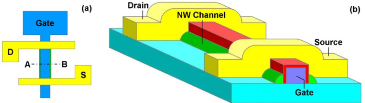
Figure 4 Cross-sectional views (along the A-B direction in Figure 4 (a)) of the device
structure after S/D implant (a) and NW etching (b)
Figure 5 (a) Top-view layout, and (b)stereo-view of the fabricated poly-SiNW FETs
Advantages of this approach include: (1) feature size of NWs is related to the thickness of deposited film and could be well controlled; (2) NWs can be positioned accurately; (3) formation of NW channel, source and drain simultaneously, (4) no metal contamination concerns; (5) S/D to channel contacts are reliable through the use of implant process; (6) both p- and n-channel devices are easily fabricated, and thus CMOS integration is easy to achieve.
12
-One of the potential issues for the proposed approach is probably the structural defects contained in the channel layer of the device which could be the noise source and contribute to the leakage current. Two strategies are adopted to cope with the issue:
(I) Promotion of the crystallinity of the NWs
Many studies have shown that metal-induced lateral crystallization (MILC)22, 23 and excimer laser annealing (ELA)24 can dramatically improve crystallinity of the Si layer and enlarge grain size to about one micrometer. MILC employs metal as precursor to lead amorphous Si to re-crystallize at lower temperatures because of lower energy barrier of recrystallization resulting from the reaction between metal and Si. Excimer laser can provide sufficient energy for amorphous Si to recrystallize, and hence the recrystallization could proceed at a lower temperature. In this subprogram both methods will be investigated to develop a practical recrystallization skill for improving the crystallinity of the NWs.
(II) Structure modification
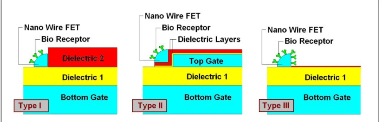
In addition to the structure mentioned in Figure 4 and 5, (denoted as “Type I” in Figure 6), two alternative types of devices will also be studied in this subprogram, as shown in Figure 6 (denoted as “Type II” and “Type III”). All these types use the substrate as the bottom control gate during operation. The modified Type II scheme possesses an additional “side-gate” which could be used to set the Vth to an appropriate value for sensing. With this design, the channel doping step for Vth adjustment could be skipped. In addition, the side-gate bias could also potentially screen part of the channel defects and improve the device sensing performance. Type III is to increase the NW sensing surface area by removing the dielectric 2. The sensitivity is consequently raised due to higher NW surface to volume ratio.
In addition to the completion of high-sensitivity biosensor devices, our research team plans to further exploit the proposed SiNW fabrication method by designing a locally-intensified electrical field to analyze the relationship between biological molecular activity and its structure. The schematic is as illustrated in Figure 7. In the loading window area, the
upperπ 13 upperπ
-shaped poly-Si electrode (light blue color) and the lower SiNW electrode (green color) with immobilized biological molecules will form a locally-intensified electrical field, with the electric field being intensified on the narrow SiNW.
Figure 6 Three types of poly-SiNW FETs for biosensing
By utilizing the electric field to induce the conformational change of the enzyme already immobilized on the SiNW, we can further quantitatively regulate its bioactivity to obtain the information regarding field-regulated enzyme activity. In this way, we can establish a theoretical model and provide a microscopic view regarding the sequence mutation on the conformation and activity of enzyme. This scheme can open a brand new frontier in the research of bio-protein engineering.
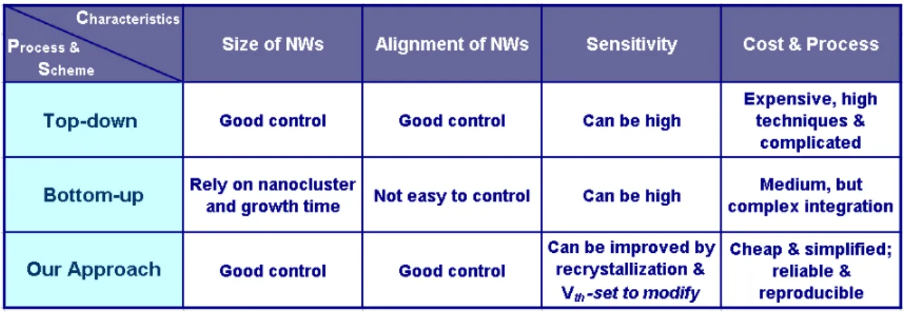
Overall, a novel technique for fabrication of SiNWs and FETs that is superior to conventional “top-down” and “bottom-up” methods is proposed. Table 1 highlights the major advantages of the proposed scheme. This technique is manufacturable and economic, and is capable of providing a reliable high-performance poly-SiNW FET not only for biosensors but also for investigating nano-scale semiconductor physics. It is also compatible with the low-temperature poly-Si (LTPS) technologies, and could be integrated on low-cost and flexible substrates (such as glass or plastic), making the fabrication of system-on-panel (SOP) for biologic sensing and regulating purpose possible. In short, we believe our proposed technique
14
-of SiNWs fabrication indeed represents a breakthrough development, and will be crucial for supplying cheap and credible biosensing chips.
Figure 7 The bioregulator device layout designed to establish a locally-intensified electric
field. It shows both the π–shaped poly-Si electrode (light blue) and the poly-SiNW (bright green) with the immobilized enzymes – sulfotransferases.
Table 1 Comparison of SiNWs formation among top-down,12, 13, 15 bottom-up 10, 16, 17, 20, 21and
15
-1.3 Highly Sensitive Detection for Trace Specific Biomolecules
and a Novel Bioregulator Controlled by a Locally-intensified
Electric Field
1.3.1 Perspective and background
Bioelectronics, a novel interdisciplinary research field, involves the integration of biomolecules with electronic transducers, such as electrochemical (EC) and field-effect transistors (FET). The application of biomolecules, such as enzymes, antigen-antibodies, or DNA, on the electronic transducers dominates the characteristics of the biomolecules-transducer interface and enables the electronic transduction of biorecognition or biocatalytic events to occur on the transducer. A couple of interesting instances include, but not limited to, a variety of biosensors, biofuel cells, and bionic utilities, such as electronic noses and tongues. Nowadays, biosensor is a relatively well-organized device that is designed and fabricated by assembling the biorecognition matrices with diverse transducers.
The employment of semiconductor-based devices, such as ion-sensitive field-effect transistors (ISFETs) and enzyme-based field-effect transistors (ENFETs), as transduction elements for biocatalytic transformations and biological recognition events is a crucial and successful subject in bioelectronics and analysis. Enzyme-based field-effect transistors (ENFETs) are based on the biocatalytic reaction, specifically between immobilized enzyme and substrate occurring on the functionalized gate surface of a well-prepared ISFET, altering the charge at the gate surface and yielding an electronic signal dependent on the substrate concentration. The sensing performance of the ENFET is generally affected by the integration method of the enzyme and the ISFET device. Several investigations reported in literatures have illustrated enzyme-immobilized in thick-film manners, including the immobilization of the desired enzymes in thick polymer films (e.g., polyvinylchloride, polyacrylamide hydrogels, or polyurethane) and membrane (e.g., Nafion or polyvinylpyridine)
16
-covered crosslinked enzyme matrices on the ISFET devices. This type of sensors, nevertheless, demonstrated a high diffusion resistance of the substrates through the polymer membranes, leading to a long response time and a moderate sensitivity. An alternative configuration organizing monolayer (or multilayer) enzyme-based ISFET, therefore, was developed and substantially improved the response time and sensitivity.
Apart from the modification of the biocatalytic matrices, the performance of a FET-based device could be enhanced by a re-construction of the FET element. A promising approach is toward a reduction of gate-dimension resulting in a highly sensitive device. Nano-dimensioned gate materials such as nanotubes or nanowires11, 29 have been illustrated in recent reports; nevertheless, these studies suffer from the limitations of reproducibility, reliability, and stability. The present project proposes a novel fabrication process that reproducibly and reliably produced a silicon nanowire and the associated FET device.
It is well understood that the conformation of the biomolecules shall be altered by the surrounding physical parameters such as temperature and pH, sequentially resulting in the change of function or biocatalytic activity. Previous researches have controlled and optimized the biocatalytic properties and functions by altering temperature, pH, concentration of cofactor, etc. These regulation methods are usually executed in batch manner, difficult to perform precisely and locally. In contrast, a few methods handling the distribution, mobility, conformation, and conjunction of biomolecules by electronic and magnetic forces are currently being investigated and are interesting. These methods demonstrated numbers of advantages over the traditional manners particularly in the ability to regulate biomolecules in a confined area. Furthermore, with respect to the viewpoint of application, the electric field is preferable over the magnetic field, as the facilities of forming a magnetic field are relatively complicated that hampers the approach toward the miniaturization. On the other hand, both fabrications of a miniaturized electronic contact and signal conduction across the interface between biomolecules and electronic transducer are quite simple and doubtlessly
17
-accomplishable by the current technology. Study associated with the regulation and coordination of biomolecules, their corresponding functions, and diverse properties by electric field is a core issue in the field of bioelectronics and is crucial.
1.3.2 Specific aims and research approaches
Many trace but important elements, of which the concentrations range from nM to pM, are well known to participate in the variously crucial homeostatic objectives in human beings. For example, neuropeptide Y (NPY; 29.2+3.6 pM) and cholecystokinin (CCK; 5.2+0.9 pM) are some of the important peptides which play vital biological roles in various degenerative disorders (Qureshi et al., 2000). Those neuropeptides are widely distribution throughout the brain and are believed to participate in several physiological and pathophysiological processes, including pain sensation, memory, neuroendocrine functions, and regulation of release of monoamine transmitters.
Furthermore, the infinitesimal absence of those minor elements could lead to enormous effects on series of physiological reactions, so how to develop a rapid, highly sensitive, reliable, and low-cost detecting system is an imperative issue in the biosensor research field insofar.
At first research phase of this subprogram, the goal is just to research and develop a highly sensitive biosensor for detecting specific trace biomolecules, especially for the continuous measurement of disintegrated rate of disparate drugs in serum and accurate detection of those microelements for the physiological disorders. We will first use avidin-biotin matching system to check and improve the performance of the Si nanowire device developed by the subprogram 1, and examine the transport function of microfluidic channel made by the
18
-subprogram 3. In the initial research period, we will immobilize biotin on the Si nanowire, which is locally and selectively modified by a monolayer of APTES in advance.
Besides bioelectronics, having been focusing on the study of the enzymes -
sulfotransferases (STs) for many years, our recent research also has aimed at the vital
relationship between sulfotransferase and neuroscience, and the intratissue distribution of sulfotransferases. After having mastered the spacially-immobilized and mon olayer-modified technologies for the previous Si nanodevices, we plan to use these technologies to detect those biomolecules involved in a cascade of bioreactions catalyzed by well-known sulfotransferases in our laboratory. This approach not only provides the advanced improvement and the technology spreading for the biosensor developed in this project, but also warms up the following research phases.
Figure 8 The structure of hSULT1A1 (or hSULT1A3) shows the simulated electrostatic
potential. The red meshes and blue ones stand for locally negative and positive charge distributions, respectively.
To research and develop a novel bioregulator controlled by a locally-intensified electric field is the core subject at the second research phase. The enzymes – STs will be immobilized on the bioregulator device developed by the subprogram 1. The structures of STs are
19
-consisting of PAPS binding site which conserved among STs and the related substrate binding site. The regulation of STs can be achieved by redox of flexible loop,30 metal ion effect,31 cofactor/substrate catalysis,32 and so on.
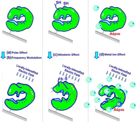
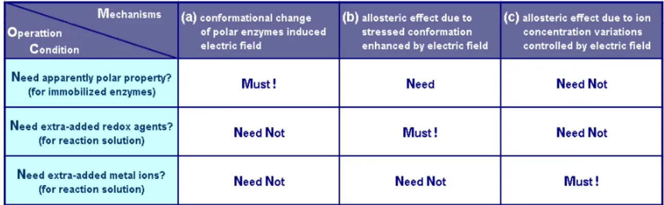
The electrostatic potentials of STs are positive around PAPS binding site and are negative near the surface sandwiched PAPS binding site. Figure 8 shows the structure and the simulated electrostatic potential distribution for STs (hSULT1A1 or hSULT1A3); the red meshes and blue ones stand for locally negative and positive charge distributions, respectively. It is apparently that STs are polar biomolecules, at least for some specific zones. The conformations of STs immobilized on the developed bioregulator device should be evidently effected under an applied electric field, so the related catalysis could be modulated. It had been reported that Mn2+ ion efficiently stimulates the specific activity of dopa/tyrosine sulfotransferase to several hundred folds.33 It is a rational inference that if we can change the local metal ion concentration around the immobilized enzymes, in about hundred-nanometer range, then we can modulate the activity. This project also plans to practice this regulating mechanism not by changing the overall concentration, but just the local concentration around the immobilized enzymes. This subprogram attempts to study and utilize these conformational variations, which modulate the activity of enzymes STs, to develop an artificial bioregulator controlled by a locally-intensified electric field. Three applicable regulating mechanisms and the related bioreactions are proposed and will be designed, respectively. These mechanisms could be classified as: (a) conformational change of polar enzymes induced by electric field, (b) turnover rate modulated by frequency of electric field, (c) allosteric effect due to conformational change enhanced by electric field, and (d) allosteric effect due to ion concentration variations controlled by electric field. Figure 9 schematically shows the operation mechanisms of the proposed bioregulator in this project, and Table 2 briefly illustrates the corresponding operation conditions to the different mechanisms.
20
-Figure 9 Three applicable regulating mechanisms induced by a locally-intensified electric
field are schematically shown. The working enzymes (the deformable green ones) on the bioregulator are immobilized on SiNWs (the inclined planes) in this project. The conformational changes of working polar enzymes under the applied electric field, are mainly caused by (a) only the polar issue, (b) turnover modulated by matching frequency of electric field, (c) the allosteric effect enhanced by the relative position change (between two cysteines; the formation of disulfide bond leads to an allosteric effect.), and (d) allosteric effect induced by the concentration change of metal ion. (light blue balls; the deeper blue one is captured by Asp86, and then an allosteric effect is induced.)
In both of the mechanisms (a) and (c), the conformational changes are induced by locally-intensified electric fields; while in the mechanism (c), this conformational change enhances an intra-molecular bond formation, which could further changes and stabilizes the conformation.
21
-Clearly, this proposed bioregulator is operated by a locally-intensified electric field around the immobilized enzymes, so it could sensitively and efficiently regulate a biochemical reaction under a mild condition, like in vivo. Even in the mechanism (d), it just changes the local metal ion concentration around the immobilized working enzymes in the bioreaction solutions. In fact, the turbulent flow in the microfluidic channel (schematically shown as Figure 11) will guarantee the well-mixed and uniform concentration of overall bioreactions except the spot, on which the working enzymes are immobilized, focused by the locally intensified electric field. Besides, by site-directed mutagenesis, this bioregulator scheme will also provide the bioresearch group a novel skill to study the allosteric mechanisms and the real conformational change in bioreactions.
Table 2 The comparison between the operation conditions of different regulating modes by
the proposed regulators.
Integrating the highly sensitive SiNW-FET biosensor and the highly efficient bioregulator, both developed at the first two phases respectively, to a multi-functioned bionic system is the goal of the third phase of this project. Subprogram 2 at this research phase will be responsible for designing the cascade of bioreactions and studying the dynamic bioregulation.
The transfer of the charged sulfonate moiety to an acceptor steroid catalyzed by human dehydroepiandrosterone sulfotransferase (DHEAST; SULT2A1; EC 2.8.2.2) decreases the biological activity of the steroid. In fact, steroid sulfates resulting from this reaction are not
22
-capable of binding to or activating steroid receptors. Steroid sulfonation has been recognized as an important means for maintaining steroid hormone levels in their metabolism. In humans, dehydroepiandrosterone sulfate (DHEAS) is the most prodigious steroid precursor and one of the major secretory products of both adult and fetal adrenals.
Figure 10 A bionic system, which includes two biosensors and one bioregulator (shown as
three green boxes), is set up to detect the concentration variations of testosterone and estradiol and to modulate the catalysis of PST. There are at least five enzymes included in this cascade of bioreactions; they are PST, DHEA-ST, 3b-HDS, 17b-HDS, and aromatase, respectively. The direction of blue line shows the commanding from the sensors, through signal processor,
23
-and then to the regulator. (The signal processor deals with the weighting between signals or other commanding inputs.)
An example of overall experimental design for bionic cascade reactions is shown as Figure 10. First, using methylumbellifery sulfate (MUS) as sulfuryl group donor to biosynthesize the universal sulfuryl group donor, 3’- phosphoadenosine 5’-phosphosulfate (PAPS), the reaction is catalyzed by phenol sulfotransferase (PST). This step has been well-controlled and used to couple to another sulfotransferase for measuring its activity in our laboratory.34 Many different enzymes involved in this system are to maintain the bionic cascade reactions. For example, dehydroepiandrosterone sulfotransferase (DHEA-ST) is used to transfer the sulfuryl group of PAPS to dehydroepiandrosterone (DHEA). The product, DHEA sulfate (DHEAS) had been demonstrated to act as sex hormone precursor and neurosteroid.35 Therefore, by supplying the enzymes involved in both catalyzing sex steroid hormone and steroid biosynthesis, such as 3β-hydroxysteroid dehydrogenase (3β-HSD), 17β-hydroxysteroid dehydrogenase (17β-HSD), and aromatase, it will be easy to obtain testosterone (T) and estradiol (E2) in this system.
There are two biosensors and one regulator involved in this case of bionic system; the related antibodies against T and E2 will be immobilized on SiNWs of FET biosensors, respectively, and PST immobilized on a SiNW of the electrically-controlled bioregulator. Clearly, these two biosensors are set up to detect the dynamic variations of T and E2, respectively, and then the system will command the catalysis of PST on the regulator after weighting these two signals from Sensor 1 and Sensor 2. These sensors will be developed at the phase 1 of this project. To our anticipation, this bionic system can provide the further research of dynamic bioreactions and the simulation study of biomedical engineering. In fact, this bionic system will be exactly a highly efficient, but mild, artificial reactor, so it has very
24
-great potentiality in medical diagnosis, pharmaceutical or ferment industry, environmental defense and national security.
25
-1.4 Interface Study and System Integration between
Nanoelectronic Devices and Biomolecules
1.4.1 Perspective and background
The biggest hurdle to bio-electronics development is the biosensor technique. This is because any complex system requires a very sensitive sensor to characterize the status. For any bio-detective system, it is almost impossible to fully understand the status of the system by simply only obtaining 1 or 2 parameters. Instead, a few tens of determinative parameters are required to describe the performance of a complicated bio-system. In the past, scientists have developed the smell and taste sensors, while the function of these sensors is still limited.
Mirkin’s group has developed the DNA array sensors which are based on the conductivity changes through the binding of oligonucleotides and gold nanoparticles. The gold nanoparticles are assembled within the electrodes and the silver deposition is facilitated by these nanoparticles bridging the gap and lead to readily measurable conductivity changes. Using this method, they can detect the target DNA concentration down to 500 fM. Star et al. have fabricated the nanoscale field effect transistor (FET) devices with carbon nanotubes (CNTs) as the conducting channel to detect protein binding. In order to avoid the alternation of physical properties of the FET due to the covalent binding of biomolecule with the CNTs, they propose a new technique of overcoating polyethylene imine and poly (ethylene glycol) onto the nanotubes. Prior to recognizing the streptavidin molecule, a biotin (molecular receptor) is immobilized onto the polymer’s surface. This sensor is very sensitive and the detection level is estimated to be of the order of 10 streptavidin molecules.
Lieber’s group fabricated the 20nm p-type silicon nanowire electronic devices in the channel of 500μm by 3mm, which functions as ultrasensitive (10fM) and selective detectors of DNA. The surfaces of the silicon nanowire devices were modified with peptide nucleic acid receptors designed to recognize wild type versus in the mutation site in the cystic fibrosis
26
-transmembrane receptor gene. This nanowire-based approach represents a step forward for direct, label-free DNA detection with extreme sensitivity and good selectivity, and could provide a pathway to integrated, high-throughput, multiplexed DNA detection for genetic screening and biothreat detection.
The electronic device has much better sensitivity than optical method, and the application of electrical sensing to biomolecule will become the major role in the future. Hence, this interdisciplinary technology attracts much attention from academic and industry. There still exist a couple of problems that need to be overcome. For examples, how to set up a fluid channel in an electrical device? How to transport biosamples? How to immobilize the biomolecules onto the sensing channel? The integration of nanodevice and biomolecule involves the research on heterogeneous interface. The interdisciplinary cooperation shall soon become the main issue of bioelectronics field.
1.4.2 Specific aims and research approaches
The 20nm poly-Si NWs fabricated from the spacer of gate line from subprogram 1 is intended to immobilize the biomolecules offered from subprogram 2 through the linker molecule. This subprogram is aimed to study the interface and system integration. It is essential for surface chemistry to occur to clean the FET surface from the contaminants. The use of acid wash solution, a mixture of H2O2 and H2SO4, simultaneously removes organic and metal contamination from the poly-Si NWs. There are two ways to immobilize the surface linker molecule, i.e. monolayer and multilayer, and the spatial structure exhibits various effects on the conductance of FET. We predict the monolayer method has better signal reliability on biomolecule sensing due to the lower thickness variation of surface electrical double layer, but exhibits poor sensitivity. On the contrary, the multilayer structure has better
27
-sensitivity, but demonstrates the poor reliability. Hence, we need to compromise the deposition method. Considering the sensitivity issue, the multilayered immobilization is a possible means to reach the single molecule detection capability with the FET.
Figure 11 This schematically shows the set-up of a bionic system developed in this project.
The bionic system integrates three major subsystems, they are: (1) the bio-system, which includes bioreactions, bio-recognized pairs for biosensors and bioregulators; (2) the nano-electronics system, which comprises SiNW biosensor devices, SiNW bioregulator devices, and the control system; (3) the nano-tech system, which includes the microfluidic channel and the related fluid system, immobilized zones, the related packing parts, etc.
We will utilize the liquid immersion method to assemble multilayered structure, while gas phase silanization under vacuum environment to achieve monolayer molecule. In order to specific assembly the linker, the polymeric soft mask is spin-coated, exposed and development to open the area of interest. Prior to lift-off the polymer by organic solvent, the
28
-linker molecule is assembled onto the NWs. In the multilayer structure, the sample is immersed in the linker solution. Then, the sample is washed and drying. In the monolayer assembly, the sample is putted in a vacuum chamber. After waiting a moment, the linker is deposited onto the NWs through the inert gas carrier. We will study the thickness and surface morphology by ellipsometer and atomic force microscope.
As we known our system should meet the purpose of sensing and regulation, Figure 11 illustrates the schematic diagram of our model system. The covering plastic material is made with poly (dimethylsiloxane) (PDMS). The silicone elastomer base and curing agent are mixed at a ratio of 10:1, and connect the tubes for latter solution delivery. After drying the plastic mold at 65oC for 1 hour, the cover material and the FET device are activated with oxygen plasma and then bonding together. We don’t known whether the plasma damages the linker molecule in the proposal stage. If it happens, we will use the flow system to re-immobilize the linker onto the NWs for sensing. Using the on-line flow system, we will study the biomolecular reaction, immobilization, electrical detection, washing, re-immobilization, detection and so on under 37oC. The system will apply to control and regulate the biomolecular reaction once the reliability of the system has no problem. The barriers, such as (1) signal interference from micro/nano scale bubbles, defect particles and memory effect, and (2) poor sensitivity from low immobilization efficiency, assembling molecular instability and contaminated element diffusion into the sensing gate, will be studied carefully.
29
-1.5 Bottle-neck Technologies on Bio-interface for Bio-sensor and
Bio-regulator on Silicon-based Material
1.5.1 Spatially Controlled Immobilization of Biomolecules on Silicon
Surface
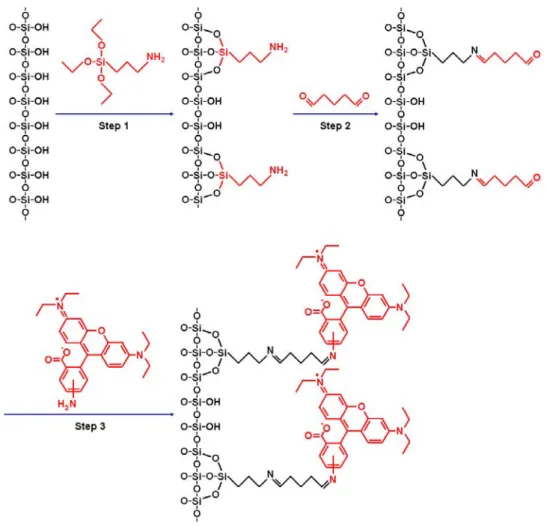
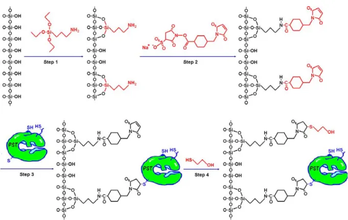
Immobilization of biomolecules on silicon-based material is the common crucial technology for the research and development of bio-sensor and bio-regulator. However, how to control the positioning, orientation, and conformation of immobilized biomolecules is the key. Chapter 2 will show our strategy and preliminary result of enzyme immobilization.
1.5.2 An Approach to Analyzing and Modeling Immobilized Enzyme
Kinetics
Many biomolecules, such as membrane proteins, perform their specific biorecognition or biocatalytic activities while they are immobilized on the surface of cells or organelles.36, 37 Artificial technologies also take advantage of immobilized enzymes for a variety of applications such as bioconversion, bioremediation, and biosensors.38-42 Methods developed for the analysis of the function of biomolecules that move and rotate freely in the homogeneous solution may not be adequate to describe the behavior of immobilized biomolecules. The immobilized enzyme is not distributed evenly and freely in the solution and may have a unique microenvironment for each enzyme molecule. For example, the specific orientation is important for the function of proteins.43, 44 However, free protein in solution can have many orientations, while the rotation for an immobilized protein is restricted. Unique kinetic behaviors of membrane-bound enzymes have also been reported.45, 46
30
-The leading-edge semiconductor devices47, 48 have attracted researches into the emerging fields of biomolecular sensors,3, 4, 49, 50 hybrid (biotic-abiotic) nanomaterials,51 lab-on-a-chip platform for further biomedical applications,52 enzyme-coupled biosensor,53 and novel enzyme-based devices.54-56 The immobilization of a variety of biomolecules including proteins and nucleic acids onto silicon-based material is essential for specific biorecognition and electronic signal manipulation required for the bio-silicon hybrid devices. How to characterize the reaction kinetics for immobilized enzyme on silicon-based supporting material has become the object of study.57-61 Kinetic properties of immobilized alkaline phosphatase have been determined in a microfluidic reaction with packed bed.62 The kinetics of immobilized horseradish peroxidase has been studied using a microfluidic packing reaction63 and modeling system.64 Using batch-assay method, the kinetic parameters of immobilized glutathione S-transferase has been determined in porous silicon.65 The kinetics and transport of this immobilized enzyme has also been simulated by an embedding method in sessile hydrogel drops.66 Fractal and jamming effects were also used to model the kinetics of heterogeneous enzymatic catalysis.67 The batch method used for the analysis of immobilized enzyme described above is similar to conventional method used for the analysis of free enzyme in solution. However, the apparent kinetic values determined may differ significantly from the intrinsic values for the factors that affect diffusion of product and substrate cannot be ignored in a non-homogeneous solution. Enzyme kinetics determined in a microfluidic system must consider mass transfer factors that may be significantly affected by the model system used.
To embed biomolecules onto the technology of standard IC (integrated circuit)68 and MEMS (Micro Electro Mechanical Systems), the planar surface of Si or SiO2 is frequently used as the substrate of immobilization. It is very important to be able to determine the kinetics of enzyme immobilized on these planar surfaces within the microfluidic system in order to evaluate the function of the whole system. So far, there is no such method reported.