Original Paper
5
,19-Epoxycucurbitane triterpenoids from Momordica
charantia and their anti-inflammatory and cytotoxic activity
Chia-Ching Liaw 1,# , Hui-Chi Huang 2,#, Ping-Chun Hsiao 3,4, Li-Jie Zhang 3, Zhi-Hu Lin 3, Feng-Lin Hsu 4,*, Yao-Haur Kuo 3,5,6,*
Affiliation
1 R&D Department, Starsci Biotech Co. Ltd., Taipei 112, Taiwan
2 Department of Chinese Pharmaceutical Sciences and Chinese Medicine Resources, China Medical University, Taichung 404, Taiwan
3 Division of Chinese Materia Medica Development, National Research Institute of Chinese Medicine, Taipei 112, Taiwan
4 Graduate Institute of Pharmacognosy, Taipei Medical University, Taipei 110, Taiwan
5 Graduate Institute of Integrated Medicine, College of Chinese Medicine, China Medical University, Taichung 404, Taiwan
6 Ph.D Program for the Clinical Drug Discovery from Botanical Herbs, School of
Pharmacy, Taipei Medical University
Correspondence
Prof. Dr. Yao-Haur Kuo. (1) Division of Chinese Material Medica Development, National Research Institute of Chinese Medicine, Taipei 112, Taiwan. (2) Graduate Institute of Integrated Medicine, College of Chinese Medicine, China Medical 1 2 3 4 5 6 7 8 9 10 11 12 13 14 15 16 17 18 19 20 21 22 23 24
University, Taichung, Taiwan. E-mail: kuoyh@ nricm. edu.tw. Tel.: +886-2-28201999 ext. 7061; fax: +886-2-28206150; Prof. Dr. Feng-Lin Hsu, Graduate Institute of Pharmacognosy, Taipei Medical University, Taipei 110, Taiwan. E-mail: [email protected]; Tel: +886-2-27361661.
# Equal contribution as first author 1
2 3 4 5
Abstract
Five new 5,19-epoxycucurbitane triterpenoids, taikugausins A-E (1-5), together with 5,19-epoxy-25-methoxycucurbita-6,23-diene-3,19-diol (6), have been isolated and characterized from the 70% EtOH extract of the fresh fruits of Momordica charantia. The chemical structures of compounds 1-6 were elucidated on the basis of extensive spectroscopic analyses, especially 2D NMR (HMQC, HMBC, and NOESY) experiments and HRESIMS data. The relationship between NMR chemical shifts and the configuration of C-19 with an OMe group in 5,19-epoxycucurbitane are described. Among them, compounds 3 and 4 exhibited remarkable anti-inflammatory activities by the inhibition of nitric oxide production at the concentration of 10 g/mL. In addition, 3 and 4 also showed moderate cytotoxicity against WiDr, Hep G2, MCF-7, and HEp-2 human tumor cell lines.
Key words
Cucurbitaceae, Momordica charantia, 5,19-epoxycucurbitane, taikuguasins, Anti-inflammatory activity, cytotoxicity
1 2 3 4 5 6 7 8 9 10 11 12 13 14 15 16
Introduction
The Cucurbitaceae plant Momordica charantia, which is a climbing annual vine and widely cultivated as a vegetable crop in the tropical and subtropical regions of world, is commonly called as bitter melon, goya in Japanese, and kugua in China and Taiwan. The fruit of the plant has been used as folk medicine for stomachic and the remedy of diabetes in Asia, India, and South America [1-2]. Pharmacological studies have demonstrated the extracts of M. charantia to possess wide medicinal properties, such as antiviral [3], antitumor [4,5], diabetic [6,7], antiulcer [8], anti-inflammatory [3, 9], and immunomodulatory [10] activities. In phytochemistry reports, various triterpenoids containing cucurbitane-type, oleanane-type, and ursane-type, together with polypeptide and sterols have been isolated from the fruits, leaves, and stems [11-13]. So far, more than 110 cucurbitane-type triterpenoids or their saponins have been isolated from M. charantia showing anti-diabetic, anticancer, anti-obesity, and hypoglycemia activity [14]. We report herein that bioassay-directed fractionations led to the isolation and characterization of five new cucurbitane-type triterpenoids, taikuguasins A-E (1-5) and one known cucurbitane, 5,19-epoxy-25-methoxycucurbita-6,23-diene-3,19-diol (6) (Fig. 1) [15]. The chemical structures of the isolates were elucidated through detailed spectroscopic analyses, including 2D 1 2 3 4 5 6 7 8 9 10 11 12 13 14 15 16 17 18 19
NMR (1H-1H COSY, HMQC, and HMBC) and HRMS experiments, as well as in comparisons with those of related compounds. The stereochemistries of the isolated cucurbitanes (5) were determined by 1D nOe and NOESY analyses. Compounds
1-6 were also evaluated for cytotoxicity, as well as anti-inflammatory activities by
LPS-induced NO production on RAW264.7 macrophage cell.
Results and Discussion
The EtOH extract yielded from the fresh fruits of M. charantia was suspended in H2O and successively subjected to Diaion HP-20 column chromatography to yield five fractions. A preliminary screening assay showed that 70% EtOH fraction possess inhibition of LPS-induced NO production (IC50 = 16.5 g/mL) and cytotoxicity against human tumor cell lines (IC50 = ca 30.0g/mL). Therefore, the active fraction was further fractionated by sequential chromatography methods, by using silica gel column, solid-phase extraction (SPE) column, and reverse phase HPLC, to afford six 5,19-epoxy cucurbitane-type triterpenes, compounds 1-6 (Fig. 1).
Compound 1 was isolated as a white amorphous powder, having a molecular formula of C37H58O9 calculated from the high-resolution ESIMS data ([M+Na]+ m/z 669.4006). The IR spectrum showed the absorption bands at 3385, 1733, and 1447 cm-1, indicating hydroxyl, ketone, and olefinic functional groups, respectively. The 1H 1 2 3 4 5 6 7 8 9 10 11 12 13 14 15 16 17 18 19 20
NMR spectrum (Table 1) of compound 1 revealed the presence of six tertiary methyls (δH 0.80, 0.83, 0.89, and 1.53 × 3, Me-18, Me-30, Me-28, Me-26, Me-27, Me-29), one secondary methyl [δH 0.93 (br d, J = 4.4 Hz, Me-21)], one methoxy [δH 3.58 (s)], one acetal protons [δH 4.65 (s, H-19)], and four olefinic protons [δH 6.30 (br d, J = 9.6 Hz, H-6), 5.48 (dd, J = 9.6, 3.2 Hz, H-7), 5.89 (m, H-24), and 5.93 (m, H-23)], as well as a sugar moiety including one anomeric proton [δH 4.88 (d, J = 7.6 Hz, H-1’)] and five oxygenated protons [δH 4.98 (m, H-4’), 4.68 (br d, J = 7.6 Hz, H-2’), 4.55 (br d, J = 11.6 Hz, H-6’), 4.46 (br d, J = 11.6, 4.8 Hz, H-6’), 3.84 (m)]. The 13C NMR and DEPT spectra (Table 2) clearly showed 37 carbon signals, including eight methyls, eight methylenes, fourteen methines, and seven quaternary carbons. These spectroscopic data, together with the reported constituents from M. charantia [12, 16-19] suggested that compound 1 was a 5,19-epoxycucurbitane triterpene with a sugar moiety and a methoxy group. HMBC correlations (Fig. 2) between OMe (δH 3.58, s) and acetal carbon (δC 113.1, 19) indicated that the methoxy group was located at C-19. The structure of sugar moiety was determined by 1H-1H COSY (1’/2’; H-3’/H-4’/H2-6’) and HMBC (H-2’/C-3; H-4’/C-3) correlations and further confirmed to be D-ribo-hexos-3-ulose (3-keto-Glu) by comparing the NMR data with the reported literature [20]. The D-form was determined by specific rotation value of its sugar from acid hydrolysis of compound 1 [21]. According to the coupling constant (J = 7.6, H-1 2 3 4 5 6 7 8 9 10 11 12 13 14 15 16 17 18 19
1’), the configuration of the sugar moiety was determined as -D-3-keto-glucose. Furthermore, HMBC spectrum showed a long-range correlation between H-1’ and C-3, suggesting that the sugar moiety was attached at C-3 in 1. The planar structure of 1 was fully established and assigned by 1H-1H COSY, HMQC, and HMBC experiments. The stereochemistry of 1 was determined by NOESY correlations (Fig. 3), indicating that H-3, H-10, H-17, Me-21, and Me-30 are on -orientation, while H-8 and Me-18 are on -orientation. The NOESY correlation between H-19 and H-8 unambiguously evidenced that acetal carbon (C-19) should be S conformation. On the basis of above observation, compound 1 was undoubtedly established to be 19S-methoxy-5,19-epoxycucurbita-6,23-dien-3,25(E)-diol 3-O--D-3-ketoglycopyranoside, and has been named taikuguasin A.
The positive-ion HRESIMS of taikuguasin B (2) displayed a quasimolecular ion at
m/z 685.4304 ([M+Na]+, calcd 685.4294), indicating a molecular formula of C38H62O9. The IR spectrum revealed the presence of hydroxyl (3408 cm-1) and olefinic (1463 cm-1) moieties. The 1D and 2D NMR spectroscopic data (Tables 1 and 2, and Fig. 2) revealed that the aglycone of compound 2 possessed a 5,19-epoxycucurbitane triterpene structure, similar to that of compound 1, except for the presence of an ethoxy group (δC 65.9, C-1’’; 15.6, C-2’’) at C-19 (δC 111.1) in 2, rather than a methoxy group in 1. This was confirmed through 1H-1H COSY correlation between 1 2 3 4 5 6 7 8 9 10 11 12 13 14 15 16 17 18 19
H2-1’’ [δH 3.53 (m); 3.95 (m)]/Me-2’’ [δH 1.10 (t, J = 7.2 Hz)] and HMBC correlations between H-19 (δH 4.98)/C-1’’. The sugar moiety of compound 2 was determined to be a D-alloside on the basis of the 13C NMR data and comparison with the reported literature of the sugar in acid hydrolysis [22]. The relative configuration of 2 was determined by NOESY experiment (Fig. 3) and comparison with the published NMR spectroscopic data of cucurbitane-type triterpenoids. The NOESY spectrum showed the correlations of Me-18/H-8, H-20, required that H-8, Me-18, H-20 being -orientation, while H-10, Me-28, and Me-30 are -oriented due to the correlations of Me-28/H-3, H-10, and Me-30/H-10, H-17. The configuration of C-19 was decided to be R basing on the lack of correlations between H-19/H-8 in NOESY spectrum of 2. Hence, the structure of compound 2 was determined as 19R-methoxy-5,19-epoxycucurbita-6,23-dien-3,25(E)-diol 3-O--D-allopyranoside, has been named taikuguasin B.
Compound 3 was isolated as a white amorphous powder, possessing a molecular formula of C37H60O9 (m/z 671.4142, [M+Na]+) calculated from the high-resolution ESIMS data. The IR spectrum displayed absorption bands of hydroxyl (3403 cm-1) and olefinic (1447 cm-1) functional groups. The 1H and 13C NMR spectroscopic data (Tables 1 and 2) showed compound 3 possesses six singlet methyls [δC 15.0 (C-18); 18.2 (C-26); 25.8 (C-27); 24.4 (C-28); 21.0 (C-29); 20.1 (C-30)], one doublet methyl 1 2 3 4 5 6 7 8 9 10 11 12 13 14 15 16 17 18 19
[δH 1.17 (d, J = 6.0 Hz); δC 19.3 21)], one methoxy group [δH 3.32; δC 57.2 (C-1’’)], one trisubstituted olefin [δH 5.62 (br d, J = 9.0 Hz, H-24); δC 129.0 (C-24); 132.3 (C-25)], a cis double band [δH 6.19 (dd, J = 9.6, 1.8 Hz, H-6); 5.51 (dd, J = 9.6, 3.6 Hz, H-7); δC 133.5 (C-6); 130.5 (C-7)], six methines (including two oxymethines), an acetal group (δH 4.52; δC 114.8, C-19), seven methylenes, six quaternary carbons (including two oxygenated), and a sugar moiety [δC 104.0 (C-1’); 75.6 (C-2’); 78.9 (C-3’); 71.8 (C-4’); 78.3 (C-5’); 62.9 (C-6’)]. Based on the similar 1H and 13C NMR spectroscopic data as compounds 1 and 2, together with the HMBC correlations (Fig.
2) between H-19 (δH 4.52)/C-5 (δC 85.3) and OMe (δH 3.32)/C-19 (δC 114.8), compound 3 was suggested as a 5,19-epoxycucurbitane-type triterpene with an OMe group at C-19 and a fused furan. The HMBC correlations between Me-26, Me-27 and C-25, C-24, together with 1H-1H COSY correlations of Me-21/H-19/H2-22/H-23/H-24 suggested that the side chain formulated as –CH(CH3)CH2CH(OH)CH=C(CH3)2. This type of the side chain has been described previously in momordicosides U and charantoside II isolated from the same title plant, M. charantia [17,19]. The sugar moiety of compound 3 was determined to be a D-glucose on the basis of the 13C NMR data and comparison with the reported literature of the sugar in acid hydrolysis. Moreover, the HMBC correlations of H-1’/C-23 unambiguously assigned the sugar moiety at the C-23. The relative stereochemistry of 3 was determined by NOESY 1 2 3 4 5 6 7 8 9 10 11 12 13 14 15 16 17 18 19
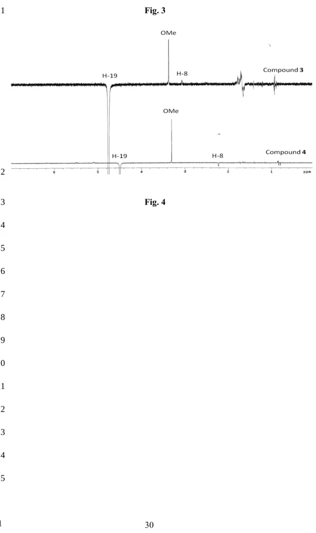
experiments and biogenetic consideration, indicating that compound 3 has similar configurations with the fused ring moiety as compounds 1 and 2. Moreover, NOESY correlations (Fig. 3) of H-21/Me-18, 22, Me-21/H-22, H-24, H-23/H-22, H-22/H-23, H-24, and H-23/Me-27, as well as correlation between H-8 and H-19, suggested that the stereochemistry of C-23 and C-19 adopted R- and S-configuration, respectively. Based on the above evidences, taikugausin C (3) was identified as 19S-methoxy-5,19-epoxycucurbita-6,24-dien-3,23R-diol 23-O--D-glucopyranoside. Compound 4 showed a quasi-molecular ion at m/z 671.4135 ([M+Na]+) in the HRESIMS spectrum and had a same molecular formula C37H60O9 as 3. Their IR, 1H-, and 13C NMR spectroscopic data (Tables 1 and 2) indicated that both compounds 3 and 4 possessed the 5,19-epoxycucurbitane-type triterpene with a sugar moiety and methoxy group. Detailed comparisons of its 1H- and 13C- NMR data with those of compound 3 revealed strong resemblance in all signals except that both the C-8 and H-8 signals in the spectra of 4 were shifted upfield to δC 42.1 and δH 3.04 (t, J = 3.0 Hz), respectively. The 1H-1H COSY and HMBC correlations were similar as well, suggested both compounds 3 and 4 have a similar planer chemical structure. Moreover, their nOe spectra (Fig. 4) showed the different correlations between H-19 and H-8, confirmed the R-configuration of C-19 on compound 4. Thus, compound 4 was established as a C-19 epimer derivative of compound 3, named taikuguasin D. 1 2 3 4 5 6 7 8 9 10 11 12 13 14 15 16 17 18 19
Compound 5 was assigned the molecular formula, C31H50O4, on the basis of a quasimolecular ion peak at m/z 509.3619 ([M+Na]+, Δ = 18) in HRESIMS. Similar to compounds 1 and 2, compound 5 showed absorptions of IR spectrum at 3305 and 1638 cm-1, indicating hydroxyl and olefinic group functionalities, respectively. Detailed inspection of 1H and 13C-NMR spectroscopic data (Tables 1 and 2) indicated the presence of the key structural features of a 5,19-epoxycucurbitane-type triterpene skeleton with a methoxy group (δH 3.20). Furthermore, compound 5 had very close 1H-NMR spectroscopic data with those of 5,19-epoxy-25-methoxycucurbita-6,23-diene-3,19-diol (6) [15]. However comparison of 13C NMR spectra with 5 and 6, the different chemical shifts (C6D5N) of C-19 (δC 107.8, 5; 105.1, 6), C-8 (δC 50.0, 5; 31.6, 6), and C-10 (δC 38.7, 5; 41.2, 6), together with HMBC correlation (Fig. 2) of OMe/C-25 in 5, assigned the OMe group at C-25 in 5 and C-19 in 6, respectively. The NOESY correlation (Fig. 3) between H-19/H-8 in 5 confirmed the S configuration of C-19; whereas it was the R-configuration of the OMe group at C-19 in 6. Based on the above observation, taikuguasin E (5), 19-epimer of 6, was determined as 19S-5,19-epoxy-25-cucurbita-6,23-diene-3,19-diol.
In previous studies, the configurations of C-19 in the known 5,19-epoxycucurbitane compounds was the most assigned as R. From the characteristics of 1H and 13C NMR chemical shifts, it was known the configurations of C-19 in 5,19-1 2 3 4 5 6 7 8 9 10 11 12 13 14 15 16 17 18 19
epoxycucurbitane derivatives. In attempt to understand the relationship between NMR data and the configurations of 19, the chemical shifts for H-8, 8, 9, 10, 11, and 19 are summarized as shown (Table 3 and Fig. 5). The chemical shift of 8 and 11 clearly appear at δC 41-43 ppm and 23-24 ppm, respectively, when the C-19 existed by R form. Furthermore, due to a correlation between H-C-19/H-8 in the NOESY spectrum, the C-19 was determined as S configuration, and the 13C NMR chemical shift of C-8 should be shifted downfield to δC 49-50 ppm. Moreover, the chemical shift of H-8 (δH 2.13-2.40) in 19S-cucurbitane was more upfiled (Δ = 0.5-1.0 ppm) than those of 19R-cucurbitane. Based on the above findings, the C-19 stereochemistry of 5,19-epoxy-19-methoxycucurbita-6,23-diene-3,25-diol and 5,19-epoxy-19,25-dimethoxycucurbita-6,23-diene-3-ol isolated from the leave of
M. foetida may be considered as 19S configuration [15].
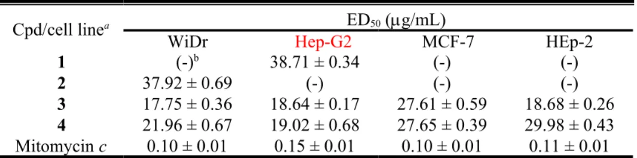
The isolated compounds 1-4 were evaluated for anti-inflammatory activity by their inhibition of LPS-induced nitric oxide production in RAW264.7 macrophages. As shown in Fig. 6, the isolated cucurbitanes 3-4 significantly decreased NO production in a dose-dependent manner and exhibited potent inhibition activities with IC50 of 5.58 and 3.9 g/mL, respectively. In addition, the 70% ethanol extract exhibited potent inhibitory activity against several human tumor cell lines. Therefore, compounds 1-4 were further examined against MCF-7 (breast adenocarcinoma), Hep G2 1 2 3 4 5 6 7 8 9 10 11 12 13 14 15 16 17 18 19
(hepatocellular carcinoma), HEp-2 (laryngeal carcinoma), and WiDr (colon adenocarcinoma) human tumor cell lines. Cytotoxicity results (Table 4) showed that compounds 3-4 had moderate cytotoxicity, while compounds 1-2 were invalid for above tumor. Interestingly, the tumor cytotoxicity results also respond to the cytotoxic effects at a concerntration of 20 g/mL in RAW264.7 cell line. The bioassay results were speculated that the side chain of 3 and 4 could play a crucial role, compared with compounds 1-2.
Materials and Methods
General
Optical rotations were recorded on a JASCO P-2000 polarimeter. Infrared (IR) spectra were measured on a Mattson Genesis II spectrophotometer. The 1D (1H, 13C and nOe) and 2D (COSY, HMBC, HMQC, and NOESY) spectra were recorded on Bruker Advance 400 MHz and/or Varian VMNRS-600 using C5D5N as solvent for measurement. Electrospray ionization mass spectrometry (ESIMS) data were obtained on a Finnigan ion trap tandem mass mass spectrometer. High resolution electronic ionization mass spectrometry (HREIMS) data were measured on a Finnigan MAT-95XL mass spectrometer. Diaion HP-20 (Mitsubishi Chemical Co.), Sephadex LH-20 (GE), and silica gel 60 (Merck, 70-230 and 230-400 mesh) were used for column 1 2 3 4 5 6 7 8 9 10 11 12 13 14 15 16 17 18 19 20
chromatography, and precoated silica gel (Merck, Kieselgel 60 F-254, 1 mm) plates were used for TLC. The spots on TLC were detected by spraying with an anisaldehyde-sulphuric acid solution and then heating at 100 °C. HPLC separations were performed on a Shimadzu LC-6AD series apparatus with a SPD-10A UV detector and Varian 380-LC ELSD detector, equipped with a 250 × 20 mm or 250 × 4.6 i.d. preparative Cosmosil 5C18 AR-II column (Nacalai Tesque, Inc.).
Plant Material
The fruits of Taiwanese M. charantia were provided from Starsci Biotech Co. Ltd. and purchased in Nantou County Taiwan in August 2009. A voucher specimen (NRICM, No. 20090901) has been deposited in the National Research Institute of Chinese Medicine, Taipei, Taiwan.
Extraction and Isolation.
The fresh fruits of M. charantia (wet, 3.6 kg) were sliced and extracted three times with 70% EtOH (7.0 L) at 50 °C for 24 hr, and concentrated under reduced pressure. The EtOH extract (75 g) was subjected to Diaion HP-20 column chromatography (9 × 50 cm) eluting with H2O, 40% EtOH, 70% EtOH, 95% EtOH, and 100% EtOAc (each 2.5 L), respectively, to obtain five fractions (Fr. A-E). The Fr. C (9.8 g) was chromatographed by silica gel column (12 × 36 cm) eluting with a gradient of 1 2 3 4 5 6 7 8 9 10 11 12 13 14 15 16 17 18 19 20
CHCl3/MeOH solvent system to afford four subfractions (Fr. C1 to Fr. C4). Fr. C2 (3.3 g) was subjected by a C18 solid phase extraction (SPE) column with MeOH/H2O solvent system and further purified by reverse phase (RP) HPLC with ELSD detector eluting with 60% MeOH to yield compounds 1 (3.4 mg), 2 (9.4 mg), 3 (50.8 mg), and
4 (27.6 mg). By applying SPE column and RP-HPLC eluting chromatography with
60% MeOH, compounds 5 (1.5 mg) and 6 (7.6 mg) were obtained from Fr. C3 (1.4 g).
Taikuguasin A (1): white amorphous powder. []Com
b i n -68 (c 0.40, MeOH); IR (KBr)
νmax 3385, 2967, 2872, 1733, 1616, 1447, 1372, 1155, 1111, 1081, and 1047 cm-1; 1 H-NMR (400 MHz, pyridine-d5) spectroscopic data see Table 1; 13C-NMR (100 MHz, pyridine-d5) spectroscopic data see Table 2; ESIMS m/z 669 [M+Na]+; HRESIMS
m/z 669.4006 [M+Na]+ (calcd for C37H58O9Na, 669.3981).
Taikuguasin B (2): white amorphous powder. []Com
b i n -88 (c 0.68, MeOH). IR (KBr)
νmax 3408, 2968, 2875, 1639, 1463, 1375, 1260, 1151, 1114, 1085, and 1029 cm−1; 1 H-NMR (400 MHz, pyridine-d5) spectroscopic data see Table 1; 13C-NMR (100 MHz, pyridine-d5) spectroscopic data see Table 2; ESIMS m/z 685 [M+Na]+; HRESIMS
m/z 685.4304 [M+Na]+ (calcd for C38H62O9Na, 685.4294).
Taikuguasin C (3): white amorphous powder; []Comb i n -47 (c 0.10, MeOH); IR (KBr) νmax 3403, 2943, 1447, 1380, 1304, 1274, 1184, 1151, 1082, and 132 cm−1; 1H-NMR 1 2 3 4 5 6 7 8 9 10 11 12 13 14 15 16 17 18 19 20 21 22
(400 MHz, pyridine-d5) spectroscopic data see Table 1; 13C-NMR (100 MHz,
pyridine-d5) spectroscopic data see Table 2; ESIMS m/z 671 [M+Na]+; HRESIMS
m/z 671.4142 [M+Na]+ (calcd for C37H60O9Na, 671.4135).
Taikuguasin D (4): white amorphous powder; []Comb i n -59 (c 0.10, MeOH); IR (KBr) νmax 3396, 2943, 2875, 1449, 1414, 1380, 1157, 1113, 1081, and 1049 cm−1; 1H-NMR (400 MHz, pyridine-d5) spectroscopic data see Table 1; 13C-NMR (100 MHz, pyridine-d5) spectroscopic data see Table 2; ESIMS m/z 671 [M+Na]+, HRESIMS
m/z 671.4135 [M+Na]+ (calcd for C37H60O9Na, 671.4135).
Taikuguasin E (5): white amorphous powder; []Comb i n -38 (c 0.20, MeOH); IR (KBr) νmax 3305, 2987, 1638, 1372, 1224, 1164, and 1089 cm−1; 1H-NMR (400 MHz, pyridine-d5) spectroscopic data see Table 1; 13C-NMR (100 MHz, pyridine-d5) spectroscopic data see Table 2; ESIMS m/z 509 [M+Na]+; HRESIMS m/z 509.3619 [M+Na]+ (calcd for C31H50O4Na, 509.3601).
Measurenmt of nitric oxide production
The mouse macrophage cell line RAW 264.7 was obtained from ATCC (Rockville, MD, U.S.A.) and cultured in Dulbecco’s modified Eagle’s medium (DMEM) containing 5% heat-inactivated fetal calf serum, 100 U/mL penicillin and streptomycin and grown at 37 °C with 5% CO2 in fully humidified air. Cells were plated at a density of 5 × 10⁴ cells/well in 96-well culture plate and stimulated with LPS (1.0 g/mL) in the presence or absence of different concentrations of tested compounds (1~20 μg/mL) for 24 hr simultaneously. Compounds 1-4 were dissolved in dimethyl sulfoxide (DMSO) and further diluted with sterile phosphate buffered saline (PBS). Nitrite (NO2-) accumulation in the medium was used as an indicator of 1 2 3 4 5 6 7 8 9 10 11 12 13 14 15 16 17 18 19 20 21 22 23 24 25
5% phosphoric acid and 0.1% naphthylenediamine in D.D. H2O). NaNO2 was used to generate a standard curve, and azo production was determined by measuring optical density at 540 nm. All experiments were performed in triplicate. NO production by LPS stimulation was designated as 100% for each experiment. Quercetin was used as a positive control.
Cytotoxicity assay
The cytotoxicity of the isolated 5,19-epoxycucurbiranes 1-4 were tested against MCF-7 (human breast adenocarcinoma), Hep G2 (human hepatocellular carcinoma), HEp-2 (human laryngeal carcinoma), and WiDr (human colon adenocarcinoma) cell lines by using the MTT [3-(4,5-dimethylthiazol-2-yl)-2,5-diphenyltetrazolium bromide] colorimetric method based on the described procedures [23]. The cells were cultured in MEM medium. After seeding of cells in a 96-well microplate for 4 hr, 20 µL of sample was placed in each well and incubated at 37 ° C for 72 hr, and then 20 µL MTT was added for 4 h. After removing the medium and putting DMSO (200 µL/well) into the microplate with shaking for 10 min, the formazan crystals were redissolved and their absorbance was measured on a microtiter plate reader (Dynatech, MR 7000) at a wavelength of 550 nm. The IC50 value was defined by a comparison with the untreated cells as the concentration of test sample resulting in 50% reduction of absorbance. Mytomycin c was used as a positive control.
1 2 3 4 5 6 7 8 9 10 11 12 13 14 15 16 17 18 19 20 21
Supporting information
The 1H and 13C NMR spectroscopic of compounds 1-5 are available as Supporting Information.
Acknowledgments
The grants from the National Research Institute of Chinese Medicine and Starsci Biotech CO. LTD. are gratefully acknowledged. The Instrument Center of National Taiwan University, for the HR-ESI-MS measurements, and the National Center for High-Performance Computing, for checking the database, are also appreciated.
References
1 Grover JK, Yadav SP. Pharmacological actions and potential uses of Momordica
charantia: a review. J Ethnopharmacol 2004; 93: 230-132.
2 Abascal K, Yarnell E. Using bitter melon to treat diabetes. J. Altern. Complement Med 2005, 1: 179-184.
3 Bourinbaiar AS, Lee-Huang S. Potentiation of HIV activity of anti-inflammatory drugs, dexamethasone and indomethacin, by MAP30, the antiviral agent from bitter melon. Biochem Biophys Res Commun 1995. 208: 779-785. 4 Jilka C, Strifler B, Fortner GW, Hays EF, Takemoto DJ. In vivo antitumor activity
of the bitter melon (Momordica charantia). Cancer Res 1983. 43: 5151-5155. 5 Ray RB, Raychoudhuri A, Steele R, Nerurkar P. Bitter melon (Momordica 1 2 3 4 5 6 7 8 9 10 11 12 13 14 15 16 17 18 19 20 21
charantia) extract inhibits breast cancer cell proliferation by modulating cell cycle
regulatory genes and promotes apoptosis. Cancer Res 2010. 70: 1925-1931. 6 Ali L, Khan AK, Mamun MI, Mosihuzzaman M, Nahar N, Nur-e-Alam M, Rokeya
B. Studies on hypoglycemic effects of fruit pulp, seed, and whole plant of Momordica charantia on normal and diabetic model rats. Planta Med 1993. 59:
408-412.
7 Kumar R, Balaji S, Uma TS, Sehgal PK. Fruit extracts of Momordica charantia potentiate glucose uptake and up-regulate Glut-4, PPAR gamma and PI3K. J Ethnopharmacol 2009. 126: 533-537.
8 Gurbuz I, Akyuz C, Yesilada E, Sener B. Anti-ulcerogenic effect of Momordica
charantia L. fruits on various ulcer models in rats. J Ethnopharmacol 2000. 71:
77-82.
9 Li CK, Chen HW, Yun WT, Liu KL. Suppressive effects of wild bitter gourd (Momordica charantia Linn. var. abbreviata ser.) fruit extracts on inflammatory responses in RAW264.7 macrophages. J Ethnopharmacol 2009. 122: 227-233. 10 Leung SO, Yeung HW, Leung KN. The immunosuppressive activities of two
abortifacient proteins isolated from the seeds of bitter melon (Momordica
charantia). Immunopharmacology 1987. 13: 159-171.
11 Lee SY, Eom SH, Kim YK, Park NI, Park SU. Cucurbitane-type triterpenoids in
Momordica charantia Linn. J Med Plants Res 2009. 3: 1264-1269.
1 2 3 4 5 6 7 8 9 10 11 12 13 14 15 16 17 18 19 20
12 Murakami T, Emoto A, Matsuda H, Yoshikawa M. Medicinal foodstuffs. XXI. Structures of new cucurbitane-type triterpene glycosides, goyaglycosides-a, -b, -c, -d, -e, -f, -g, and -h, and new oleanane-type triterpene saponins, goyasaponins I, II, and III, from the fresh fruit of Japanese Momordica charantia L. Chem Pharm Bull 2001. 49: 54-63.
13 Begum S, Ahmed M, Siddiqui, BS. Triterpenes, a sterol and a monocyclic alcohol from Momordica Charantia L. Phytochemistry 1997. 44: 1313-1320.
14 Chen JC, Chiu MH, Nie RL, Cordell GA, Qiu SX. Cucurbitacins and cucurbitane glycosides: structures and biological activities. Nat Prod Rep 2005. 22: 386-399. 15 Mulholland DA, Sewram V, Osborne R, Pegel KH, Connolly JD. Cucurbitane
triterpenoids from the leaves of Momordica foetida. Phytochemistry 1997. 45: 391-395.
16 Li QY, Chen HB, Liu ZM, Wang B, Zhao YY. Cucurbitane triterpenoids from
Momordica charantia. Magn Reson Chem 2007. 45: 451-456.
17 Akihisa T, Higo N, Tokuda H, Ukiya M, Akazawa H, Tochigi Y, Kimura Y, Suzuki
T, Nishino H. Cucurbitane-type triterpenoids from the fruits of Momordica charantia and their cancer chemopreventive effects. J Nat Prod 2007. 70:
1233-1239.
18 Chen JC, Liu WQ, Lu L, Qiu MH, Zheng YT, Yang LM, Zhang XM, Zhou L, Li ZR. Kuguacins F–S, cucurbitane triterpenoids from Momordica charantia. 1 2 3 4 5 6 7 8 9 10 11 12 13 14 15 16 17 18 19 20
Phytochemistry 2009. 70: 133-140.
19 Nguyen XN, Phan VK, Chau VM, Ninh KB, Nguyen XC, Lee MH, Bui HT, Tran
HQ, Nguyen HT, Kim YH. Cucurbitane-type triterpene glycosides from the fruits
of Momordica charantia. Magn Reson Chem 2010. 48: 392-396.
20 Pan ZM, Cheng JT, He J, Wang YY, Peng LY, Xu G, Sun WB, Zhao QS, Splendidins A-C, three new clerodane diterpenoids from Salvia splendens. Helv Chim Acta 2011. 94: 417-422.
21 Fukui S, Hochster RM. D-ribo-Hexos-3-ulose, a new dicarbonyl-sugar. J Am Chem Soc 1963. 85:1967-1968.
22 Harinantenaina L, Tanaka M, Takaoka S, Oda M, Mogami O, Uchida M,
Asakawa Y. Momordica charantia constituents and antidiabetic screening of the
isolated major compounds. Chem Pharm Bull 2006. 54: 1017-1021.
23 Zhang LJ, Chiou CT, Cheng JJ, Huang HC, Kuo-Yang LM, Liao CC, Bastow KF,
Lee KH, Kuo YH. Cytotoxic polyisoprenyl benzophenonoids from Garcinia subelliptica. J Nat Prod 2010. 73: 557-562.
1 2 3 4 5 6 7 8 9 10 11 12 13 14 15
Legends for Tables and Figures
Table 1 1H-NMR spectroscopic data of compounds 1-5 (Pyridine-d
5, 400 MHz).
Table 2 13C-NMR spectroscopic data of compounds 1-5 (Pyridine-d
5, 100 MHz).
Table 3 NMR chemical shifts and C-19 configurations for 5,19-epoxycucurbitane.
Table 4 The cytotoxic activities of the isolated compounds from the fruits of M.
charantia.
Fig. 1 The Structures of compounds 1-6 from the fruits of M. charantia. Fig. 2 1H-1H COSY and key HMBC correlations for compounds 1-4.
Fig. 3 Key NOESY correlations for compounds 1-5. Fig. 4 The NOE difference spectra of compounds 3 and 4.
Fig. 5 Difference between several carbon chemical shifts of 5,19-epoxycucurbitanes.
Fig. 6 NO production Inhibition effect of compounds 1-4 on RAW 264.7 macrophage
cells induced by LPS. 1 2 3 4 5 6 7 8 9 10 11 12 13 14 15 16
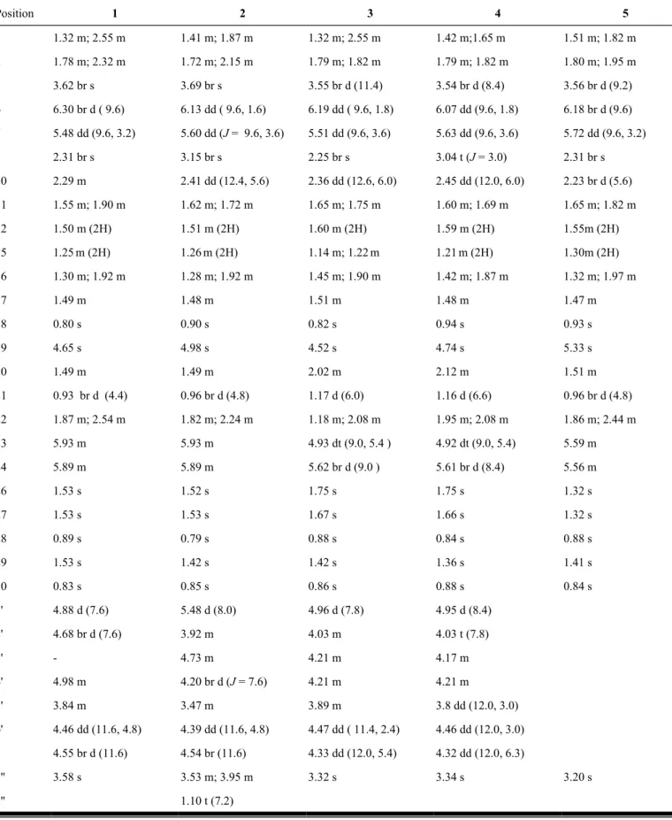
Table 1. 1H-NMR spectroscopic data of compounds 1-5 (Pyridine-d 5, 400 MHz). Position 1 2 3 4 5 1 1.32 m; 2.55 m 1.41 m; 1.87 m 1.32 m; 2.55 m 1.42 m;1.65 m 1.51 m; 1.82 m 2 1.78 m; 2.32 m 1.72 m; 2.15 m 1.79 m; 1.82 m 1.79 m; 1.82 m 1.80 m; 1.95 m 3 3.62 br s 3.69 br s 3.55 br d (11.4) 3.54 br d (8.4) 3.56 br d (9.2) 6 6.30 br d ( 9.6) 6.13 dd ( 9.6, 1.6) 6.19 dd ( 9.6, 1.8) 6.07 dd (9.6, 1.8) 6.18 br d (9.6) 7 5.48 dd (9.6, 3.2) 5.60 dd (J = 9.6, 3.6) 5.51 dd (9.6, 3.6) 5.63 dd (9.6, 3.6) 5.72 dd (9.6, 3.2) 8 2.31 br s 3.15 br s 2.25 br s 3.04 t (J = 3.0) 2.31 br s 10 2.29 m 2.41 dd (12.4, 5.6) 2.36 dd (12.6, 6.0) 2.45 dd (12.0, 6.0) 2.23 br d (5.6) 11 1.55 m; 1.90 m 1.62 m; 1.72 m 1.65 m; 1.75 m 1.60 m; 1.69 m 1.65 m; 1.82 m 12 1.50 m (2H) 1.51 m (2H) 1.60 m (2H) 1.59 m (2H) 1.55m (2H) 15 1.25m (2H) 1.26m (2H) 1.14 m; 1.22m 1.21m (2H) 1.30m (2H) 16 1.30 m; 1.92 m 1.28 m; 1.92 m 1.45 m; 1.90 m 1.42 m; 1.87 m 1.32 m; 1.97 m 17 1.49 m 1.48 m 1.51 m 1.48 m 1.47 m 18 0.80 s 0.90 s 0.82 s 0.94 s 0.93 s 19 4.65 s 4.98 s 4.52 s 4.74 s 5.33 s 20 1.49 m 1.49 m 2.02 m 2.12 m 1.51 m 21 0.93 br d (4.4) 0.96 br d (4.8) 1.17 d (6.0) 1.16 d (6.6) 0.96 br d (4.8) 22 1.87 m; 2.54 m 1.82 m; 2.24 m 1.18 m; 2.08 m 1.95 m; 2.08 m 1.86 m; 2.44 m 23 5.93 m 5.93 m 4.93 dt (9.0, 5.4 ) 4.92 dt (9.0, 5.4) 5.59 m 24 5.89 m 5.89 m 5.62 br d (9.0 ) 5.61 br d (8.4) 5.56 m 26 1.53 s 1.52 s 1.75 s 1.75 s 1.32 s 27 1.53 s 1.53 s 1.67 s 1.66 s 1.32 s 28 0.89 s 0.79 s 0.88 s 0.84 s 0.88 s 29 1.53 s 1.42 s 1.42 s 1.36 s 1.41 s 30 0.83 s 0.85 s 0.86 s 0.88 s 0.84 s 1' 4.88 d (7.6) 5.48 d (8.0) 4.96 d (7.8) 4.95 d (8.4) 2' 4.68 br d (7.6) 3.92 m 4.03 m 4.03 t (7.8) 3' - 4.73 m 4.21 m 4.17 m 4' 4.98 m 4.20 br d (J = 7.6) 4.21 m 4.21 m 5' 3.84 m 3.47 m 3.89 m 3.8 dd (12.0, 3.0) 6' 4.46 dd (11.6, 4.8) 4.39 dd (11.6, 4.8) 4.47 dd ( 11.4, 2.4) 4.46 dd (12.0, 3.0) 4.55 br d (11.6) 4.54 br (11.6) 4.33 dd (12.0, 5.4) 4.32 dd (12.0, 6.3) 1'' 3.58 s 3.53 m; 3.95 m 3.32 s 3.34 s 3.20 s 2'' 1.10 t (7.2) 1 2 3 4 5
Table 2. 13C-NMR
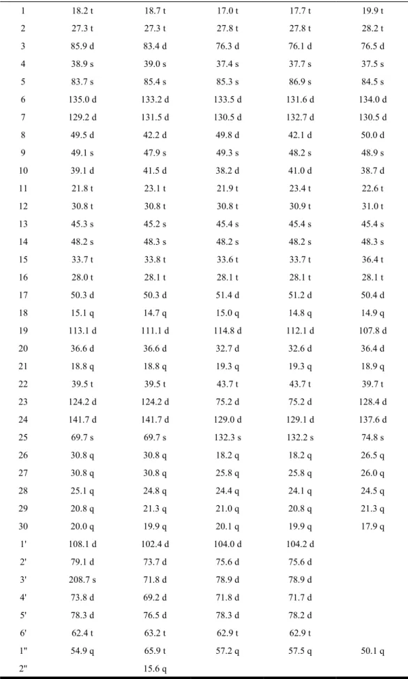
spectroscopic data of compounds 1-5 (Pyridine-d5, 100 MHz).
Position 1 2 3 4 5 1 18.2 t 18.7 t 17.0 t 17.7 t 19.9 t 2 27.3 t 27.3 t 27.8 t 27.8 t 28.2 t 3 85.9 d 83.4 d 76.3 d 76.1 d 76.5 d 4 38.9 s 39.0 s 37.4 s 37.7 s 37.5 s 5 83.7 s 85.4 s 85.3 s 86.9 s 84.5 s 6 135.0 d 133.2 d 133.5 d 131.6 d 134.0 d 7 129.2 d 131.5 d 130.5 d 132.7 d 130.5 d 8 49.5 d 42.2 d 49.8 d 42.1 d 50.0 d 9 49.1 s 47.9 s 49.3 s 48.2 s 48.9 s 10 39.1 d 41.5 d 38.2 d 41.0 d 38.7 d 11 21.8 t 23.1 t 21.9 t 23.4 t 22.6 t 12 30.8 t 30.8 t 30.8 t 30.9 t 31.0 t 13 45.3 s 45.2 s 45.4 s 45.4 s 45.4 s 14 48.2 s 48.3 s 48.2 s 48.2 s 48.3 s 15 33.7 t 33.8 t 33.6 t 33.7 t 36.4 t 16 28.0 t 28.1 t 28.1 t 28.1 t 28.1 t 17 50.3 d 50.3 d 51.4 d 51.2 d 50.4 d 18 15.1 q 14.7 q 15.0 q 14.8 q 14.9 q 19 113.1 d 111.1 d 114.8 d 112.1 d 107.8 d 20 36.6 d 36.6 d 32.7 d 32.6 d 36.4 d 21 18.8 q 18.8 q 19.3 q 19.3 q 18.9 q 22 39.5 t 39.5 t 43.7 t 43.7 t 39.7 t 23 124.2 d 124.2 d 75.2 d 75.2 d 128.4 d 24 141.7 d 141.7 d 129.0 d 129.1 d 137.6 d 25 69.7 s 69.7 s 132.3 s 132.2 s 74.8 s 26 30.8 q 30.8 q 18.2 q 18.2 q 26.5 q 27 30.8 q 30.8 q 25.8 q 25.8 q 26.0 q 28 25.1 q 24.8 q 24.4 q 24.1 q 24.5 q 29 20.8 q 21.3 q 21.0 q 20.8 q 21.3 q 30 20.0 q 19.9 q 20.1 q 19.9 q 17.9 q 1' 108.1 d 102.4 d 104.0 d 104.2 d 2' 79.1 d 73.7 d 75.6 d 75.6 d 3' 208.7 s 71.8 d 78.9 d 78.9 d 4' 73.8 d 69.2 d 71.8 d 71.7 d 5' 78.3 d 76.5 d 78.3 d 78.2 d 6' 62.4 t 63.2 t 62.9 t 62.9 t 1'' 54.9 q 65.9 t 57.2 q 57.5 q 50.1 q 2'' 15.6 q 1 2 3 4
Table 3. NMR chemical shifts and C-19 configurations for 5,19-epoxycucurbitane. Compound C-19 Configuratio n R Group NMR Data (ppm) H-8 C-8 C-9 C-10 C-11 C-19 Taikaguasin A (1)a S OMe 2.31 49.5 49.1 39.1 21.8 113.1 Taikaguasin B (2) a R OEt 3.15 42.2 47.9 41.5 23.1 111.1 Taikaguasin C (3) a S OMe 2.25 49.8 49.3 38.2 21.9 114.8 Taikaguasin D (4) a R OMe 3.04 42.1 48.2 41.0 23.4 112.1 Taikaguasin E (5) a S OH 2.31 50.0 48.9 38.7 22.6 107.8 Kuguacin R a S OH 2.31 50.1 50.0 38.7 28.2 107.8 Goyaglycoside-a a R OMe 3.12 42.3 48.2 41.7 23.3 112.4 Goyaglycoside-b a R OMe 3.13 42.3 48.3 41.7 23.4 112.5 Goyaglycoside-c a R OMe 3.13 42.2 48.1 41.7 23.2 112.3 Goyaglycoside-d a R OMe 3.14 42.2 48.1 41.6 23.3 112.2 Goyaglycoside-g a R OMe 3.12 42.3 48.2 41.6 23.3 112.5 Charantoside I a R OMe 3.13 42.2 48.2 41.7 23.3 112.4 Charantoside II a R OMe 3.13 42.2 48.2 41.6 23.4 112.4 5,19-Epoxy-25-methoxycucurbita-6,23-diene-3,19-diol b R OH 2.82 41.4 48.5 40.6 30.5 105.4 5,19-Epoxyycucurbita-6, 23-diene-3,19,25-triol b R OH 2.82 41.4 48.5 40.6 30.5 105.4 5,19-Epoxy-19-methoxycucurbita-6,23-diene-3,25-diol b S OMe 2.13 49.8 48.9 37.9 30.4 114.7 5,19-Epoxy-19,25-dimethoxycucurbita-6,23-diene-3-ol b S OMe 2.32 49.8 48.9 36.1 30.4 114.7
19R-n-Butanoxy-5,19-epoxycucurbita-6,23-diene-3,25-diol 3-O--glucopyranoside a R OMe 3.10- 42.3 48.1 41.6 23.3 111.3
Momordicoside U b R OMe 2.86 41.7 47.4 41.0 23.0 112.6
Momordicoside O a R OH 3.37 41.8 45.6 41.8 23.3 105.1
Momordicoside W c R OH 2.89 42.7 46.2 42.6 24.0 106.0
(19R,23E)-5,19-Epoxy-19-methoxycucurbita-6,23,25-trien-3-ol a R OMe 2.89 41.7 48.0 40.5 23.2 112.1
aSpectra recoded in pyridine-d
5; bSpectra recoded in CDCl3; cSpectra recoded in CD3OD.
1
Table 4. The cytotoxic activities of the isolated compounds from the fruits of M.
charantia.
Cpd/cell linea ED50 (g/mL)
WiDr Hep-G2 MCF-7 HEp-2
1 (-)b 38.71 ± 0.34 (-) (-)
2 37.92 ± 0.69 (-) (-) (-)
3 17.75 ± 0.36 18.64 ± 0.17 27.61 ± 0.59 18.68 ± 0.26
4 21.96 ± 0.67 19.02 ± 0.68 27.65 ± 0.39 29.98 ± 0.43 Mitomycin c 0.10 ± 0.01 0.15 ± 0.01 0.10 ± 0.01 0.11 ± 0.01
aCell lines: WiDr (Human colon adenocarcinoma), Hep-G2 (Human hepatocellular
carcinoma), MCF-7 (Human breast adenocarcinoma), and HEp-2 (Human laryngeal carcinoma) tumor cell lines. b(-): ED50 > 40 g/mL. Mitomycin c was as positive
control. 1
2
3 4
Fig. 1
1 2
Fig. 2
1 2
Fig. 3 Fig. 4 1 2 3 4 5 6 7 8 9 10 11 12 13 14 15
Fig. 5 1 2 3 4 5 6 7
Fig. 6
1