ANALYTICAL BIOCHEMISTRY 132,342-344 (1983)
Polyamide Thin-Layer
Chromatography
of Phosphorylated
Tyrosine, Threonine,
and Serine
WEN-CHANG CHANG,* MUH-LIN LEE,* CHEN-KUNG CHOU,~ AND SHENG-CHUNG LEE*,’
*Institute of Biological Chemistry, Academia Sinica, *Institute of Biochemical Sciences, National Taiwan University, and TDepartment of Medical Research, Veterans General Hospital Taipei, Taiwan, Republic of China
Received January 3, 1983
One-dimensional thin-layer chromatography on polyamide plates offers an easy and rapid identification of U-phosphotyrosine. The thin-layer plate is developed for 30 min in 5% propionic acid containing 0.013%0.025% sodium dodecyl sulfate. O-Phosphotyrosine, with Rf = 0.54, can be well separated from O-phosphothreonine and O-phosphoserine, which comigrate at R, = 0.72.
KEY WORDS: polyamide; TLC, phosphoamino acids.
Phosphorylation of tyrosine residues has been reported in polyoma T antigens (1) the Abelson murine leukemia virus protein (2), ~~60”” of Rous sarcoma virus (3), the insulin receptor (4), and a membrane protein in Swiss mouse 3T3 cells stimulated by platelet-derived growth factor and epidermal growth factor (5). It appears that phosphorylation of the tyrosine side chain might be a very early step in the transformation by tumor viruses as well as in the expression of some hormonal functions. The phosphotyrosine has been detected by high-voltage electrophoresis on paper (4) or cellulose thin layers (1,3,5), both requiring special apparatus and taking hours to complete an analysis. Here we describe a simple and rapid thin-layer chromatography for the anal- ysis of phosphotyrosine.
MATERIALS AND METHODS
O-Phosphorylated threonine, serine, and tyrosine, 5’-UMP, and 3’UMP were purchased from Sigma Chemical Company (St. Louis,
MO.). Polyamide thin-layer plates were the
’ TO whom correspondence should be addressed: In- stitute of Biological Chemistry, Academia Sinica, P.O. Box 23-106, Taipei, Taiwan, Republic of China.
product of Cheng-Chin Company (Taiwan, R. 0. C.). Propionic acid and sodium dodecyl sulfate (SDS)2 were from E. Merck (Darm- stadt, West Germany). [T-~~P]ATP was pur- chased from New England Nuclear Company (Boston, Mass.).
Polyamide plates, 5 X 15 cm, were loaded with 0.4 pg each of these three phosphoamino acids and developed in 5% propionic acid containing various amounts of SDS for 30 min. After drying, the plates were sprayed with 0.2% ninhydrin solution and dried in an oven (approx 50°C) for 1 min. The spots of amino acids should be located within 5- 10 min; oth- erwise these spots cannot be differentiated from the background color.
RESULTS AND DISCUSSION
The chromatogram of authentic com- pounds is shown in Fig. 1. Phosphotyrosine could be easily separated from the other two phosphorylated amino acids. However, phos- phothreonine and phosphoserine comigrated in this simple system. Several solvents con- taining formic, acetic, and butyric acids in various proportions have been tried and did not offer better results.
* Abbreviation used: SDS, sodium dodecyl sulfate. 0003-2697/83 $3.00
Copyright 0 1983 by Academic Press. Inc. All rights of reproduction in any firm reserved.
POLYAMIDE THIN-LAYER CHROMATOGRAPHY OF PHOSPHOAMINO ACIDS 343
000
0
0
. .
A 6 c 0
FIG. I. Thin-layer chromatogram of phosphoamino ac- ids in 5% propionic acid containing 0.025% SDS. (A) P- Tyr; (B) P-Thr; (C) P&r; (D) mixture of the three.
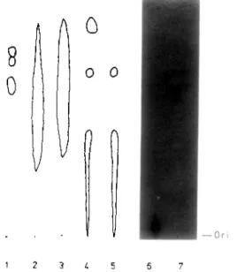
In order to check the potential effect of re- sidual acid and the separation of phospho- tyrosine from other phosphorylated com- pounds commonly found in the hydrolyzed phosphoprotein samples, ATP, hydrolyzed phosphotyrosine, phosphothreonine, phos- phothreonine, S-UMP, and 3’-UMP were run in this system. The results are summarized in Fig. 2. Furthermore, we have isolated a phos- photyrosine-containing polypeptide from mouse thymoma cell EL-4 (S.-C. Lee, manu- script in preparation). The phosphotyrosine from the hydrolyzed sample could be detected in this simple system (Fig. 2). It is clear that phosphotyrosine could be separated from many potentially interfering substances in this system. Inorganic phosphate ran with the sol- vent front (data not shown). Within 30 min, the solvent should reach 1-2 cm from the end of the plate in our system.
The presence of SDS significantly improves the resolution of phosphotyrosine from other
8
0
1 2 3 L 5 6 7
FIG. 2. Thin-layer chromatogram of hydrolyzed phos-
photyrosine, phosphoserine, and phosphoserine (lane I), 3’-UMP (lane 2) 5’-UMP (lane 3) hydrolyzed 3’-UMP (lane 4). hydrolyzed 5’-UMP (lane 5) [y-32P]ATP (lane 6), and hydrolyzed phosphotyrosine-containing phospho- protein from EL-4 (lane 7). Lanes 2-5 were visualized by short-wavelength uv while lanes 6-7 were detected by autoradiography (Kodak XAR-2 X-ray film, -70°C. 20 h exposure). S.F. is solvent front. Ori is origin.
phosphoamino acids in polyamide thin-layer chromatography. The optimal concentration was 0.013-0.025%, as shown in Table 1. Two other detergents, Tween 20 (E. Merck) and
TABLE 1
THE EFFECT OF SDS CONCENTRATION ON RESOLUTION
OF PHOSPHOAMINO ACIDS
SDS (%I
Rj values (average of two experiments)
P-Tyr P-Thr P-Ser 0 0.66 0.75 0.77 0.0063 0.55 0.71 0.7 1 0.013 0.53 0.70 0.71 0.025 0.56 0.73 0.75 0.05 0.59 0.75 0.73
Note. SDS was dissolved in 5% propionic acid as the developing solvent. See Materials and Methods for details. P-Tyr, phosphotyrosine: P-Thr, phosphothreonine; P-Ser, phosphoserine.
344 CHANG ET AL.
NP-40 (Sigma Chemical Co.), were found to cause extensive tailing of the amino acid spots (data not shown). We believe that the ionic detergent nature of SDS makes it useful in this system. However, we have not investigated this point further. Ninhydrin (0.2%) was used for the localization of phosphoamino acids. The amino acid spots could easily be located within 5 min of spraying. Phosphotyrosine appeared to be a light yellowish-brown spot on a blue background even after several days. Polyamide thin-layer chromatography of- fers a simple, fast, economical, and easy-to- perform method for the detection of phos-
photyrosine. Moreover, the study of tyrosine kinase could be facilitated by using this tech- nique.
REFERENCES
I. E&hart, W., Hutchinson, M. A., and Hunter, T.
(1979) Cell 18, 925-933.
2. Witte, 0. N., Dasgupta, A., and Baltimore, D. (1980) Nature (London) 283, 826-83 I.
3. Hunter, T., and Se&on, B. M. (1980) Pm. Nat. Acad. Sci. USA 77, 1311-1315.
4. Kasuga, M., Zick, Y., Blithe, D. L., Crettaz, M., and Kahn, C. R. (1982) Nature (London) 298, 667-
669.
5. Nishimura, J., Huang, J. S., and Deuel, T. F. (1982) Pm. Nat. Acad. Sri. USA 79,4303-4307.