臺灣地區女子划船選手膝關節肌力特徵分析
陳文和* 淡江大學體育組事務處 * 作者姓名:陳文和 通訊地址:251 新北市淡水區英專路 151 號 淡江大學體育事務處 E-mail:[email protected] DOI:10.6167/JSR/2015.24(1)1 投稿日期:2014 年 6 月 接受日期:2015 年 5 月 摘 要 目的:划船傷害特別是腰椎,往往歸因於不良的技術。划船技術包括一系列的在背部,上肢和 下肢,並在這些可能導致異常傷害之間的協調動作。本研究的目的是針對臺灣地區優秀女子划 船選手膝關節的測量與評定,探討其左右腳之肌力是否有不平衡之狀況。。方法:以 Cybex 6000 等速測力系統,針對臺灣地區優秀划船女選手 21 名(平均年齡 20.7 ± 1.0 歲,平均身高=166.1 ± 1.8 公分,平均體重 58.3 ± 4.2 公斤)分三組不同測試速度進行測試,並以單因子變異數分 析與 Tukey’s 事後考驗進行分析。結果:(1)我們觀察到左右腳伸肌和屈肌間最大力矩的顯著 差異,但是伸/屈肌比在不同速度之間的差異不顯著,因此左右腳的肌肉力量是均衡的(2)在不 同腳之間的伸肌和屈肌產生最大力矩時的關節角度分別為約 60 度,這顯示今後的訓練可以使 選手在一個特定的角度,使腿部達到最大的力量。(3)在雙腳的伸肌和屈肌的延遲時間,並未 因角速度不同而有顯著差異,這意味著選手於划船不能延長的力的作用時間。因此,每次划槳 的效率不能被累積增加划程。此外在角速度 60°/s 時具有較長時間的等長肌力,但在划船最後 的快速衝刺階段,無法提供更長時間的力量,因此也必須加強肌肉耐力。結論:划船選手伸肌 最大力矩顯著比屈肌強,且左右腳呈現平衡狀態。此外在力量耐力上,也需加強比賽最後階段 肌耐力,維持快速伸屈肌轉換時間,不因疲勞而減緩作用時間。其次在速度力量的特徵方面並 無特別顯著,也無法將有效用力的時間拉長,顯示出國內選手在速度力量的訓練上明顯不足。 關鍵詞:等速肌力、西式划船、不對稱I. Introduction
Competitive rowing is an extremely technical and physically demanding ac-tivity. When competing in similar events, the rowing time for women on the water is approximately 10% longer than that for men (Secher, 2000). Based on the world records for 2000-m rowing on an ergometer, the winning time for female rowers is approximately 16% slower than that for male rowers. It is known that nearly all muscles are involved in rowing (Secher, 2000), and rowing performance is related to the size of the leg muscles (Yoshiga, Yashiro, Higuchi & Oka, 2002). Female athletes are generally lighter than their male counterparts (Ingjer, 1991; Jensen, Johansen, & Secher, 2001), and the rowing performance of women is in-fluenced by their small body size. All other factors remains equal for rowers, who sustain greater net propulsive forces (or strength), achieve faster boat speeds. (Hofmijster, Landman, Smith, & Knoek van Soest, 2007). The increased partici-pation of women in athletics has prompt-ed a greater awareness of their musculo-skeletal performance characteristics.
The activated muscle mass during rowing is larger than during leg exercise, because rowing engages both the upper and lower body musculature(Secher,
2000). The agonist-antagonist disparity was explained as being due to the repet-itive load demanded on the quadriceps group during the power phase of rowing stroke, in which a fast leg extension is initiated (Nolte, 2011). However, the repetitive cyclic action of rowing may predispose the rower to low back injury. In this context, few studies have inves-tigated the relationship between imbal-anced muscle strength and occurrence of injuries. Kramer, Leger, & Morrow (1991) identified an asymmetry in the isokinetic strength of quadriceps muscle group in oarsmen. Later on, Parkin, Nowicky, Ru- therford, & McGregor (2001) used the EMG of rowers to investigate the asym-metric patterns of muscle activity be-tween left and right erector spinae mus-cles during extension, which was sub-stantially related to the rowing side. These observations could relate to the high incidence of low back pain (LBP) in oarsmen. Koutedakis, Frischknecht, & Murthy, (1997) noted a low hamstring to quadriceps strength ratio, suggesting weakness of the hamstring muscle groups in rowers with LBP. This study further suggested that the abnormal hamstring- quadriceps ratio might interfere with lumbo-pelvic rhythm, leading to an in-creased stress on the spine. Holcolmb (2007) found that H/Q ratio and injury
to the hamstrings and anterior cruciate ligament (ACL) were inversely linked. The dynamic strength of quadriceps mus-cle is crucial for the stability, movement of human body, and for effective sports activity. Reduced dynamic strength of the quadriceps muscle has been associated with knee injury in athletes (Ekstrand & Gillquist, 1983; Thomee, Renstrom, Kar- lsson, & Grimby, 1995), and strengthening exercises of quadriceps muscle is crucial for preventing and rehabilitating the knee injuries (Grimby, 1992).
Studies of Impellizzeri (2008) and Hewett (2006) were similarly explored the strength imbalance ratio as a means to monitor and rehabilitate the injury. In contrast, Parkin (2001) reported no sig-nificant lower extremity muscle imbal-ance, whilst studying on elite national and junior oarsmen. The authors prof-fered the benefits of prophylactic as-sessment and rehabilitation as a reason for alternate findings. Strength ratio and injury have shown variability across gender in sportsmen and women. The detailed studies on whether one gender has a greater tendency to develop mus-cle imbalance was undertaken. Hewett (2006) indicated that at higher speeds i.e. approaching those naturally seen in sport, women showed a greater signifi-cant dip in H/Q ratio. This was
attribut-ed to the hamstring component, which showed lack of adaption in females. At low speeds, no significant difference was ditected between the sexes. Differ-ences across gender have been attributed to a gender specific physiology, training regimes and ability to recover(Parkin, 2001).
The efficiency, safety and effec-tiveness of strength-training programs are paramount for the sports condition-ing. Therefore, identifying the optimal doses of training variables elicits maxi-mal gains in muscular strength per time unit, and reduces the risks of overtrain-ing or overuses injuries. Athletic trainers and sports physical therapists emphasize injury prevention by identifying under-lying the deficits in strength and bilat-eral and reciprocal muscle-group strength relationships. Rowing injuries, particular-ly of the lumbar spine, are often attributed to poor technique. Rowing technique comprises a series of coordinated move-ments between the back upper limbs and lower limbs, and abnormalities in these may lead to injury. Therefore, we pro-vided information whether the asym-metric strength of leg musculature is more prominent for Taiwanese female rowing athletes than other female row-ing athletes. The aim of this study was to test the hypothesis that strength of leg
musculature is symmetrical with respect to knee isokinetic. Therefore, we ex-plored the information whether the asymmetric strength of leg musculature is more prominent for Taiwanese female rowing athletes.
II. Methodology
A. Participants
Twenty-one elite female college athletes (aged 20.7 ± 1.0 years, height= 166.1 ± 1.8 cm, weight=58.3 ± 4.2 kg), who had 7.0 ± 1.8 years rowing experience were participated in this study. Rowers were selected from the national or youth team, had a history of winning medals at the Asian Games, Asian Championships, Asian Youth Championships or other in-ternational competitions. Informed con-sent with no known knee pathologies was obtained from all participants prior to their participation.
B. Dynamometer set up
Knee extensor and flexor isokinetic peak torque assessments were conducted using an isokinetic dynamometer (Cybex 6000 model, Division of Lumex, Inc. Ronkonkoma, NY, USA). Subjects were positioned and stabilized in accordance with the manufacture’s recommenda-tions (Cybex 6000, 1993). (Fig. 1). The torque measurements obtained with
iso-kinetic devices not only reflect the ten-sion a muscle is generating, but also re-flect mechanical factors (such as the moment arm) at the point in the range of motion where torque is being measured. Each subject's functional range of motion was set electronically between 0-110° of knee flexion to prevent hyperextension and hyperflexion (Table 1). Gravity correction was made for limb weight on torque measurement. The participant was instruct- ed to grab stabilization handles during the test, and fully extend the leg and then flex it as hard and fast as possible (one maxi-mal extension followed immediately by a reciprocal maximal flexion). Torque, position and angular velocity data were recorded from the isokinetic dynamom-eter with a sampling rate of 100 Hz. For concentric and eccentric strength trials, the software calculated a large number of parameters, but we retained only those commonly used in isokinetic studies, name- ly the peak torque, the average work, the average power and the angle of peak torque (Brown, 2000; Wrigley & Strauss, 2000).
Table 1
The range of motion in different legs (unit: degree)
ROM 60./s 120./s 180./s Right Left 109.00 ± 11.72 109.57 ± 8.46 111.14 ± 8.36 110.86 ± 10.68 109.29 ± 7.14 116.43 ± 8.10 C. Test protocol
Maximal concentric quadriceps and hamstring muscle contraction strength data was obtained by measuring the maximal force and moment (torque) during isoki-netic knee extension and flexion move-ments set at a fast and slow angular veloc-ity. The mean peak force ± SD was sub-sequently calculated from the repetitions.
The participants performed five con-tinuous repetitions at 60°/s, 120°/s, and 180°/s. Participants were instructed to take a 5-min rest between each test speed, and a 30-min rest between each test con-dition to minimize the effect of fatigue on the test muscle (Perrin, 1993). Ob-tained data were gravity-corrected to account for the weight of each subject’s leg and the dynamometer input arm
(Fillyaw et al., 1986).
Following a warm-up session in-volving submaximal exercise, concen-tric measurements involved three con-tinuous, reciprocal (maximal) knee ex-tensions–flexions, were performed by participants at three preset constant an-gular velocities in the following order 60°/s, 120°/s and 180°/s (slow to fast) (Wilhite et al., 1992) with their hips and knees flexed to 90 and their backs sup-ported. Participants dominant lower ex-tremities were secured to the arm of the dynamometer with straps at both the thighs and ankles, and their trunks were fixed to the chair with hook-and-loop straps (Fig. 2). Whatever the action mode and the velocity, subjects recov-ered passively for 60s between series of measurements.
D. Operative Definitions 1. Peak Torque (PT)
Peak torque is the maximum torque production during an extension/flexion. Peak torque values are the isokinetic pa-rameters most frequently used to assess the human muscle performance and constitute the greatest force or torque produced in the range of motion. This can be determined within each repetition or the entire set. Peak torque indicates the muscle’s maximal capability of de-veloping force, which is equivalent to a one-repetition maximal isotonic-strength test. Peak torque is an absolute value.
2. Joint Angle (JA)
The joint angle at peak torque is the point in the range of motion where the peak torque first occurs.
3. Time to Peak Torque (TPT)
The time to peak torque is the time from the beginning of the torque devel-opment until the point where peak torque is first developed. TPT data from electri-cally stimulated isometric muscle actions have been used to examine the influence of muscle contractile properties in streng- th development (Blimkie, 1989).
4. Time Peak Torque Held (TPTH)
The time peak torque held is the time that the peak torque is maintained.
5. Reciprocal Delay (RD)
Reciprocal delay is the time re-quired to reverse the limb direction, which may be associated with prolonged contraction of the agonist at the end of movement in each direction (Sahrmann, 1977). Watkins, Harris, & Kozlowski (1984) defined the reciprocal delay time as the time from the end of motion in one direction to the initiation of motion in another direction.
6. Delay Time (DT)
Delay time is the time from the be-ginning of a motion until the bebe-ginning of torque development, and may be an-other manifestation of slowed and irreg-ular recruitment patterns (Rosenfalck, 1980; Angel, 1975).
E. Data analysis
Peak torque was determined for the knee extensors and flexors at each test velocity, and peak torque was identified as the highest torque value among the sampled repetitions. Data presented as the mean (standard deviation) with the range. SPSS was used for all statistical calculations (Version 10.0 for Windows, SPSS, Chicago, IL, USA). Comparisons were performed using a one-way analy-sis of variance with Tukey’s post hoc validation in different leg and speed. Independent-samples t-tests were used
to compare the differences in H/Q ratio between extensor and flexor muscle peak torque ratio in different leg. All tests were two-sided and the level of significance was set at p <.05.
III. Results
Isokinetic dynamometry enables the rapid and reliable quantification of force or torque. If the force and distance of a given muscle contraction are known, then the amount of tension produced by the muscle may be expressed as work. If the required time to produce work is known, then the ability of the muscle to generate power may be determined. Following is a schematic representation of the torque and angle curves of the
performance of the quadriceps and ham-strings, based on the torque, joint-angle, and time data obtained at the test speeds of 60°/s, 120°/s and 180°/s.
A. Torque data 1. Peak Torque
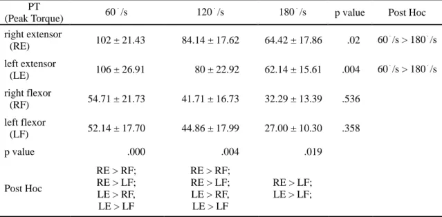
Table 2 shows the peak torque (PT)data of the extensors (quadriceps) and flexors (hamstring) of the two legs in various speeds. The result shows that the peak torque of the extensors mark-edly decreases as the angular velocity increases. However, this does not occur for the flexors, and at the same speed the peak torque of the extensors was larger than that of the flexors.
Table 2
Extensor and flexor muscle peak torque for right and left legs at different speeds
PT
(Peak Torque) 60 .
/s 120./s 180./s p value Post Hoc right extensor (RE) 102 ± 21.43 84.14 ± 17.62 64.42 ± 17.86 .02 60 . /s > 180./s left extensor (LE) 106 ± 26.91 80 ± 22.92 62.14 ± 15.61 .004 60 . /s > 180./s right flexor (RF) 54.71 ± 21.73 41.71 ± 16.73 32.29 ± 13.39 .536 left flexor (LF) 52.14 ± 17.70 44.86 ± 17.99 27.00 ± 10.30 .358 p value .000 .004 .019 Post Hoc RE > RF; RE > LF; LE > RF, LE > LF RE > RF; RE > LF; LE > RF, LE > LF RE > LF; LE > LF; *p <.05 (unit: newton-meter)
There are a variety of ways of as-sessing thigh agonist-antagonist muscle balance, of which hamstring/quadriceps (H/Q) ratio comparison was one. The Coombs and Garbutt (2002) review of methods found that the most commonly quoted protocol was the concentric ham- string/quadriceps (Hcon/Qcon) peak moment ratio. Limitations suggested by this review were that the peak moments did not account for the function of the antagonist component in a contraction. Furthermore, as various prospective and retrospective studies have shown a rela-tionship between lower limb strength imbalance and hamstring or knee inju-ries (Dvorak & Junge, 2000; Croisier et al., 2002; Dauty et al., 2003; Devan et al., 2004), some studies proposed the use of preseason screening of unilateral and bilateral strength imbalance among healthy subjects is a valid approach to identify athletes at increased risk of in-curring lower limb injuries during train-ing and competition (Croisier, 2004).
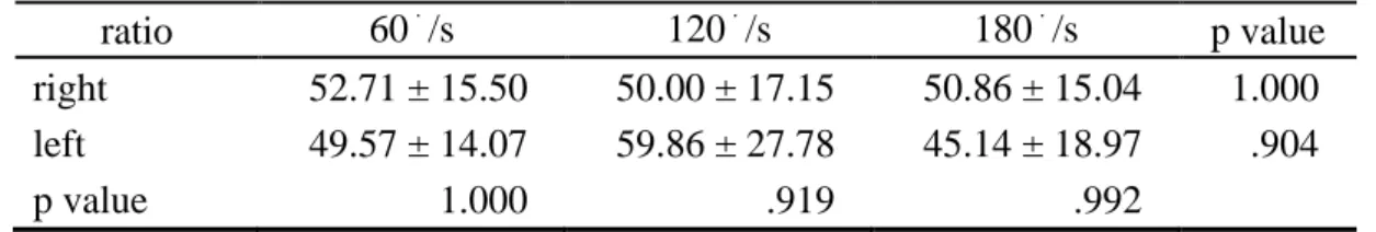
Table 3 shows the ratio of the peak torque of the extensors and flexors of both legs. The peak torque ratio produced
during flexion to the peak torque was produced during extension. The require-ment of this ratio varies in different sports. Previous studies have observed that the peak torque ratio of the ham-strings to the quadriceps increases as the speed increases (Davies, Heiderscheit, & Brinks, 1985). Coombs and Garbutt (2002) hypothesized that the H/Q ratios would show a greater correlation at higher angular velocities than lower. This was because, it would be more rep-resentative of the velocities achieved during the bulk of rowing training, thereby appropriately conditioning the muscles. In addition the level of ham-string co-activation was dependant on angular velocity and linked to type of training; thereby implying the hamstring component increased with increasing speed in order to provide joint protection. However, in this study, the ratio of the extensors to the flexors did not increase as the angular velocity increased. We also observed no obvious differences between the ratios of the extensors to the flexors at various speeds. The muscle strength of both legs was balanced.
Table 3
Extensor and flexor muscle peak torque ratio for right and left legs
ratio 60./s 120./s 180./s p value
right 52.71 ± 15.50 50.00 ± 17.15 50.86 ± 15.04 1.000 left 49.57 ± 14.07 59.86 ± 27.78 45.14 ± 18.97 .904
p value 1.000 .919 .992
B. Joint-angle data
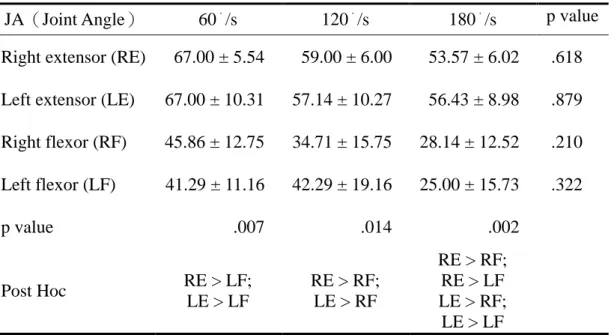
The angle of peak torque was found to be highly variable in individual subjects. When producing the peak torque, the body position is crucial. For instance, the work of treading on a foot stretcher is op-timal for a rowing athlete, when the seat slides forward and the knee is bent for-ward to generate the peak torque of the lower extremity, thereby forming a joint angle that approximates the angle of the lower extremity after the stroke is per-formed. Table 4 indicates that the peak torques of both legs appeared to be similar
degrees at any angular velocity. However, the joint angle at the peak torque of the extensor was larger than that of the flexor at the same speed.
C. Time data
1. Time to Peak Torque
Table 5 shows the time from the start of muscular contraction to the point of highest torque development. This value is an indicator of the muscle’s functional ability. The shorter the time is, the quicker the peak torque appears, and vice versa. The optimal time to peak torque is the same as the force-generating time.
Table 4
Extensor and flexor muscle Joint Angle at Peak Torque for right and left legs
JA(Joint Angle) 60./s 120./s 180./s p value
Right extensor (RE) 67.00 ± 5.54 59.00 ± 6.00 53.57 ± 6.02 .618 Left extensor (LE) 67.00 ± 10.31 57.14 ± 10.27 56.43 ± 8.98 .879 Right flexor (RF) 45.86 ± 12.75 34.71 ± 15.75 28.14 ± 12.52 .210 Left flexor (LF) 41.29 ± 11.16 42.29 ± 19.16 25.00 ± 15.73 .322 p value .007 .014 .002 Post Hoc RE > LF; LE > LF RE > RF; LE > RF RE > RF; RE > LF LE > RF; LE > LF *p <.05 (unit: degree)
Table 5
Extensor and flexor muscle Time to Peak Torque for right and left legs at different speeds
TPT (Time to Peak Torque)
60./s 120./s 180./s p value Post Hoc
Right extensor (RE) 0.4400 ± 0.1901 0.3586 ± 0.0564 0.2871 ± 0.0275 .872 Left extensor (LE) 0.5257 ± 0.2405 0.2857 ± 0.1351 0.1957 ± 0.0830 .025 60./s > 180./s Right flexor (RF) 0.5057 ± 0.2259 0.4100 ± 0.0396 0.2957 ± 0.0550 .482 Left flexor (LF) 0.5743 ± 0.3574 0.4071 ± 0.1830 0.2071 ± 0.0907 .007 60./s > 180./s p value .943 1.000 .997 *p <.05 (unit: second)
When comparing the extensor and the flexor of the same leg, the time to peak torque was decreased when the an-gular velocity was increased. In the right flexor and left flexor, the time to peak torque at 60°/s was longer than that at 180°/s. When comparing the extensors and flexors of both legs under the same angular velocity, no significant differ-ences were existed among the statistics.
2 .Time Peak Torque Held
Table 6 shows the time of the peak torque data. When comparing the ex-tensor and flexor of the same leg, the time of the peak torque was decreased as the angular velocity increased. The time peak torque held at 60°/s was longer than that at 180°/s for left flexor. Besides, no significant differences were observed among the statistics of exten-sors and flexors of both legs under the same angular velocity.
Table 6
Extensor and flexor muscle Time Peak Torque Held for right and left legs at different speeds
TPTH (Time Peak Torque Held)
60./s 120./s 180./s p value Post Hoc
Right extensor (RE) 0.0529 ± 0.0189 0.0243 ± 0.0054 0.0214 ± 0.0069 . 372 Left extensor (LE) 0.0657 ± 0.0276 0.0329 ± 0.0111 0.0214 ± 0.0076 .051 Right flexor (RF) 0.0571 ± 0.0180 0.0171 ± 0.0125 0.0143 ± 0.0079 .051 Left flexor (LF) 0.0757 ± 0.0663 0.0343 ± 0.0190 0.0100 ± 0.0100 .000 60./s > 180./s p value .813 1.000 .997 *p <.05 (unit: second) 3 .Reciprocal Delay
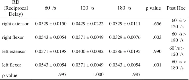
Table 7 shows data of the time needed for the body to shift from sion to flexion or from flexion to exten-sion. It is defined that smaller number indicates a better score. When comparing
the extensor and the flexor of the same leg, the time was decreased as the angu-lar velocity increased. The difference was not large, but the reciprocal delay at 60°/s was longer than those at 120°/s and 180°/s for the right flexor and left flexor. Table 7
Extensor and flexor muscle Reciprocal Delay for right and left legs at different speeds
RD (Reciprocal
Delay)
60./s 120./s 180./s p value Post Hoc
right extensor 0.0529 ± 0.0150 0.0429 ± 0.0222 0.0329 ± 0.0111 .656 60 . /s > 120./s right flexor 0.0543 ± 0.0054 0.0371 ± 0.0049 0.0329 ± 0.0076 .003 60 . /s > 180./s left extensor 0.0571 ± 0.0198 0.0400 ± 0.0082 0.0386 ± 0.0195 .990 60 . /s > 120./s left flexor 0.0543 ± 0.0054 0.0371 ± 0.0049 0.0343 ± 0.0054 .001 60 . /s > 180./s p value .997 1.000 .987 *p <.05 (unit: second)
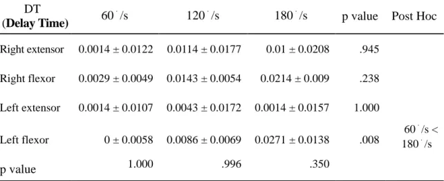
4. Delay Time
Table 8 shows the time interval between the onset of motion into exten-sion and the initiation of the quadriceps torque curve. A smaller number indi-cates a better score. When comparing the extensor and the flexor of the same leg, the time was increased as the angu-lar velocity increased, but the difference was not large. For the left flexor, the de-lay time at 60°/s was shorter than that at 180°/s.
Direct feedback controls the ath-lete’s muscles directly and the delay time should be in a range of tenths or
hundredths of a second. According to Watkins et al., (1984) terminology we consider the quadriceps femoris muscle force delay time, the reciprocal delay time, the delay time before the ham-string muscle contraction reaches the selected speed of the isokinetic motion, and the time to peak torque of the ham-string muscles. The increased delay time must be accounted for predicting the muscle response times during standing or else total collapse of the knee may result. Clearly, different activation con-stants should be used when modeling a wide variety of activities.
Table 8
Extensor and flexor muscle Delay Time for right and left legs at different speeds
DT
(Delay Time) 60 .
/s 120./s 180./s p value Post Hoc
Right extensor 0.0014 ± 0.0122 0.0114 ± 0.0177 0.01 ± 0.0208 .945 Right flexor 0.0029 ± 0.0049 0.0143 ± 0.0054 0.0214 ± 0.009 .238 Left extensor 0.0014 ± 0.0107 0.0043 ± 0.0172 0.0014 ± 0.0157 1.000 Left flexor 0 ± 0.0058 0.0086 ± 0.0069 0.0271 ± 0.0138 .008 60./s < 180./s p value 1.000 .996 .350 *p <.05 (unit: second)
IV. Discussion
The specialized strength of rowing athletes involves neuromuscular capa-bilities to overcome the drag force of stroke completion. Specialized strength training is critical in rowing because it primarily influences the achievement of the athletes, and consequently valued in Taiwan and other countries. Although the training methods are varing, the the-ory remains the same to develop muscle capability to overcome the drag.
Isokinetic dynamometry objective-ly represents the function of the meas-ured muscle and is valuable in the eval-uation of rehabilitation, sports medicine and the application of the study in exer-cise training. Both the position (degree) of the measured body and the time range influence the result of isokinetic dyna-mometry. The following sections clearly explained the results of this study.
A. Analysis of the peak torque pa-rameter
Numerous studies have proven that peak torque decreases as speed increases. Sucdder (1980) indicated that it takes a certain amount of time for the muscle fibers to produce tension. A faster exer-cise speed is the quicker the muscle contracts, which causes fewer muscle fibers to be collected, and generates less
force. From our study, we proved that the peak torque was decreased as the angular velocity was increased.
Rowing requires a high standard of athletic physical characteristics. The net result of the system equations of motion is that system velocity is determined by the difference between the propulsive force applied and the drag force acting on the system (Baudouin, Hawkins, & Seiler,, 2002). Therefore, rowers, both as individuals and members of a crew should attempt to maximize their force input to the system, while minimizing their contribution to drag. The average power of athletes in boar race was re-ported to be between 250 and 550 W (Steinacker, 1993). Certain well-trained rowers could generate considerably more power than athletes in other endurance athletics. The average power measured using an ergometer in a 6-min test of world-class rowers’ was 420 W. Besides, the average power measured in another 40-s test was between 550 and 780 W (Steinacker, 1993). Rowing is a sport that requires a high level of force and muscle endurance. We emphasized that long-term training enables the extensors of both legs to develop quickly. Moreo-ver, because both legs must generate the force simultaneously, the muscle strength of the left and right leg was balanced.
In speed athletics, athletic trainers and sports medicine specialists focused on the peak torque ratio of the quadri-ceps and hamstrings. Steindler (1955) suggested that knee extensor force should exceed flexor force on a 3:2 scale i.e. 66% threshold. The implications were higher the H/Q ratio above this threshold, the greater the balance and stability; and vice versa. Interestingly, subsequent novel research findings re-vealed a variety of quoted values rang-ing from 0.43 to 0.9, dependrang-ing on test population, angular velocity, type of training undertaken and test position (Nolte, 2011; Wyatt and Edwards, 1989; Heiser et al., 1984; Coombs and Garbutt, 2002; Kannus, 1994).
Previous studies have linked bilat-eral strength imbalance with injury. Knapik et al., (1991) found that athletes had a higher injury rate with a knee flexor or hip extensor imbalance of 15% or more on either side of the body. Side-to-side strength imbalance sug-gested as a risk factor for anterior cruci-ate ligament injury in female athletes (Cowley, Ford, Myer, Kernozek & He- wett, 2006; Hewett, Stroupe, Nance, & Noyes, 1996; Myer, Ford & Hewett, 2004). While hamstring injuries were shown to be associated with low ham-string muscle side-to-side peak torque
ratio at 60 deg•s-1 (Orchard, Marsden, Lord, Garlick, 1997). Parkin et al., (2001) measured H/Q ratio in elite rowers and reported no significant findings of patho- logical ratio compared to injury rates, which could be explained by the regular use of testing as a means of adapting training schedules to prevent these im-balances.
While acknowledging the limita-tions with past research, strength train-ing has been used to correct the muscle imbalances, which in specific cases ap-peared to be useful in the reduction of pain associated with rowing. For exam-ple, the number of training days lost due to low back pain was reduced once the relatively excessive quadriceps strength (i.e. a knee flexion to extension ratio less than 45%) was addressed by a specific hamstring strengthening program over 6-8 months (Koutedakis & Frischknecht, 1997). However, it is unclear whether changes in muscle imbalances led a subsequent improvement in rowing technique, or whether similar outcomes could have been achieved by allocating an equivalent time to practice of a re-vised rowing technique. Other predis-posing factors for back injury, including a low hamstring-to-quadriceps strength ratio (Koutedakis et al., 1997) and strength asymmetries in the left and
right erector spinae muscles during ex-tension (Parkin et al., 2001). Conse-quently, strengthening the extensor/ flexor ratio of the knee joint may effec-tively prevent rowers from experiencing LBP.
In the combined extension of the hip and knee, either supine or upright, the quadriceps insertion and origin change their roles, the tibial tuberosity becomes the stable anchor, and the pel-vic and femoral origins of the quadri-ceps move in an approximating mode. For instance, in stair climbing, the leg on the higher step becomes stable end of the chain, while the thigh, which is the next link, turns in an extensory mode from the knee. This configuration affects the shear loading at the knee joint. The quadriceps pull up the femoral condyles that roll forward, but the tibia cannot extend because the foot is planted on the step. This force system may result in a posterior shear. The extensor/flexor ratio deficiency may also be a crucial factor leading to knee injury. When comparing the peak torque of extensors and flexors of both legs, we observed a marked dif-ference among the statistics, but no ob-vious difference between the ratio of the extensors to the flexors at various speeds, and muscle strength of both legs was balanced.
B. Analysis of the joint-angle pa-rameter
In the study of concentric isokinet-ic speed and contracting speed of row-ing, Davies (1987) divided the speed of knee joint measurement into three cate-gories: 60°/s as slow exercise, 180°/s as mid-speed exercise and 300°/s as fast exercise. Zhang and Zhang (2003) indi-cated that fast speed (typically approxi-mately 150°/s) plays a crucial role in the first stage of rowing. In an intact human, the angle at which the maximum torque is generated around a joint is likely a function of both the length-tension rela-tionship and the muscle’s mechanical advantage. For example, maximum knee extension torque typically occurs at ap-proximately 60° of knee flexion (con-sidering knee extension as being 0°). In this study, the extensor and flexor mus-cle joint angles at peak torque in differ-ent legs were approximately 60 degrees, suggesting that future training can ena-ble athletes to implement the greatest strength at a specific degree such that the leg force achieves the greatest torque.
Assuming that the resting length of the hamstrings occurs when the hips and knees are at 0°, this decrease in torque as knee flexion progresses from 30° to 90° is consistent with the muscle
length-tension relationship. However, the improved hamstring mechanical ad-vantage when the knee is flexed is in-sufficient to compensate the decreased force production. Tsai (1997) considered that the peak torque of excellent athletes to be larger than that of average athletes. The peak torque of excellent athletes appears earlier than it does in average athletes, and the angular velocity of the knee joint is larger in excellent athletes than average athletes.
C. Analysis of the time parameter
The peak torque of the extensors and flexors of both legs appears simul-taneously, and the time quickens as the angular velocity increases. Our study results indicated that the degree of the knee joint at the peak torque, and the time to reach the peak torque was de-creased as the speed inde-creased. During concentric contraction of the extensors, the peak torque appears earlier as the speed increases. Regarding the flexors, the speed influences only the time to peak torque. The rate of force develop-ment is related to the contractile speed of a muscle, which is highly depends on the degree of motor unit activation (Asai & Aoki, 1996). Therefore, numerous researchers have suggested the rate of torque development or acceleration time to be synonymous with the time
re-quired to reach peak torque during iso-kinetic actions (Blimkie, 1989; Davies et al., 2000). TPT can also be used to estimate the rate of energy turnover and fiber-type composition (Hosking et al., 1978).
The extended TPT and time peak torque held recorded from the quadri-ceps femoris muscles of rowers may be related to slowed and irregular recruit-ment patterns (Rosenfalck, 1980; Angel, 1975). Green and Wilson (2000) deter-mined the possibility of using magnetic resonance imaging (MRI) to define the pattern of muscle recruitment in a spe-cific sport (rowing), and investigated the possible differences in this pattern among athletes with various experiences. Their study further suggested that trained ath-letes recruit selected muscle groups to perform a given task, which they con-duct more efficiently than untrained or less experienced athletes. If the exten-sors and flexors shift quickly in a com-petitive long-term rowing competition, the muscles perform efficiently in the last stage of a 2000-m rowing competi-tion. In our study, for the delay time of extensors and flexors of both legs, the fast speed was not significantly different from the slow speed, which means row-ers cannot prolong the time of force. Hence, the efficiency of each stroke
cannot be accumulated to increase the distance of the stroke.
Isometric strength of the quadri-ceps and hamstrings was measured in order to demonstrate fatigue of the knee musculature following the fatigue pro-tocol. As previously mentioned, an ath-lete began to display a reduction in mo-tor control as they become fatigued (Worrell, 1994). This results in a greater role of the stabilizer muscles, of which the hamstrings plays a primary role for the lower body (Baratta, Solomonow, Zhou, Letson, Chuinard & D’Ambro- sia, 1988). The greater loading, along with the damage that has occurred from numerous eccentric contractions may result in a soft tissue injury (Thelen, Chumanov, Best, Swanson & Heider- scheit, 2005). To overcome the effect of fatigue on injury, we have to reduce the level of fatigue that the athlete encoun-ters. While this could theoretically be done by reducing the playing time or implementing more breaks in play, and the optimal method would be to ensure the higher fitness levels of the players. This would not only reduce their risk of injury, but also enhance their sporting performance. In this study, the time peak torque held at 60°/s was longer than that at 180°/s, but unable to provide long-er-isometric strength at the final sprint
stage of rowing. Thus, muscular endur-ance must be strengthened in the final stage of a competition.
V. Conclusion
A rowing stroke involves the move-ment of the extremities and trunk. Ap-proximately 70% of human muscles are involved in the work-generating phase (Steinacker, 1993). The explosive force of the lower extremity is the immediate power of rowing. The extremities and trunk function as the support. In this study we showed that (1) The peak torque of extensors and flexors of both legs were significantly different among the statistics, but no obvious difference between the ratios of the extensors to the flexors at various speeds, and muscle strength of both legs was balanced. (2) The extensor and flexor muscle joint angles at peak torque for both legs were approximately 60 degrees, which sug-gesting that future training can enable athletes to implement the greatest strength at a specific degree such leg force may achieves the greatest torque. (3) For the delay time of extensors and flexors of both legs, the fast speed was not signif-icantly different from the slow speed, which means rowers cannot prolong the time of force. The characteristics of speed strength were not obvious, and the
time of force cannot be prolonged. This data indicating that the Taiwanese fe-male rowers do not have sufficient training in the speed strength. Hence, the efficiency of each stroke cannot be accumulated to increase the distance of the stroke. In addition, 60°/s (slow speed) has longer isometric strength, but unable to provide longer-isometric strength in the final sprint stage of rowing. There-fore, muscular endurance must be streng- thened in the final stage of a competition. Rowers’ performance can be improved to a higher level only by advancing the strength and endurance of rowing ath-letes. This study concludes that the training principles are (a) increase drag and maintain speed; (b) maintain drag and increase speed; and (c) increase drag and increase speed.
Reference
Angel, R. W. (1975). Electromyograp- hic patterns during ballistic move-ment of normal and spastic limbs.
Brain research, 99, 387-392. doi:
10.1016/0006-8993(75) 90042-6 Asai, H. and Aoki, J. (1996). Force
de-velopment of dynamic and static contractions in children and adults.
International Journal of Sports Medicine, 17, 170-174. doi: 10. 10
55/s-2007-972827
Baratta, R., Solomonow, M., Zhou, B. H., Letson, D., Chuinard, R., & D'ambrosia, R. (1988). Muscular coactivation The role of the antag-onist musculature in maintaining knee stability. The American
jour-nal of sports medicine, 16(2), 113-
122. doi: 10.1177/0363546588 01600205
Baudouin, A., D. Hawkins, Seiler, S. (2002). A biomechanical review of factors affecting rowing perfor-mance* Commentary. British jour- nal of sports medicine, 36(6), 396.
doi: 10.1136/bjsm.36.6.396
Blimkie, C. J. R. (1989). Age-and sex- associated variation in strength dur-ing childhood: Anthropometric, mor- phologic, neurologic, biomechanical, endocrinologic, genetic, and physical activity correlates. Perspectives in
exercise science and sports medi-cine, 2, 99-163. doi: 10.1002/ ajhb.
Brown, L. E., & Applegate, B. (2000).
Isokinetics in human performance
(Vol. 1). Champaign, IL: Human Ki-netics.
Coombs, R., & Garbutt, G. (2002). De-velopments in the use of the ham-string/quadriceps ratio for the as-sessment of muscle balance.
Jour-nal of sports science & medicine, 1(3), 56-62.
Cowley, H. R., Ford, K. R., Myer, G. D., Kernozek, T. W., & Hewett, T. E. (2006). Differences in neuromus-cular strategies between landing and cutting tasks in female basket-ball and soccer athletes. Journal of
athletic training,41(1), 67-73.
Croisier, J. L. (2004). Factors associated with recurrent hamstring injuries. Sports medicine, 34(10), 681-695. doi: 10.2165/00007256-20043410 0-00005
Croisier, J. L., Forthomme, B., Namurois, M. H., Vanderthommen, M., & Crie- laard, J. M. (2002). Hamstring mus-cle strain recurrence and strength performance disorders. The
Amer-ican Journal of Sports Medicine, 30(2), 199-203.
Cybex 6000 (1993). Testing and rehabilit- ation user’s guide. Ronkonkoma,
NY: Cybex, Divis- ion of Lumex. Dauty, M., & Rochcongar, P. (2001).
Reproducibility of concentric and eccentric isokinetic strength of the knee flexors in elite volleyball play- ers. Isokinetics and exercise
sci-ence, 9(2), 129-132.
Davies, G. J. (1987). A Compendium of
Isokinetics in Clinical Usage and Rehabilitation Techniques. S & S
publishers, La Crosse, USA. doi: 10.1016/B978-1-4377-2411-0.00025-3 Davies, G.J. ,Heiderscheit, B. and Brinks, K. (2000). in: Isokinetics in human
performance, L. Brown, ed.,
Hu-man Kinetics, Illinois, pp. 3-24. doi: 10.1097/00005768-20001200000034 Davies, G.J., Kirkendall, D. T., Leigh, D. H., Lui, M. L., Reinbold, T. R., & Wilson, P. K. (1985). Isokinetic characteristics of professional foot- ball players: normative relationships between quadriceps and hamstrings muscle groups and relative strength: Normal valsues. Archives of
Phys-ical Medicine and Rehabilitation, 66, 384-386. doi: 10.1249/0000576
8-198101320-00041
Devan, M. R., Pescatello, L. S., Faghri, P., & Anderson, J. (2004). A pro-spective study of overuse knee in-juries among female athletes with muscle imbalances and structural abnormalities. Journal of athletic
training, 39(3), 263–267.
Dvorak, J., & Junge, A. (2000). Football Injuries and Physical Symptoms A Review of the Literature. The
Amer-ican Journal of Sports Medicine, 28(suppl5), S3–S9.
Ekstrand, J. and J. Gillquist (1983). The avoidability of soccer injuries.
In-ternational Journal of Sports Med-icine, 4(2): 124-128. doi: 10.1055/
Fillyaw, M., Bevins, T., Fernandez, L. (1986). Importance of correcting isokinetic peak torque for the effect of gravity when calculating knee flexor to extensor muscle ratios.
Physical therapy, 66(1), 23-31. doi:
10.2519/jospt.1987.8.10.480 Green, R. and D. Wilson (2000). A pilot
study using magnetic resonance imaging to determine the pattern of muscle group recruitment by row-ers with different levels of experi-ence. Skeletal radiology, 29(4): 196-203. doi: 10.1007/ s00256005 0593
Grimby, G. (1992). Clinical aspects of strength and power training. Strength
and Power in Sport, 338-354. doi:
10.1002/978047075 7215.ch22 Heiser, T. M., Weber, J., Sullivan, G.,
Clare, P., & Jacobs, R. R. (1984). Prophylaxis and management of hamstring muscle injuries in inter-collegiate football players. The
Amer-ican Journal of Sports Medicine, 12(5), 368-370. doi: 10.1177/03635
4658401200506
Hewett, T. E., Myer, G. D., & Zazulak, B. T. (2008). Hamstrings to quad-riceps peak torque ratios diverge between sexes with increasing iso-kinetic angular velocity. Journal of
Science and Medicine in Sport, 11(5),
452-459. doi: 10.1016/j.jsa- ms. 20 07.04.009
Hewett, T. E., Stroupe, A. L., Nance, T. A., & Noyes, F. R. (1996). Plyom-etric training in female athletes
de-creased impact forces and inde-creased hamstring torques. The American
Journal of Sports Medicine, 24(6),
765-773. doi: 10.1177/0363546596 02400611
Hickey, G. J., Fricker, P. A. & McDon-ald, W. A. (1997). Injuries to elite rowers over a 10-yr period.
Medi-cine and Science in Sports and Ex-ercise, 29(12), 1567-1572. doi: 10.
1097/00005768-199712000-00004 Hofmijster, M. J., Landman, E. H.,
Smith, R. M., & Knoek van Soest, A. J. (2007). Effect of stroke rate on the distribution of net mechani-cal power in rowing. Journal of
sports sciences, 25(4), 403-411. doi:
10.1080/02640410600718046 Holcomb, W. R., Rubley, M. D., Lee, H.
J., & Guadagnoli, M. A. (2007). Effect of hamstring-emphasized re-sistance training on hamstring: quadriceps strength ratios. The Jour-
nal of Strength & Conditioning Research, 21(1), 41-47. doi: 10.15
19/00124278-200702000-00008 Hosea, T. M., Boland, A. L., McCarthy,
K. & Kennedy, T. (1989). Row-ing injuries. Postgraduate Advance
Sports Medicine, 3(9), 1-16. doi: 10.
2165/00007256-200535060-00005 Hosking, G.P., Young, A., Dubowitz, V.
and Edwards, R.H.T. (1978). Tests of skeletal muscle function in chil-dren. Archives of Disease in
Child-hood, 53, 224–229. doi: 10. 1136/ad
Howell, D. W.(1984). Musculoskeletal profile and indicence of musculo-skeletal injuries in light weight common rowers. American Journal
of Sports Medicine, 12(4), 278-282.
doi: 10.1177/036354658401200407 Impellizzeri, F. M., Bizzini, M., Rampinini, E., Cereda, F., & Maffiuletti, N. A. (2008). Reliability of isokinetic strength imbalance ratios measured using the Cybex NORM dynamom-eter. Clinical physiology and
func-tional imaging, 28(2), 113-119. doi:
10.1111/j. 1475-097X.2007. 00786.x Ingjer F. (1991). Maximal oxygen uptake
as a predictor of performance ability in women and men elite cross-country skier. Scandinavian journal of
med-icine & science in sports, 1, 25–30.
doi: 10.1111/j. 1600-0838. 1991. tb 00267.x
Jensen, K., Johansen, L., Secher, N. H. (2001). Influence of body mass on maximal oxygen uptake: effect of sample size. European journal of
applied physiology, 84, 201–205. doi:
10.1007/s004210170005
Kannus, P. (1994). Isokinetic evaluation of muscular performance.
Interna-tional Journal of Sports Medicine, 15(S1), S11-S18. doi: 10.1055/s-
2007-1021104
Knapik, J. J., Bauman, C. L., Jones, B. H., Harris, J. M., & Vaughan, L. (1991). Preseason strength and flex-ibility imbalances associated with athletic injuries in female collegiate athletes. The American Journal-
of Sports Medicine, 19(1), 76-81.
doi: 10.1177/036354659101 900113 Koutedakis, Y., Frischknecht, R., & Murthy,
M. (1997). Knee flexion to exten-sion peak torque ratios and low-back injuries in highly active individu-als.International journal of sports
medicine, 18(4), 290-295. doi: 10.
1055/s-2007-972636
Kramer, J. F., Leger, A., & Morrow, A. (1991). Oarside and non-oarside knee extensor strength measures and their relationship to rowing ergometer performance. Journal of
Orthopaedic and Sports Physical Therapy, 14(5), 213-219. doi: 10.
25 19/jospt.1991.14.5.213
Myer, G. D., Ford, K. R., & Hewett, T. E. (2004). Rationale and clinical tech-niques for anterior cruciate liga-ment injury prevention among fe-male athletes. Journal of athletic
training, 39(4), 352.
Nolte, V. (Ed.). (2011). Rowing faster. Human Kinetics.
Orchard, J., Marsden, J., Lord, S., & Garlick, D. (1997). Preseason ham-string muscle weakness associated with hamstring muscle injury in Australian footballers. The
Ameri-can Journal of Sports Medicine, 25(1), 81-85. doi: 10.1177/0363546
59702500116
Parkin, S., Nowicky, A. V., Rutherford, O. M., & McGregor, A. H. (2001). Do oarsmen have asymmetries in the strength of their back and leg muscles?. Journal of sports
scienc-es, 19(7), 521-526. doi: 10.1080/02
6404101750238971
Perrin, D.H. (1993). Isokinetic Exercise
and Assessment. Human Kinetics,
IL, Champaign. doi: 10.1249/0000 5768-199401000-00022
Rosenfalck, A., Andreassen, S. (1980). Impaired regulation of force and firing pattern of single motor units in patients with spasticity. Journal
of neurology, neurosurgery, and psychiatry, 43, 907-916. doi: 10. 11
36/jnnp.43.10.907
Sahrmann, S. A., Norton, B. J. (1977). The relationship of voluntary move- ment to spasticity in the upper mo-tor neuron syndrome. Annals of
neurology, 2, 460-465. doi: 10.1002
/ana.410020604
Secher, N.H. (2000). Rowing. In Endu- rane in Sports (edited by R.J. Shep- hard and P.-O. A˚ strand), pp. 836- 843. Oxford: Blackwell Science. doi: 10.1002/9780470694930.ch56 Steinacker, J. M. (1993). Physiological
aspects of training in rowing.
In-ternational journal of sports medi-cine, 14, Suppl 1:S3-10. Review.
doi: 10.1055/s-2007-1021214 Sucdder, G. N. (1980). Torque curves
produced at the knee during iso-metric and isokinetic exercise.
Ar-chives of physical medicine and rehabilitation, 61(2), 68-73. doi:
10.1016/S0003-9993(98)90204-0
Thelen, D. G., Chumanov, E. S., Best, T. M., Swanson, S. C., & Heiderscheit, B. C. (2005). Simulation of biceps femoris musculotendon mechanics during the swing phase of sprinting.
Medicine and science in sports and exercise, 37(11), 1931-1938. doi:
10.1249/01.mss.0000176674.42929.de Thomee, R., Renstrom, P., Karlsson, J.
& Grimby, G. (1995). Patellofemoral pain syndrome in young women. II. Muscle function in patients and healthy controls. Scandinavian jour-
nal of medicine & science in sports , 5(4): 245.
Tsai (1997). The Dynamic Analysis of
Rowling Movement. Unpublished
master's thesis. National College of Physical Education and Sports, Taoyuan, Taiwan. doi: 10. 1111/j. 1600-0838.1995.tb00041.x
Watkins, M. P., Harris, B. A. & Ko-zlowski, B. A. (1984). Isokinetic testing in patients with hemiparesis: A pilot study. Physical Therapy,
64,184-189. doi: 10.1016/j.gaitpost.
2007.12.043
Watkins, M. P., Harris, B. A., & Ko-zlowski, B. A. (1984). Isokinetic Testing in Patients with Hemipare-sis A Pilot Study. Physical therapy,
64(2), 184-189.
Wilhite, M. R., Cohen, E. R., & Wilhite, S. C. (1992). Reliability of concen-tric and eccenconcen-tric measurements of quadriceps performance using the KIN-COM dynamometer: the ef-fect of testing order for three dif-ferent speeds. Journal of
Ortho-paedic & Sports Physical Therapy, 15(4), 175-182. doi: 10.2519/jospt.
1992.15.4.175
Worrell, T. W. (1994). Factors associated with hamstring injuries. Sports
Medicine,17(5), 338-345. doi: 10.
2165/00007256-199417050-00006 Wrigley, T., & Strauss, G. (2000). Strength
assessment by isokinetic dynamome-try. CJ Gore, Physiological tests for
elite athletes(pp. 155-199).
Cham-paign, Illinois: Human Kinetics. Wyatt, M. P., & Edwards, A. M. (1981).
Comparison of quadriceps and ham-string torque values during isoki-netic exercise. Journal of
Ortho-paedic & Sports Physical Therapy, 3(2), 48-56. doi: 10.2519/jospt.198
1.3.2.48
Yoshiga CC, Yashiro K, Higuchi M, Oka J. (2002). Rowing prevents muscle wasting in older men.
Eu-ropean journal of applied physiol-ogy, 88, 1–4
Zhang Xiao-bin & Zhang Jun (2003). A probe into strength training of rowing. Journal of Wuhan Institute
of Physical Education, 37(4), 72-73.
Assessment of Knee Strength Characteristics
among Taiwanese Female Rowers
Wen-Her Chen*
Office of Physical Education, Tamkang University *
Corresponding author: Wen-Her Chen
Address: No.151, Yingzhuan Rd., Tamsui Dist., New Taipei City 25137, Taiwan (R.O.C.) E-mail: [email protected]
DOI: 10.6167/JSR/2015.24(1)1
Received: June, 2014 Accepted:May, 2015
Abstract
Rowing injuries, particularly of the lumbar spine are often attributed to poor technique. Rowing tech-nique comprises a series of coordinated movements between the back upper limbs, and lower limbs, and abnormalities in these movements may lead to injury. The aim of this study was to test the hy-pothesis whether strength of leg musculature is symmetrical with respect to knee isokinetic. We further explored whether the asymmetric strength of leg musculature is more prominent for Taiwanese female rowing athletes. In this study twenty-one elite female college athletes (aged 20.7 ± 1.0 years, height= 166.1 ± 1.8 cm, weight=58.3 ± 4.2 kg) were performed the Cybex 6000 test at 3 test speeds, and comparisons were performed using one-way analysis of variance with Tukey’s post hoc validation. The main findings of the study were: (1) When comparing the peak torque of extensors and flexors of both legs, we observed significant difference among the statistics, but no obvious difference was found be-tween the ratios of the extensors to the flexors at various speeds, and muscle strength of both legs was balanced. (2) The extensor and flexor muscle joint angles at peak torque for both legs were approxi-mately 60 degrees, which suggesting that future training can enable athletes to implement the greatest strength at a specific degree, and such leg force may achieves the greatest torque. (3) For the delay time of extensors and flexors of both legs, the fast speed was not significantly different from the slow speed, which means rowers cannot prolong the time of force. Hence, the efficiency of each stroke cannot be accumulated to increase the distance of the stroke. In addition, 60°/s has longer isometric strength, but unable to provide longer isometric strength in the final sprint stage of rowing. Thus, the muscular endurance must be strengthened in the final stage of a competition. This study concludes that the peak torque of the extensors of Taiwanese female rowers was larger than that of the flexors, and the states of two legs were balanced. The characteristics of high-speed strength were not obvious, and the time of the force could not be prolonged, which indicates that Taiwanese rowers do not have suffi-cient strength training in the speed.