檢測人類子宮內膜癌的典型鈣黏蛋白並與正常子宮內膜作一比較; Identification of the classical cadherin subtypes present in the endometrial adenocarcinoma and in comparison with the normal endometrium
120
0
0
全文
(2) 收集 12 例在月經中期因子宮肌瘤而手術的正常子宮內膜,利用半定 量(semiquantitative) RT-PCR 來比較子宮內膜腺癌與正常子宮內膜之 間不同鈣黏素和 catenins 的 mRNA 的表現差異。. 結果. 我們發現在子宮內膜癌的病灶當中有六種的典型 cadherin. 存在,它們是 E-cadherin、 P-cadherin、N-cadherin、cadherin-6、 cadherin-9、和 cadherin-11。而藉由半定量 RT-PCR 比較子宮內 膜腺癌組織與正常子宮內膜組織中 cadherin 的 mRNA 的表現差異時 發現,E-cadherin 在宮內膜癌的表現明顯減少而 P-cadherin 的表 現在子宮內膜癌中明顯增加。 而其他 cadherin 及 α-catenin, β-catenin, γ-catenin 的表現則無明顯差異。. 結論. 我們確認有六種典型 cadherin 存在子宮內膜癌組織,與正常. 子宮內膜比較之下 P-cadherin 在腺癌的表現增加而 E-cadherin 的表 現減少。因此我們推測這兩種鈣黏蛋白的一消一長是導致正常子宮內 膜轉變成子宮內膜癌的主要機轉之一。. -2-.
(3) Abstract Background In Taiwan, the endometrial cancer is not the most common malignancy in female reproductive tract. However, the incidence is increasing possibly due to the increasing of obesity women, Tamoxifen users, hormone replacement therapy users. Previous studies have demonstrated that endometrial cancers are associated with sex hormones, especially estrogen. However, the mechanisms by which sex steroids mediate the formation of endometrial cancers remain unknown. Cadherins are a family of integral membrane glycoproteins that mediate cell-cell interactions in a homophilic manner. Cadherins have been shown to be involved in not only cell-cell adhesion bot also cell differentiation, tissue formation, tumorigenesis and cancer invasion. In addition, catenins are intracellular proteins that link cadherins to the cytoskeleton to form a functional complex and promote the functions of cadherins. Previous studies have showed that catenin expressions are associated with various cancers. To date, only a few reports have studied this topic and all focused on E-cadherin. In view of these observations, it is valuable to study the differential expressions of cadherins and catenins other than E-cadherin between endometrial cancers and normal endometria.. Methods In this study, we will collect twelve specimens of poor-differented (grade-3) adenocarcinoma of the endoemtrium (five have been analyzed in this thesis) from patients who are undergoing staging operations. The profile of classical cadherins presented in endometrial cancers will be identified using RT-PCR with degenerate primers of classical cadherins and DNA sequencing. After the identification of the cadherin subtypes, we will then collect twelve specimens of normal endometrium of early secretory phase from patients undergoing hysterectomy for uterine myomas and compare the differential expressions of these cadherins and catenins with cancer specimens using semiquantitative RT-PCR for the mRNA levels.. Results We found six subtypes of classical cadherin in grade-3 endometrial cancers; they are E-cadherin, P-cadherin, N–cadherin, cadherin-6, cadherin-9 and cadherin-11. Among these cadherins, the expressions of -3-.
(4) E-cadherin were decreased but P-cadherin levels were increased in the endometrial cancers than normal endometria. There were no significant differences of the mRNA levels of a-catenin, ß-catenin and ?-catenin between cancer lesions and normal endometria. Conclusion We demonstrated the mRNA levels of E-cadherin were decreased and of P-cadherin were increased in grade-3 endometrial adenocarcinomas in comparison with normal endometria. As cadherins are major morphoregulators, we speculated that tumorigenesis of poor- differentiated endometrial adenocarcinoma is mediated at least in part by the down-regulation of E-cadherin and the up-regulation of the P-cadherin.. -4-.
(5) 誌謝辭 自醫學院畢業之後, 即在母校的附設醫院從事婦產科的臨床工作, 臨床的工作雖然辛苦,但每當看見病人康復出院,心中的喜悅與成就感 會使我忘卻工作過程中的煎熬, 然而,當我面對癌症末期的病患時, 我的心情卻時常異常的沉重與無奈,在與癌細胞對抗的過程中,我希望 我能參與這聖戰,期待由基礎醫學的研究來了解癌病的形成,進而治療 或預防癌症, 縱使我是如此的微不足道¸這也是當初我想要進研究所 唸書的動機。 匆匆地,兩年的時光過去了,這段日子以來要感謝的人實在太多。首 先要謝謝我的指導教授陳澤昭博士,在這二年來給予我的指導與照 顧 ,不論在實驗的原理、資料的判讀或做學問應有的態度這些方面, 我都獲益良多。也要謝謝黃鈺媚小姐在實驗過程中的鼎力協助,以及 我的同學:葉靜華、林伶娟、吳淑芬的相互打氣,還有蔡鴻德教授、林 武周主任、葉聯舜主任在工作上予以我的協助與體諒。 雖然課程已暫告一段落,但對研究這條路來說,我才跨出第一步,未 來的路還很長,希望自己能秉持理想,不畏艱難,往前邁進。 最後要感謝醫院提供給我進修的機會以及許多日子以來我摯愛 的母親、哥哥及妹妹伊迪的鼓勵與陪伴,使我論文得以完成。. -5-.
(6) 目錄 頁數 中文摘要 -----------------------------------------------------------------1 英文摘要 -----------------------------------------------------------------3 誌謝辭 --------------------------------------------------------------------5 目錄 -----------------------------------------------------------------------6 圖目錄 --------------------------------------------------------------------8 表目錄 --------------------------------------------------------------------10 符號與縮寫 --------------------------------------------------------------11 第一章 前言 第一節 研究緣起 --------------------------------------------------14 第二節 研究目的 --------------------------------------------------16 第二章 文獻探討 第一節 子宮內膜癌總論 ----------------------------------------17 第二節 癌症的形成、侵犯與移轉 ---------------------- ------24 第三節 鈣黏蛋白 --------------------------------------------------26 第四節 子宮內膜癌與鈣黏蛋白 --------------------------------37 第三章 研究假說與研究架構 第一節 研究假說 --------------------------------------------------38. -6-.
(7) 第二節 研究架構 ---------------------------------------------------38 第四章 研究材料與研究方法 第一節 確認子宮內膜組織中典型鈣黏蛋白的種類 ---------40 第二節 比較六種典型鈣黏蛋白及 a-,ß-,?-catenin 在子宮內膜 癌與正常子宮內膜組織中 mRNA 的表現差異------56 第五章 研究結果 第一節 典型鈣黏蛋白的種類 -----------------------------------59 第二節 不同的典型鈣黏蛋白在子宮內膜癌與正常子宮內 膜中的不同表現 -----------------------------------------69 第六章 討論 ------------------------------------------------------------87 第七章 結論與建議 第一節 結論 ---------------------------------------------------------91 第二節 未來研究 ---------------------------------------------------91 參考文獻 -----------------------------------------------------------------93 作者簡歷-----------------------------------------------------------------120 博碩士論文電子檔案上網授權書 ---------------------------------121. -7-.
(8) 圖目錄. Figure 1.. Cadherin structure -------------------------29. Figure 2.. Classical cadherin structure ---------------30. Figure 3.. Cadherin-Catenin complex -------------------31. Figure 4.. WNT signal ---------------------------------36. Figure 5.. PCR-II vector ------------------------------50. Figure 6.. Agar plate for the selction of transformed bacteria------------------------------------60. Figure 7.. An example ethidium bromide-stained gel of plasmid DNA and PCR products----------------62. Figure 8. An example of E-cadherin sequence----------------------63 Figure 9. An example of N-cadherin sequence ---------------------64 Figure 10. An example of P-cadherin sequence ---------------------65 Figure 1 1. An example of cadherin-6 sequence ---------------------66 Figure 1 2. An example of cadherin-9 sequence ---------------------67 Figure 1 3. An example of cadherin-11 sequence --------------------68 Figure 14. Validation of PCR cycles for GAPDH -------------70 Figure 15. Validation of PCR cycles for E -cadherin ------71. -8-.
(9) Figure 16. Validation of PCR cycles for N -cadherin ----72 Figure 1 7. Validation of PCR cycles for P -cadherin ----73 Figure 1 8. Validation of PCR cycles for cadherin-6 ----74 Figure 1 9. Validation of PCR cycles for cadherin-9 ----75 Figure 20. Validation of PCR cycles for cadherin-11 ---76 Figure 2 1. Validation of PCR cycles for alpha-catenin -77 Figure 2 2. Validation of PCR cycles for beta-catenin --78 Figure 2 3. Validation of PCR cycles for gamma-catenin -79 Figure 24. Comparisons of E-, P-, and N-cadherin in Normal endometria and grade-3 endometrial Adenocarcinomas ----------------------------81 Figure 25. Comparisons of cadherin-6, -9, and -11 in normal endometria and grade-3 endometrial adenocarcinomas ----------------------------83 Figure 26. Comparisons of α-, β-, and γ-catenin in normal endometria and grade-3 endometrial adenocarcinoma ----------------------------------85. -9-.
(10) 表目錄. Table1. 1988 FIGO surgical staging for endometrial cancer --------------------------------------------20 Table2. Analysis of cadherin cDNA clones generated from endometrial adenocarcinoma lesions-----------61 Table3. PCR primers ----------------------------------80. - 10 -.
(11) 符號與縮寫. Ala. Alanine. APC. Adenomatous polyposis coli. Cad. Cadherin. CAM. Cell adhesion molecule. CAR. Cell adhesion recognition. CNRS. Cadherin-related neural receptors. CP. Cytoplasmic. DNA. Deoxyribonucleic acid. Dscs. Desmocolin. Dsgs. Desmoglein. EC. Extracelluar. E-cad. Epithelial cadherin. FGF. Fibroblast growth factor. GAPDH. Glyceraldehyde-3-phsophate dehydrogenase. His. Histidine. LEF-1. Leukemic enfacement factor-1. mg. Milligram. - 11 -.
(12) mL. Milliliter. mM. Millimolar. MMLV. Moloney Murine Leukemia Virus. MMPS. Matrix metalloproteinase. N-cad. Neural cadherin. PBS. Phosphate-buffered saline. P-cad. Placental cadherin. Pcdh. Protocadherin. PD-ECGF. Platelet-derived endothelial cell growth factor. R-cad. Retinal cadherin. RNA. Ribonucleic acid. RT-PCR. Reverse transcription polymerase chain reaction. TCF. T cell factor. TGF. Transforming growth factor. TM. Transmembrane. Tris-HCl. Tris(hydroxymethyl)-aminomethane-hydrochloric acid. Val. Valine. - 12 -.
(13) μl. Microliter. VE-cad. Vascular-Endothelial cadherin. VEGF. Vascular endothelial growth factor. - 13 -.
(14) 第一章 前言. 第一節 研究緣起 子宮內膜癌常侵犯停經後婦女,其中有 50%具有子宮內膜癌危險 因子。在西方國家子宮內膜癌是婦女生殖器官最常見的癌症,在台灣 則次於子宮頸癌。子宮內膜癌雖然不是台灣婦女最常見的癌症,但由 於飲食生活逐漸西化,肥胖女性增多,停經後女性賀爾蒙補充治療更 普遍,以及乳癌病人使用 Tamoxifen 等因素,使子宮內膜癌的發生率 相對增加。然而較幸運地是子宮內膜癌的病人多因有不正常的子宮出 血症狀而在早期就被診斷出來,以手術為主的治療方式,也使治療有 很好的成效,但是晚期與細胞型態分化較差的子宮內膜癌,除了手術 治療之外,往往還需要其它的輔助治療,包括放射線治療、 化學治療, 或者賀爾蒙治療等方法。對於疾病,我們的策略除了預防它的發生之 外,就是早期診斷與早期治療,但是對於晚期及細胞型態較差的病人 其治療方式與成效一直都是一個棘手的問題。所以有許多的研究探討 關於癌症轉移及侵犯的機轉。癌症的發生主要是由於細胞生長調控的 基因出現異常,但是真正造成死亡的因素卻是癌症的轉移與侵犯,這 個 過 程 主 要 有 幾 個 步 驟 : loss of adhesion, degradation of surrounding stroma, migration and neovascularization.而所有. - 14 -.
(15) 參與這些步驟有關的因子都成為探討的對象。 典型鈣黏蛋白(classical cadherin)具有下列主要功能,第一: 使俱有相同 cadherin subtype 的細胞聚集在一起。第二:在胚胎發 育時期,使不同的細胞和組織分化形成。第三:促使細胞之間 junction 的形成。第四:維持上皮細胞的方位性 (polarity)。第五:與腫瘤 形成和癌細胞侵犯性有關。例如 E-鈣黏蛋白(E-cadherin)失去它本 有的功能,細胞的接合與移動便會改變,反而具有侵犯其他組織與轉 移至其他器官的能力,而這樣的現象足以影響疾病的分期和治療的模 式。 Catenins 包含 α−, β−, γ-catenin 是細胞質內的週邊細小蛋白質,它 將 cadherin 和細胞內的 cytoskeleton 聯結形成一個功能性組合,來調 節 cadherin 的功能,並涉及細胞內信號的傳遞(signal transduction) 。 例如過去的研究指出 cadherin 會與β−或 γ-catenin 中擇一聯結,而 α-catenin 再附著在其中的β-或γ-catenin 分子上面,然後α-catenin 可 直接和 cytoskeleton (如 microfilaments) 聯結,或借由 actinin 再和 cytoskeleton 聯結。這樣一個 cadherin-catenin-cytoskeleton 組合才能使 cadherin 發揮作用。 鈣黏蛋白除了影響細胞間的接合之外也與癌症訊號的傳遞有關. 鈣黏蛋白的細胞質部可與 β-catenin 接合, 而 β-catenin 本身是. - 15 -.
(16) WNT signal pathaway 中一個重要的組成分子, WNT signal 路徑中 任何一個蛋白的異常表現,例如 APC ,Axin, GSK3B 都可能導致癌症。 過去幾年來,探討有關於子宮內膜癌與典型 cadherin 與 catenin 間關 係的文獻並不多。因此,從細胞分子的層面研究子宮內膜癌與正常子 宮內膜的差異,藉此探討癌症的形成。不僅能讓我們對子宮內膜癌如 何形成有更進一步的了解,也期待這樣的研究有助於疾病的治療與預 防。. 第二節 研究目的 由於 cadherin 和 catenin 與腫瘤形成有關,加上過去在這方面的 研究很少, 多數只提到 E-cadherin 與子宮內膜癌有關,並未對 classical cadherin 做全面性的探討,因此本研究將先找出子宮內膜癌中所有的 classical cadherin subtypes,再探討 cadherin 和 catenin 在子宮內膜癌以 及在正常子宮內膜的表現差異,藉由分析其異同來找尋 cadherin 和 catenin 在子宮內膜癌形成所扮演的角色。. - 16 -.
(17) 第二章. 文獻探討. 第一節 子宮內膜癌總論 (1)導 論 子 宮 體 癌 ( Corpus cancer) 中 最 常 見 的 是 子 宮 內 膜 腺 癌 ( endometrial adenocarcinoma) 。在歐美國家中,這是婦女最常見 的骨盆腔惡性腫瘤之一。根據美國癌症醫學會( American Cancer Society)的統計,美國每年有 33,000 個新病例發生,佔所有婦女癌 症的第四位。在台灣地區子宮內膜癌的發生率並不如子宮頸癌和卵巢 癌那麼高,但是根據衛生署的統計近年來的確有升高的趨勢。. (2)流 行 病 學 與 危 險 因 子 子宮內膜癌可以發生在生育年齡及停經後的任何一個年齡層,平 均的發病年齡大約是 61 歲左右。在統計上只有 5%的病例發生在 40 歲以前[1-2]。有一些情況可能和子宮內膜癌的發生有關,目前認為過 度的雌性激素(Estrogen)刺激是造成子宮內膜增生或子宮內膜癌最 基本的因素[3-6]。 有研究顯示使用複合型口服避孕藥比單一型有較 低的危險性。另外,一些會產生大量雌性激素以及和 un-opposed estrogen 有關的疾病,例如 granulosa cell tumor 及多囊性卵巢. - 17 -.
(18) 症候群(polycystic ovary syndrome)也是危險因子[7]。最近的研究 發現,接受 Tamoxifen (40mg/day for 3-10 years, mean 4.2 years) 治療乳癌的病人發生子宮內膜癌的危險性也會增加[8-11] 。 許多研究提出三項重要的危險因子:肥胖、未生育以及過遲停經 [4,12-14]。體重增加 21 到 50 磅的婦女發生子宮內膜癌危險性增加 3 倍,超過 50 磅則增加 10 倍。沒有生產過的婦女比生過一胎的人危 險性增加 2 倍,比生過五胎以上的人增加 3 倍[4]。此外,糖尿病與 高血壓也被認為是危險因子之一。有一些研究飲食和子宮內膜癌關係 的報告則指出,日常食物中含有高蛋白質及脂肪,尤其是動物性蛋白 質及脂肪,比較容易得子宮內膜癌;素食者得到子宮內膜癌的機會較 小[13] 。. (3) 症 狀 與 診 斷 大部份的子宮內膜癌患者會有不正常陰道出血的症狀尤其是停經 後的出血以及更年期不正常出血[15]。子宮內膜癌的診斷主要靠分段 式子宮擴刮術。另外超音波檢查對子宮內膜癌的診斷有幫助,但確立 診斷還是要靠分段式子宮擴刮術的病理組織證實[16-18] 。 子宮內膜癌的預後和癌細胞的形態、分化程度、子宮肌層被侵入 的深度,腹水或腹膜沖洗液之細胞學檢查、卵巢及輸卵管是否被侵入,. - 18 -.
(19) 有無淋巴結轉移或其它器官的轉移等等有關。. (4) 分 期 與 組 織 學 分 類 A. 大 部 分 子 宮 內 膜 癌 的 病 理 是 腺 癌 。 以 細 胞 分 類 來 講 endometrioid type 佔 87%,adenosquamous 4.4%, mucinous 0.6%, papillary serous 2.9%, clear cell 2.2%, squamous cell 0.2%,其他的細胞型態佔 2.3% [19]。 B.疾病的分期 (1988 FIGO surgical staging. for. Endometrial cancer) 如 Table 1。 C.細胞分化之分級如下: (a) Grade-1: 5% or less of a nonsquamous or non-morular solid growth pattern. (b) Grade-2: 6~50% of a nonsquamous or non-morular solid growth pattern. (c) Grade-3: more than 50% of a nonsquamous or non-morular solid growth pattern.. - 19 -.
(20) Table1. 1988 FIGO surgical staging for endometrial cancer. Stage 1a. Tumor limited to endometrium. Stage 1b. Invasion to less than 1/2 myometrium. Stage 1c. Invasion to more than 1/2 myometrium. Stage 2a. Endocervical glandular involvement only. Stage 2b. Cervical stromal invasion. Stage 3a. Tumore invades serosa and /or adnexa, and /or positive peritoneal cytology. Stage 3b. Vaginal metastases. Stage 3c. Metastases to pelvic and/or paraaortic lymph nodes. Stage 4a. Tumor invasion of bladder and/or bowel mucosa. Stage 4b. Distant metastases. - 20 -.
(21) (5) 預 後 癌細胞的分化越差,則病人的預後越差[20-22]。Genest 在 1987 年發表報告指出統計 244 個分期Ⅰ的病人中, Grade1,2,3 的五年存 活率分別為 96,79,70%[20]。根據 1992 年 FIGO 發表的 Annual report on the results of treatment in gynecological cancer 中的資料 Grade 1, 2, 3 的 Stage I 子宮內膜癌病人的五年存活率分別為 91, 88, 81%。同樣的,在 GOG 的一個研究,Stage I Grade 1, 2, 3 的 復發率分別為 4, 15, 41%。子宮肌層的侵犯程度可以作為癌細胞惡 性度的指標,復發的機會也直接和子宮肌層的侵犯程度有關[23-24]。 除 此 之 外 染 色 體 的 套 數 [25-29] 、 capillary-like space involvement[21,30],腫瘤體積的大小[30],和賀爾蒙接受器的有無 [26,31-32]也都是預後的指標。. (6) 治 療 對於子宮內膜癌的治療依其侵犯程度,及病人身體狀況,對不同 的病人有不同的治療原則。 對於分期I的病人如果病人的身體狀況許可,分期手術治療(全 子宮切除及雙側卵巢輸卵管切除)應該是比較適當的方法。統計上, 單用手術治療的五年存活率大約在 87%左右,而單用放射線治療大. - 21 -.
(22) 約是 69%[33]。是否需要術後的輔助性放射線治療,則依癌細胞在 子宮的擴散情況、侵入子宮肌層的深度、及是否有腹膜後淋巴結、卵 巢及輸卵管、或腹腔內的子宮外擴散來評估。但放射線治療只能減少 局部復的機會,並不會提高存活率[34]。由於 grade 2 及 grade 3 的子宮內膜癌有比較高的淋巴結轉移的機會,所以手術時需摘除主動 脈旁及骨盆腔淋巴結[35-37]。如果 grade 2 的癌細胞已侵入子宮肌 層的中間三分之一或更多或者是 grade 3 的癌細胞開始有子宮肌層侵 入時,則需要接受整個骨盆腔的放射線照射,如果淋巴結沒有癌細胞 存在,或者只侵入內三分之一的 grade 2 或局限於子宮內膜的 grade 3 腫瘤,只要由陰道作局部的放射線治療就夠了,但如果卵巢輸卵管 有癌細胞侵入,那已是 stage 3 的腫瘤,需要接受整個骨盆腔的放射 治療[38]。 由於分期Ⅱ子宮膜癌的癌細胞如果有淋巴結細胞轉移,則術後一 定要加上放射線治療。如果癌細胞分化良好( grade 1),特別是侵入 子宮肌層不超過三分之一時,不需要再作放射線治療。 分期Ⅲ的子宮內膜癌並不多見。對這一類的病人的治療原則仍是 儘可能地切除癌細胞,包括全子宮及兩側卵巢輸卵管以及變大的淋巴 結摘除,術後再加上 4000 到 5000cGY 的放射線治療,五年存活率大 約可以達到 16-27%左右[39]。. - 22 -.
(23) 分期Ⅳ的病人大約只佔 3%左右,而且大部份都超出骨盆腔以 外,其中最常見的是肺部的轉移。在治療上, 子宮切除可以達到局部 治療的目的;再加上放射線治療或荷爾蒙治療,但是並無顯著的效果。. (7)復 發 以位置來看,大部分都復發生在骨盆腔及陰道;骨盆腔以外的復 發則以肺部、上腹部、骨骼比較常見。對於局部復發可以用手術或放 射線治療,甚至兩者合併治療,對於擴散型的復發多考慮靠荷爾蒙或 化學治療。 黃體素類製劑常被用在治療嚴重及復發的子宮內膜癌[40-41]。 在有反應的病人中,絕大部分都具有很強的黃體素受體活性,而沒有 反應的病人則幾乎沒有黃體素受體活性。分化良好的癌細胞具有較強 的黃體素受體活性,對黃體素類製劑也有比較強的反應。目前常用的 治 療 包 括 MPA(Depo-Provera),400mg intramuscular at weekly intervals,oral MPA (provera) of 150mg/day and megestrol acetate (Megace), 160mg/day。 化學治療對於子宮內膜癌的效果並不好,目前常使用的是包含 cisplatin, doxorubin, cyclophosphamide 或 5-fluorouracil 的 protocol,也有人再加上荷爾蒙治療,其效果仍有待評估[42-43] 。. - 23 -.
(24) 第二節. 癌症的形成、侵犯與轉移. 癌症是由於細胞的生長週期(cell cycle)失去調控造成細胞不 正常的快速生長,這與調控細胞生長的因子與基因產生變化有關,例 如 :控制細胞週期的 cyclin-dependent protein kinases (Cdks) dysfunction[44]、 細胞計劃死亡 (programmed cell death) 失 控 [45]、致癌基因過度表現或腫瘤抑制基因的表現降低所造成[46-47], 然而真正造成致命的原因是癌症細胞的侵入,擴散與轉移。這個擴散 轉移的過程如之前所述可分成幾個步驟 :正常細胞之間失去接合 (loss of adhesion)、細胞周圍的間質組織分解(degradation)、癌 細胞的移動(migration)、 以及血管的新生(neovascularization) [48-49]。. (1) 正 常 細 胞 失 去 接 合 (loss of adhesion) 正常的組織中, 細胞與細胞、細胞與間質間的接合有一定的排 列。在癌症中,這些接合的分子會產生改變, 破壞了組織正常恆定穩 固 的 狀 態 。 例 如 Integrin, collagen,laminin,fibronectin 和 transforming growth factor (TGF),cadherin 等蛋白在這個步驟中 扮演重要的角色[50]。. - 24 -.
(25) (2) 侵 入 和 移 動 (invasion and migration) 將基底膜和基質蛋白破壞是腫瘤細胞入侵轉移的必要程序。例 如:Matrix metalloproteinase (MMP)將細胞外基質 degradation 之 後 ,藉由 cytokine,例如:interleukin-6 及生長因子,例如:(VEGF) 促進血管的新生使腫瘤得以成長[51-53]。 除了破壞基底膜之外,癌症細胞的移動也是侵入過程中的重要因 子。一些細胞自體分泌的物質,如類胰島素生長因子(insulin-like growth factor) 就參與這個步驟[54-55]。. (3) 血 管 新 生 (Angiogenesis) 新生的癌細胞需周邊的間質提供養分,一般超過一立方厘米體積 的腫瘤便需要新生的血管,新生的血管不但提供養分,也提供癌細胞 轉移的機會。血管新生可由細胞間質素,例如 fibroblast growth factor (FGF), angiogenin, endothelial cell growth factor and vascular endothelial growth factor (VEGF),platelet-derived endothelial cell growth factor(PD-ECGF) 等來作用[56-58]。 目前一些被認為可以抑制血管生成的因子也慢慢的被發現,例如 angiostatin [59],有一些關於藉由抑制血管新生的方式來治療癌症 的研究也正進行著。. - 25 -.
(26) (4) 致癌基因(Oncogene) 與腫瘤抑制基因(Tumor suppressor gene) 以子宮內膜癌為例, Her-2/neu 接受器 tyrosine kinase 的過度 表現與疾病的 advanced stage 及不好的預後有關[60-62]。除此之 外, c-fms, K-ras 和 c-myc 可在 10-30%的子宮內膜癌病人中被發現 [63-64]。β-catenin 基因的突變可導致某些分子的活化,進而引起 癌症[65]。[註:β-catenin,cadherin 與子宮內膜癌的關係在第四節 中敘述]。 p53 基因的突變 普遍存在於人類癌症中, 當然也存在於 子宮內膜癌中,有報告指出在第三和第四期的病人可發現 41% 有 p53 基因的突變[66]。位於第十對染色體上的 PTEN 基因突變可於 30-50% 的子宮內膜癌病人中發現[67]。這些基因突變的狀況可當成 type 1 and type 2 子宮內膜癌的病理發生原因的特徵。. 第三節. (1). 鈣黏蛋白(Cadherin). 基本結構 Cadherin 本身是一種 transmembrane glycoprotein (醣蛋. 白)。在鈣離子存在的環境下(calcium dependent),cadherin 以同質 細胞親合的方式,使細胞與細胞能黏附在一起,因此它在組織的形成. - 26 -.
(27) (cell morphogenesis )、細胞移動 (motility)、 組織的分化(tissue remodeling)、 傷口癒合(wound healing )這些過程中扮演很重要的 角色,如果 cadherin 的表現失去原有的功能,會引起細胞的移動與增 殖失控,進而導致腫瘤的形成 [68-71]。 cadherin 的 主 結 構 裡有三個部分: 氮端的細胞外部份 (N-terminal extracellular domain)、細胞穿膜部分(transmembrane domain)及碳端細胞質部分(C-terminal cytoplasmic domain)。在氮 端的細胞外部份有 3 到 5 組的約 110 個左右氨基酸的重覆序列,而鈣 離子則附著於這些重覆序列之間, 穿膜細胞部分有 24 個氨基酸,而 氨基酸最高保存性的地方是碳端細胞質部分 , 而這個區域也是 cadherin 發揮功能最主要的部分 。我們可依主結構上的差異將 cadherin 分成不同的類別(subtype),他們也因結構上的差異有不同 的 特 性 與 功 能。 基 本 上 可 分 成 (A) 典 型 鈣 黏 蛋 白 (classical cadherin) : 其中包括 type 1 and type 2 (B) Desmosomal cadherin (C) Protocadherins (D) Cadherin-related proteins:其中也包括 了 truncated cadherin and unclassified family members[68-72]。. (2) 功 能 與 分 類 (A)典型鈣黏蛋白(classical cadherin):. - 27 -.
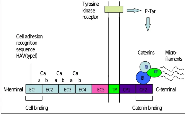
(28) classical cadherin 的結構如 Figure 1 及 Figure 2。主要由 含有五個位於 N terminal 的 extracellular domains(EC1 –EC5), 一組 transmembrane (TM),及二組 cytoplasmic (CP-1 - CP-2)所構 成。每一個 EC sub-domain 有一百個氨基酸,其中 EC1 在同質接合辨 識(homophilic recognition)的過程中扮演最重要的角色,同時在 EC1 的 C-terminal region 中有由三個氨基酸 His-Ala-Val(HAV)所 組成的 cell adhesion recognition (CAR) sequence,藉由 CAR 相 同的 cadherin 彼此之間可以結合成拉鍊狀(zipper-like)的構造 [68-69,73-74]。位於細胞質部分的 cytoplasmic domain 是結合細胞 骨骼的主要部分,cytoplasmic domain 中有一 serine rich region, 這個區域能與 α-catenin,β-catenin,γ-catenin 及 p120. cas. 這些. 蛋白結合,進而與 actin microfilaments bind 形成一完整的細胞骨 架,也就是 cadherin-catenin complex(Fig. 3.)[73-77]。. - 28 -.
(29) Fig. 1 . Cadherin structure. Tyrosine kinase receptor. P-Tyr. Cell adhesion recognition sequence HAV(typeI). Catenins γ. Ca a b N-terminal. EC!. EC2. Ca a b. Ca a. EC3. β. b EC4. Cell binding. EC5. TM. CP1. CP2. Microfilaments. α. C-terminal. Catenin binding. - 29 -.
(30) Fig. 2. The homogeneous amino acid sequences in the second cytoplasmic domain of all classical cadherins were used for designing the degenerate primers.. - 30 -.
(31) Fig. 3. Cadherin-catenin complex.. From: Polakis, Curr. Opinion Genetics Dev. 1999. - 31 -.
(32) Type1 Cadherin 有 Epithelial-cadherin (E-cad) ,Neural-cadherin (N-cad), Placental–cadherin(P-cad),. Vascular-Endothelial-cadherin. (VE-cad) 及 Retinal-cadherin (R-cad)。早在 1950 年代,就有科學 家發現一些因子在調節細胞間的接合, 引導著器官的形成與胚胎的 發育。在不同的時間、不同的條件下、不同的 cadherin 會有其適當 的表現,引導不同組織的形成。例如: 在鼠胚的研究中, morula stage 時 E-cad 有關鍵性的角色, 在 blastomere stage 時,如果降低 E-cad 和 P-cad 則胚胎發育會被損壞。還有許多的研究顯示不同 cadherin 引導不同組織發育,例如:N-cad 引導神經板的形成,在 mesenchymal cell 增 加 時 N-cad 也 會 增 加 [72,78-80] 。 這 些 現 象 都 顯 示 著 cadherin 在組織器官形成中重要的地位。cadherin 在維持細胞的方 向有重要的地位 ,例如:表皮細胞的 E-cad dysfunction,就會造成失 去表皮細胞的 phyenotype 及 enhacement of motility 和 removal of contact suppression of growth 最後造成細胞增生失去控制及造成 腫瘤的侵犯性。 有許多研究指出 cadherin 與腫瘤的關係,例如: E-cad 降低與肺癌、食道癌、胃癌、直腸癌、膀胱癌、前列腺癌、皮 膚癌、乳癌、子宮頸癌、卵巢癌、子宮內膜癌有關[50,81-89], N-cad 與神經外胚層及中胚層有關,例如: 乳癌、星狀細胞瘤[89-91],而. - 32 -.
(33) P-cad 則與肺癌、黑色素瘤、乳癌有關[72,92]。. Type2. Classical Cadherin. 包括了 cadherin- 4 到-14 [93-94], 它與 type 1 最大的不同是 在於 type 2 並沒有 His–Ala-Val(HAV)這個 CAR。 Type 2 典型 cadherin 的功能與 Type 1 是很類似的, 都與細胞接合、組織的形成 有關,例如它與人類的胎盤形成和著床有關[95],Cad-6 和 Cad-11 與 運動神經元的分化有關也與子宮內膜的上皮細胞和基質細胞的表現 調節有關[96-97], Cad-11 受到賀爾蒙調節,可當子宮內膜基質細胞 decidualization 過程中的一個指標,同時 Cad-11 也可調控滋養層細 胞的分化[98-99],Cad-6 與腎臟及腎臟癌的形成有關[100],除此之 外,type 2 的 cadherin 也與腫瘤有關,例如:對於缺乏 E-cadherin 的 腎臟癌病人,cadherin-6 可以用來評估預後[101],cad-6 同時也和卵 巢 癌 的 進 展 有 關 [102], 在 一 些 慢 性 骨 髓 性 白 血 病 的 病 人 中 cadherin-13 的表現會降低[103]。. Desmosomal Cadherin Desmosomal cadherin 主 要 有 desmocollin(Dscs 1,2,3) 及 desmoglein (Dsgs 1,2,3) ,它和典型 cadherin 最主要的差別在. - 33 -.
(34) cytoplasmic domain 的部分, desmosome 不是與 catenin 接合,而是 與 plakoglobin,desmoplakin 及 plakophillins 結 合 [73] 。 Desmosome 可以接受外界機械性的壓力,保持細胞的完整性, 它可位 於 皮 膚 等 組 織 ,如 果 受 到 傷 害 便 有 可 能 產 生 病 變 , 例 如 天 ? 瘡 (pemphigus vulgaris)[104]。. Protocadherin 它與典型 cadherin 最大的不同是 protocadherin 細胞外的 cadherin-repeats sequence 超過五組,而且在 cytoplasmic domain 部分它也不與 catenin 接合,有一些研究發現三個 protocadherin gene, Pcadhα、Pcadhβ、Pcadhγ,而這幾個 gene 可以 encode 超過 50 個 蛋 白 質 , 其 中 Pcadhα 又 稱 為 CNRS (cadherin-related neuronal receptors),由此可知 protocadherin 在神經發育中扮演重 要的角色[105]。. Cadherin-related proteins 包括了 T-cad、H-cad、Li-cad,這些蛋白缺少細胞外的部分,事 實上它也和一些癌症有關 ,例如:皮膚癌 、 腎臟癌與卵巢癌 [106-108]。. - 34 -.
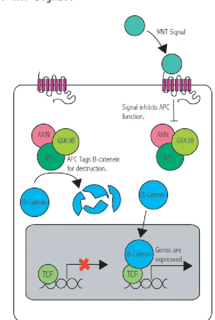
(35) (3) Cadherin 及 catenin 與 癌 症 間 的 關 係 Cadherin 的 cytoplasmic domain 與 catenin 結合形成了細胞骨 架,穩定細胞的構型,然而它的功能除了形成組織和器官之外 ﹐ 也和 訊號的傳遞 WNT signal(Wnt/wg signal)有密切的關係(如 Fig. 4.)[109-110]。WNT signal pathway 本身與胚胎的發展和訊號的傳 遞有關,而這個路徑 受到幾個重要因素的控制,包括: GSK3-β、 LEF-1/TCF(leukemic enfacement factor-1/T cell factor)、axin、 及 APC。訊號傳遞一開始是,Wnts( secreted glycoprotein) bind 到 Frizzled receptor 上, 在 Frizzled 活化了 Disheveled 之後,接著 便阻斷了 axin conduction,抑制了 APC 的功能。當 APC 和 GSK3 及 Axin 結合時可使 β-catenin 被 degraded,所以當 APC 不 能 degrade β-catenin 時, β-catenin 便會 accumulation,接著進入細胞核與 TCF 結合, upregulate c-Myc,cyclin D1 這些 oncogene 的表現。由 上述的機轉我們可以知道 β-catenin 會影響 oncogene 的表現,因此 可與 β-catenin 結合的 cadherin 同樣也會影響整個訊號的傳遞,進 而影響癌症的發生,例如: 結腸癌、黑色素瘤、前列腺癌、肝癌、子 宮內膜癌、卵巢癌等[111-114]。. - 35 -.
(36) Fig. 4. WNT signal.. From : Barker N., Clevers H. Catenins, Wnt signalling and cancer. Bioessays 2000; 22: 961-965.. - 36 -.
(37) 第四節. 子宮內膜癌與鈣黏蛋白. Risinger 從 卵 巢 癌 與 子 宮 內 膜 癌 的 組 織 中 發 現 E-cad gene mutation[50],而 Saito 則發現淋巴結被侵犯及細胞 grade3 的病人其 E-cad 基因被甲基化的比例較高[115], Sakuragi、Nomura 等皆提出 在細胞分化較差、肌肉層侵犯深度較深、主動脈旁淋巴結有轉移及預 後較差的病人,其 E-cad 的表現也較弱[116-118],N-cad 在較具侵犯 性的子宮內膜癌中表現也是降低的[119],而 E-cad 除了與疾病的嚴 重度有關,也可當細胞組織分類的標竿,例如:E-cad 在 endometrioid type 的表現比在 serous type 中強[120-122],除此之外還有許多學 者發現 cadherin 和 catenin 與子宮內膜癌之間的關係。Moreno 指出 E-cad 及 β-catenin 表現在 typeII 的子宮內膜癌中是降低的,而 P-cad 的表現是加強的[123],而另有一些研究指出 beta-catenin 在 內膜癌組織中有較強的 nuclear localization 的現象[124]。而 Desmosome 在 子 宮 內 膜 癌 的 表 現 則 是 減 弱 的 [128] 。 Kim 發 現 γ-catenin 的表現與肌肉侵犯的深度有關,而 與 α-catenin 及 β-catenin 無關[129]。. - 37 -.
(38) 第三章 研究假說與研究架構. 第一節 研究假說 從 Takeichi 研究 cadherin 在細胞、組織及器官形成中扮演 的重要角色之後,科學家們也抽絲剝繭地探討 cadherin 如何與其 它的蛋白質結合, 例如:α-catenin、β-catenin 及 γ-catenin , 進而發揮它的功能,然而不同的 cadherin 在不同的時間會有不同 的功能。Cadherin 除了與胚胎發育有關之外,在維持細胞的方向 上也有重要的地位 ,如果 cadherin dysfunction,就會造成細胞 增生失去控制,造成腫瘤的侵犯性。而我們知道 catenin 是 WNT signal pathway 中的重要調控因子之一,因此 cadherin-catenin complex 也會影響癌症的表現。所以我們基於這樣的概念,想要進 一步探討各種不同典型的 cadherin 及 catenin 在子宮內膜癌中的 角色。. 第二節 研究架構 : 我 們 的 研 究 的 研 究 分 成 二 個 部 分 ,第 一 個 部 分 是 利 用 degenerate. reverse. transcription-polymerase. chain. reaction(RT-PCR) 的方式找出典型 cadherin 在子宮內膜癌中的. - 38 -.
(39) 種類,第二部分接著再以半定量 (semi-quantitative) RT-PCR 的 方法比較存於子宮內膜癌中的典型 cadherin 與 catenin 的 mRNA 濃度與正常子宮內膜中的差異。. - 39 -.
(40) 第四章 研究材料與研究方法. 第一節 子宮內膜癌中 Cadherin 的種類 Part A. 材 料 (Materials) 實驗的檢體取自於在本院(中國醫藥學院附設醫院)接受子宮內 膜癌分期手術的病人以及子宮肌瘤接受全子宮切除手術病人的正 常子宮內膜,正常子宮內膜病人三個月內未接受賀爾蒙藥物治療, 且月經週期在早期分泌期(16-23th day)。本實驗只收集的檢體是 adenocarcinoma type with grade-III. differentiation 的檢. 體,所以從 2002 至 2003 年我們總共收集了五位病人的子宮內膜 癌症的組織檢體。在子宮一取下之後,立刻以刀片取下癌症的組織 或正常子宮內膜以液態氮急速冷凍保存。. - 40 -.
(41) Part B. 方 法 (Methods) (1).RNA 的萃取(RNA extraction) A. 儀器(Machine) 1.Source capture system (Germfree Laboraties) 2.Centrifuge 5810R (Germany) 3.Microcentrifuge(Uover Laboratories,USA) 4.Sectrafuge 16M(National LAbnet,USA) 5.TM firestek B206(Firstek Scientific) 6.Minigel Migration Trough Mupid-2(Cosmo Bio Co LTD,USA) 7.Rocker platform (Bellco Biotechnology,USA) 8.UV box TFX20M (Vukber Lourmat,France) 9.DS34 Polaroid Electrophrosis hood (UK). - 41 -.
(42) B.試劑(Reagents) 1.Trizol Reagent (Life Technologies,Inc.,Gainthersburg,MD) 2.1X TBE Buffer 3.2% Agarose 4.Chloroform 5.Isopropanal 6.Ethyl alcohol 7.Diethyl pyrocarbonate 8.Ethidium bromide(10mg/ml). C.步驟 (Procedure) 1.首先將取得的組織與 1ml 的 Trizol reagent 混合之後,接著 磨碎。 。. 2.於 4 C 下,以 8000rpm 離心 1 分鐘後,取出我們所需的上清液。 3.加入 0.2 ml chloroform 均勻混合,在室溫靜置下 5 分鐘後, 。. 接著在 4 C 下,以 12000 rpm 離心 15 分鐘,同樣取出上清液 (含 RNA)。 4.加 0.5 ml cold isopropanol 於上清液中均勻的混合,接著 。. 於室溫下靜置 10 分鐘後,在 4 C 下,以 13000rpm 離心 8 分 - 42 -.
(43) 鐘之後,留下沉澱物。 5.加 1 ml 之 100% alcohol 於沉澱物中,均勻混合。 。. 6.接著以 4 C 離心 13000rpm 5 分鐘,去除上清液,室溫靜置 5 分鐘,避免沉澱物過度乾燥。 。. 。. 7.加入 40μl 58 C 之 sterile ddwater,接著於 58 C 加熱 5 分鐘使沉澱物溶解,將所得的 RNA product run diagnostic gel,確認我們的 RNA。. - 43 -.
(44) (2)反轉錄(Reverse transcription) A.儀器(Machine) 1.TM firestek HB-100 (Fiestek Scientific) 2.TM firestek B206 (Firestk scientific). B.試劑(Reagent) 1.Random primer (Promega,Madison, WI,USA)) 2.Oligo-dT primer(Promegma) 3.M-MLVRTase (Moloney murine leukemia virus) (Promegma) 4.0.1M DDT(Dithiotheatol) 5.dNTP(Protech) 6.RNAsin(Invitrogen) 7.MMLV RT buffer. - 44 -.
(45) C.步驟(Procedure) 1.將約 5μl 之 total RNA 加 dd water 至 23μl。 2.接著加入 濃度 0.5μg/μl 之 2μl random primer and 。. 0.5μl oligo-dT primer 至總量 22.5μl,加熱 70 C, 5 分鐘後,置於冰上 2 分鐘,接著短暫離心。 3.加入 8μl MMLV RT buffer, 2μl MMLV RTase,2μl 0.1M DDT,2μl dNTP,0.5μl RNAsin,至總量 40μl 之後, 於 。. 。. 。. 37 C 放 2 小時後,於 94 C, 加熱 5 分鐘後,置於-70 C 冰箱中保存。. - 45 -.
(46) (3).degenerate-PCR (Polymerase chain reaction using degenerate primers ) A.儀器 (Machine) 1.PCR system 2400 (Perkin Elmer,USA) 2.Minigel Migration Trough Mupid-2(Cosmo Bio Co LTD,USA) 3.Rocker platform (BEllco Biotechnology,USA) 4.UV box TFX 20M (Vukber Lourmat,France) 5.DS34 Polaroid Electrophoresis hood (UK). B.試劑(Reagent) 1.10X PCR buffer 2.2.5mMdNTP 3.MgCl2 4.20μM cadherin degenerate forward primer 5.20μM cadherin degenerate reverse primer 6.Taq polymerase. - 46 -.
(47) Degenerate primers of classical cadherin are: Forward: GAATTCACNGCNCCNCCNTAYAYGA Reverse: GAATTCTCNGCNARYTTYTTRAAR R=either A or G, Y=either C or T, N=either A,C,G or T. C.步驟(Procedure) 1.2-4μl cDNA 加入 dd water 至總量 35μl。 2.接著加入 5μl 的 10X PCR buffer,5μl 的 2.5mM dNTP,2μl 的 MgCl 2, 1μl 的 20μM forward primer,1μl 的 20μM reverse primer,1μl 的 Taq polymerase 至總量 50μl。 。. 3.接著進行 PCR,而 PCR 溫度的條件是 95 C hold 5 min, 接 。. 。. 。. 著 95 C 1.5 min、45 C 2 min、72 C 3 min, run 35 cycles 接著是 72. 。. C 8 min。. 4.將 PCR product 植入 PCR-II vector。. - 47 -.
(48) (4). Cloning A.儀器(Machine) 1.Microcentrifuge tube 2.Thermocycler 3.Water bath 4.Orbital Shaking incubator OSI502 B.試劑(Reagent) 1.Luri-Bertani (LB) medium and agar 2.DMF (Dimethylformamide) 3.X-Gal in DMF (40mg/ml 5-bromo-4-chloro-3 indolyd-β-galactoside). 4.Ampicillin stock (50mg/ml) 5.IPTG (100mM isopropyl-β-D-thiogalactoside) 6.Ligation buffer 7.One shot competent cell Kit (TOP 10F’ cells) 8.TA cloning Kit (Invitrogen, San Diego,CA,如 Fig 4). - 48 -.
(49) C.步驟(Procedure). 1.Ligation (clone into PCR IIvector) ↓將 PCR II vector ( 1 vial) 簡短離心 ↓將先前 RT-PCR 之 PCR product 加上 1μl 之 10X ligation buffer 和 2μl 的 PCR II vector ↓接著加入 sterile water 和 T4 DNA ligase 1μl 至總量 10μl 。. ↓放置 14 C water bath, overnight. - 49 -.
(50) Fig. 5. PCR-II Vector.. - 50 -.
(51) 2. Transformation ↓將先前已被 PCR product clone 的 PCR II vector 簡短離 心之後,置於冰上 ↓加入 2μl TOPO cloning reaction 至 competent cells, 將它們輕輕的混合之後,置於冰上 30 分鐘 。. ↓接著放置於 42 C 水槽 30 秒,之後置於冰上 。. ↓於室溫下加入 250μl SOC medium,在 37 C shaking incubator 以 225 rpm 平行搖晃 1 小時 ↓取 50μl 及 200μl 之 solution 分別塗於含有 X-Gal 及 50μg/ml ampicillin 之 LB agar plates 上 。. ↓等乾之後,將 plate 巔倒置於 37 C 的 incubator 18 小時, 。. 之後置於 4 C 冰箱. 3. Bacterial growth ↓挑出至少 30 個白色菌落(Figure 6),分別將他們放到 30 管 2c.c.LB medium(內含 50μg/ml ampicillin), 在 400 rpm 搖,overnight 之後做 plasmid DNA 分析. - 51 -.
(52) (5).Plasmid DNA extraction and DNA sequence. A.儀器(Machine) Microcentrifuge. B.試劑(Reagent) 1.Gene-spin Miniprep Purification Kit(Beckman CEQ2000.AB1377,LICORIR2, Pharmacia A.L.F.)(Solution I,Solution II,Solution III,Wash Solution). 2.Restriction enzyme 10X buffer H (900mM Tris-HCL,500mM NaCl,100mM MgCl2). 3.EcoRI Enzymes storage buffer (10mM Tris-HCL,400mMNaCl, 0.1mM EDTA,1mM DTT,0.15% Triton X-100,0.5 mg/ml BSA,50% glycerol). - 52 -.
(53) C.步驟(Procedure) Plasmid DNA extraction ↓取 1ml 的 bacterial media,以 13000rpm 離心 30-60 秒之後, 取 pellet ↓接著加入 200μl Solution I,Vortex 5 -6 次使細胞溶解 ↓之後加入 200μl Solution II,Vortex 5-6 次 ↓加入 200μl Solution III,vortex 5-6 次 ↓13000 rpm 離心 5 分鐘 ↓將 spin column 放入 collection tube,接著將上清液小心 放入 spin column ↓離心 30 秒之後,將 spin column 移出 collection tube ↓將過濾液丟棄,加入 500μl 的 washing solution ↓離心 60 秒之後, 丟棄過濾液,再度離心 3 分鐘,去除殘餘的 ethanol 。. ↓將 spin column 放入新的 1.5ml eppendorff 中,加入 65 C 的 50μl H2O ↓離心 1 分鐘,以分離出 DNA (如果重覆最後兩個步驟就可多得 10-15%的 DNA). - 53 -.
(54) DNA 定序 (sequencing) ↓取 5μl 的 plasmid DNA,加 0.5μl 的 EcoRI restriction enzyme 和 1μl 的 buffer ↓接著加 3.5μl 的 acetylated BSA (sterile dd water),使 總量為 10μl 。. ↓於 37 C 下放置 1 小時 ↓將所得的產物以電泳分析是否含有約 170 bp 的典型鈣黏蛋 白,如果有的話,接下去做 sequencing ↓首先 prepare reaction solution ↓加入 4μl big dye,1μl plasmid DNA,2μl M-13 primer 以及 3μl 的 DW,總共 10μl 。. ↓然後將 Gene Amp 上的 cycle 設定,分別是 96 c 1 分 30 秒, 。. 。. 。. 以及 96 C 10 秒,50 C 5 秒, 60 C 4 分 共 25cycles , 。. 和4 C. 7 分鐘. ↓上面的 product 加入 10μlsequence reaction solution,10μl H2O,60μl 100%的 isopropanol,之後 vertex briefly ↓室溫下靜置 25 分鐘, ↓用最大速度離心 30 分鐘之後,去除上清液. - 54 -.
(55) ↓以 75% isopropanol 250μl rinse, vertex briefly ↓離心 5 分鐘,接著同樣去除上清液 。. ↓air dry,stock at -20C ,接著將 samples loading 到 gel ↓接著以 automated DNA sequencer 來分析 DNA sequence ↓再利用 BLAST computer program 與 Genebank/EMBL 的資料 庫與我們得到的 DNA sequence 做比對,以確認我們得到的典 型 cadherin 有那一些種類. - 55 -.
(56) 第二節. 比較六種典型 cadherin 及 α-,β-,γ-catenin 在子宮內膜癌與正常子宮內膜織中 mRNA 濃度的表 現差異. Part A 材料 在第一部分的實驗中,我們已知在子宮內膜腺癌組織中有 哪一些典型 cadherin 存在。以相同的檢體,接著我們要比較 這些典型 cadherin 在正常子宮內膜及子宮內膜腺癌的 mRNA level 的表現差異。. Part B 方法 (1)RNA 的萃取與定量. 儀器和試劑與第一節 partB 相同 。 步驟(Procedure) ↓取 2μl RNA,加入 3μldd water 及 5μl RNA denature buffer 之後 。. ↓在 58 C 下加熱 15 分鐘 ↓接著放置在冰上 2 分鐘 ↓短暫離心之後,加入 2μl 的 RNA loading buffer. - 56 -.
(57) ↓跑膠 ↓將膠放在 EBTBA 內染色 30 分鐘 ↓接著取出膠,在 UV box 照相,評估 RNA 的相對含量。. (2) RT 儀器、試劑和步驟與第一節 partB 相同。 (3) PCR 儀器、試劑和步驟與第一節 partB 相同,其中要特別注意的是, 因為我們要比較 mRNA 的量, 所以要求出每一個典型鈣黏蛋白 及 catenin 適當的 cycle 數。. 步驟(Procedure) ↓首先以 PCR 的方式得到的 GAPDH(house keeping gene), run 不同的 cycle 數,會得到一線性直線,取其中間值為 我們要的 cycle 數, GAPDH 是 22(Fig.14.) ↓用同樣的方法算出各種典型 cadherin 適當的 cycle(Fig. 15.- Fig. 20.) ↓ 接 著 以 六 種 典 型 鈣 黏 蛋 白 的 primer(Table3) 製 造 cadherin product. - 57 -.
(58) ↓將所得的 cadherin product 以電泳分析及評估 DNA 的量 ↓加入 2μl DNA loading buffer ↓跑膠 ↓將膠放在 EBTBA 內染色 30 分鐘 ↓接著取出膠,在 UV box 照相,評估 DNA 的相對含量 ↓定量是藉由 Hewlett Packer Digital scanner 將 cadherin product 相 片 掃 描 至 電 腦 之 後 , 以 UN-SCAN-IT Gel Version 5.1 system (Silk Scientific,Orm, Utah)來 分析,求得 Cadherin/GAPDH mRNA 的比值。. - 58 -.
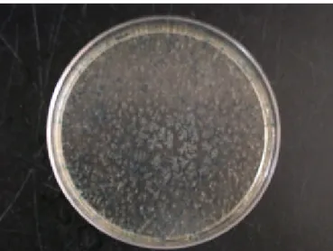
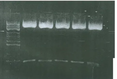
(59) 第五章 研究結果. 第一節 典型鈣黏蛋白的種類 我們將含有 cadherin product 的 PCR-II vector clone 到 E. coli 中,如果有轉植成功的話會形成白色的菌 落(Fig. 6.)。接著我? 挑出了 30 個 colonies,而各種 classical cadherin colonies 的數目如 Table 3,以萃取 DNA,. 在我們萃取出 plasmid. DNA 後 ,接著. 以 Eco RI restriction enzyme 將 plasmid 切斷, 之後將 所得產物跑電泳,結果我們在 gel 上發現二種 band ,一段的 base pair 約 3.9 Kb,這段是 plasmid 的 DNA,另一段約 180 base pairs ,這段是 cadherin 的 DNA (Fig. 7.),接著再定 序,結果發現在子宮內膜癌中有六種典型 cadherin,分別 是:E-cad、 N-cad、 P-cad、 Cad-6、Cad-9 以及 Cad-11 (如 Fig.8.- Fig. 13. )。. - 59 -.
(60) Fig. 6. Agar plate for transformed bacterial selection.如 果有被含 cadherin product 所 clone 的 PCR-II vector 轉植成功的細胞是白色的菌落,反之則無形成菌落,但是 若是被不含有 cadherin product 所 colone 的 PCR-II vector 所轉植成功的話,則會形成藍色的菌落。. - 60 -.
(61) Table2. Analysis of cadherin cDNA clones generated from Grade-3 endometrial adenocarcinoma lesion Cadherin subtype. No of clone. Clones analyzed/patient(%). Patient 1 E-cad. 2. 7. P-cad. 7. 23. N-cad. 3. 10. Cad-6. 5. 17. Cad-9. 1. 3. Cad-11. 12. 40. E-cad. 3. 10. P-cad. 8. 27. N-cad. 3. 10. Cad-6. 6. 20. Cad-9. 1. 3. Cad-11. 9. 30. Patient 2. - 61 -.
(62) Fig. 7. Plasmid DNA 約 3.9 Kb,而我們的 degenerate PCR product 約 180 bp 經過定序分析後, 發現有六種典型 cadherin。. ß plasmid DNA 3.9 Kb. ß 180-bp Degenerate PCR products for sequencing. - 62 -.
(63) Fig. 8. An example of E-cadherin sequence. - 63 -.
(64) Fig. 9. An example of N-cadherin sequence. - 64 -.
(65) Fig. 10. An example of P-cadherin sequence. - 65 -.
(66) Fig. 11. An example of cadherin-6 sequence. - 66 -.
(67) Fig. 12. An example of cadherin-9 sequence. - 67 -.
(68) Fig. 13. An example of cadherin-11 sequence. - 68 -.
(69) 第二節 不同典型鈣黏蛋白在子宮內膜癌與正常子宮內膜中 的不同表現 我們知道六種同時存在於正常子宮內膜與子宮內膜腺 癌組織中的 calssical cadherin 的種類之後, 因為我們要 比較 mRNA的量, 所以要求出每一個典型鈣黏蛋白及catenin 適當的 cycle 數,結果如 Fig. 14. - Fig. 23。 而我們所用 的 primers 如 Table 3。六種典型 cadherin 經過電腦的比較 之後,可發現 P-cad 在子宮內膜癌組織中的表現比正常子宮 內膜組織強,而 E-cad 在子宮內膜癌組織中的表現比正常子 宮內膜組織弱 ,但是 cad-6、 cad-9、cad-11 則無不同。 α-,β-,γ-catenin 在子宮內膜癌組織中的表現和正常子 宮內膜組織中相同(如 Fig. 24.-Fig. 26.)。. - 69 -.
(70) Fig. 14. GAPDH 的半定量(semiquantitative) RT-PCR 理想 cycle 數。圖 (A)是 GAPDH 在正常子宮內膜的 RT-cycle 數,圖(B)是在子宮內膜 癌組織的 RT-cycle 數。由圖中的線性增加發現,當 cycle 數為 22 時,為理想的定量 cycle 數。. (A) 正常子宮內膜. GAPDH Relative optical density. 7 6 5 4 3 2 1 0 20. 22. 24. Number of cycles. (B)子宮內膜癌組織. GAPDH. Relative optical density. 7 6 5 4 3 2 1 0 20. 22. Number of cycles. - 70 -. 24.
(71) Fig. 15. E-cad 的半定量(semiquantitative) RT-PCR 理想 cycle 數。圖 (A)是 E-cad 在正常子宮內膜的 RT-cycle 數,圖(B)是在子宮內膜 癌組織的 RT-cycle 數。由圖中的線性增加發現,當 cycle 數為 28 時,為理想的定量 cycle 數。. (A) 正常子宮內膜. E-cad. Relative optical density. 6 5 4 3 2 1 0 26. 28. 30. 28. 30. Number of cycles. (B)子宮內膜癌組織. E-cad. Relative optical density. 6 5 4 3 2 1 0 26. Number of cycles. - 71 -.
(72) Fig. 16. N-cad 的半定量(semiquantitative) RT-PCR 理想 cycle 數。圖 (A)是 N-cad 在正常子宮內膜的 RT-cycle 數,圖(B)是在子宮內膜 癌組織的 RT-cycle 數。由圖中的線性增加發現,當 cycle 數為 28 時,為理想的定量 cycle 數。. (A)正常子宮內膜. N-cad Relative optical density. 6 5 4 3 2 1 0 26. 28. 30. Number of cycles. 子宮內膜癌組織. N-cad 5. Relative optical density. (A). 4 3 2 1 0 26. 28. Number of cycles. - 72 -. 30.
(73) Fig. 17. P-cad 的半定量(semiquantitative) RT-PCR 理想 cycle 數。圖 (A)是 P-cad 在正常子宮內膜的 RT-cycle 數,圖(B)是在子宮內膜 癌組織的 RT-cycle 數。由圖中的線性增加發現,當 cycle 數為 28 時,為理想的定量 cycle 數。. (A)正常子宮內膜. P-cad Relative optical density. 4. 3. 2. 1. 0 26. 28. 30. Number of cycles. (B)子宮內膜癌組織. P-cad. Relative optical density. 7 6 5 4 3 2 1 0 26. 28. Number of cycles. - 73 -. 30.
(74) Fig. 18. Cad-6 的半定量(semiquantitative) RT-PCR 理想 cycle 數。圖 (A)是 Cad-6 在正常子宮內膜的 RT-cycle 數,圖(B)是在子宮內膜 癌組織的 RT-cycle 數。由圖中的線性增加發現,當 cycle 數為 28 時,為理想的定量 cycle 數。. (A). 正常子宮內膜. Cad-6. Relative optical density. 6 5 4 3 2 1 0 26. 28. 30. Number of cycles. 子宮內膜癌組織. Cad-6. Relative optical density. 6 5 4 3 2 1 0 26. 28. Number of cycles. - 74 -. 30.
(75) Fig. 19. Cad-9 的半定量(semiquantitative) RT-PCR 理想 cycle 數。圖 (A)是 Cad-9 在正常子宮內膜的 RT-cycle 數,圖(B)是在子宮內膜 癌組織的 RT-cycle 數。由圖中的線性增加發現,當 cycle 數為 30 時,為理想的定量 cycle 數。. (A). 正常子宮內膜. Cadherin-9 Relative optical density. 5 4 3 2 1 0 28. 30. 32. Number of cycles. 子宮內膜癌組織. Cadherin-9 5. Relative optical density. (B). 4 3 2 1 0 28. 30. Number of cycles. - 75 -. 32.
(76) Fig. 20. Cad-11 的半定量(semiquantitative) RT-PCR 理想 cycle 數。 圖(A)是 Cad-11 在正常子宮內膜的 RT-cycle 數,圖(B)是在子宮 內膜癌組織的 RT-cycle 數。由圖中的線性增加發現,當 cycle 數 為 28 時,為理想的定量 cycle 數。. (A). 正常子宮內膜. Cad-11 Relative optical density. 5 4 3 2 1 0 26. 28. 30. Number of cycles. 子宮內膜癌組織. Cad-11 6. Relative optical density. (B). 5 4 3 2 1 0 26. 28. Number of cycles. - 76 -. 30.
(77) Fig. 21. Alpha-catenin 的半定量(semiquantitative) RT-PCR 理想 cycle 數。圖(A)是 Alpha-catenin 在正常子宮內膜的 RT-cycle 數,圖 (B)是在子宮內膜癌組織的 RT-cycle 數。由圖中的線性增加發現, 當 cycle 數為 26 時,為理想的定量 cycle 數。. (A). 正常子宮內膜. Alpha-catenin. Relative optical density. 6 5 4 3 2 1 0 24. 26. 28. Number of cycles. 子宮內膜癌組織. Alpha-catenin. 5. Relative optical density. (B). 4 3 2 1 0 24. 26. Number of cycles. - 77 -. 28.
(78) Fig. 22. Beta-catenin 的半定量 (semiquantitative) RT-PCR 理想 cycle 數 。 圖 (A) 是 Beta-catenin 在 正 常 子 宮 內 膜 的 RT-cycle 數,圖(B)是在子宮內膜癌組織的 RT-cycle 數。由 圖中的線性增加發現,當 cycle 數為 26 時,為理想的定量 cycle 數。. (A). 正常子宮內膜. Beta-catenin. Relative optical density. 5 4 3 2 1 0 24. 26. 28. Number of cycles. 子宮內膜癌組織. Beta-catenin. 5. Relative optical density. (B). 4 3 2 1 0 24. 26. Number of cycles. - 78 -. 28.
(79) Fig. 23. Gamma-catenin 的半定量(semiquantitative) RT-PCR 理想 cycle 數 。 圖 (A) 是 Gamma-catenin 在 正 常 子 宮 內 膜 的 RT-cycle 數,圖(B)是在子宮內膜癌組織的 RT-cycle 數。由 圖中的線性增加發現,當 cycle 數為 26 時,為理想的定量 cycle 數。. (A). 正常子宮內膜. Gamma-catenin. Relative optical density. 5 4 3 2 1 0 24. 26. 28. Number of cycles. 子宮內膜癌組織. Gamma-catenin. 6. Relative optical density. (B). 5 4 3 2 1 0 24. 26. Number of cycles. - 79 -. 28.
(80) Table 3. Primers used for PCR in these studies Gene. Primer. Cadherin-degenerate. 5’ 3’ 5’ 3’ 5’ 3’ 5’ 3’ 5’ 3’ 5’ 3’ 5’ 3’ 5’ 3’ 5’ 3’ 5’ 3’. E-Cadherin P-cadherin N-cadherin Cadherin-6 Cadherin-9 Cadherin-11 α-catenin β-catenin γ-catenin. Sequence. Size. 5’-GAATTCACNGCNCCNCCNTAYGA-3’* ∼180 bp 5’-GAATTCTCNGCNARYTTYTTRAA-3’* 5’-TCCATTTCTTGGTCTACGCC-3’ 361 bp 5’-CACCTTCAGCCAACCTGTTT-3’ 5’-GACCAACGAGGCCCCTTTTGTGCTG-3’ 357 bp 5’-GTGGTGGGAGGGCTTCCATTGTCCA-3’ 5’-GTGCCATTAGCCAAGGGAATTCAGC-3’ 373 bp 5’-GCGTTCCTGTTCCACTCATAGGAGG-3’ 5’-TTCTTGCTGCTCTTTTGGGT-3’ 275 bp 5’-CCTGCTCCATCTCCTGAAAG-3’ 5’-CAAAACCTGGGCAGTTGATT-3’ 416 bp 5’-CCTCTTCAATGCAGCAAACA-3’ 5’-ACCAGATGTCTGTGTCAGA -3’ 742 bp 5’-GTCATCCTTGTCATCTGCA-3’ 5’-CAGAGGGAGCATGACTTCGG-3’ 290 bp 5’-CTACAGCAGCCACCAACTCT-3’ 5’-AAGGTCTgGAGGAGCAGCTTC-3’ 668 bp 5’-TGGACCATAACTGCAGCCTT-3’ 5’-ATGGAGGTGATGAACCTGATGG-3’ 284 bp 5’-CCTGACACACCAGGGCACAT-3’. - 80 -.
(81) Fig. 24.. E-,P-and N-cadherin 在子宮內膜癌與正常子宮內膜中 的比較。圖中 a 表示 negative control, b 為正常子宮 內膜,而 c 是子宮內膜癌組織。DNA marker 在 gel 的最 左邊。ethidium bromide-stained 的 gel 上都有顯示 我們用 specific 的 primers(E-cad, P-cad , N-cad) 所 amplified 的 PCR 產物。而 E-cad,P-cad 和 N-cad 分 別位在 361, 357,373 base pairs 的位置,而 GAPDH 則 位 於 359 base pairs 的地方。 而每一個 cadherin subtype 的 absorbance value ,都會以相對應的 GAPDH 標準化,算出他們的比值。結果發現 P-cad mRNA levels 在子宮內膜癌組織中表現增強 ,E-cad 則減弱 ,但 是 N-cad 兩種組織中的表現無差別。. - 81 -.
(82) E-,P-, and N-cadherin 在子宮內膜癌與正常子宮內膜 中的比較。 M. E-cadherin. a. b. c. P-cadherin. a. b. c. N-cadherin. a. b. c. Cadherins. GAPDH. E-cadherin. P-cadherin. N-cadherin. 1.0. Ratio of cadherin/GAPDH. Fig. 24.. 0.8 0.6 0.4 0.2 0.0 a. b. c. a. - 82 -. b. c. a. b. c.
(83) Figure 25.. Cadherin-6,-9,and11 在子宮內膜癌與正常子宮內膜中 的比較。圖中 a 表示 negative control, b 為正常子宮 內膜,而 c 是子宮內膜癌組織。DNA marker 在 gel 的最 左邊。ethidium bromide-stained 的 gel 上都有顯示 我們用 specific 的 primers(Cad-6, Cad-9 , Cad-11) 所 amplified 的 PCR 產物。而 Cad-6,Cad-9 和 Cad-11 分 別位在 275, 416,和 742 base pairs 的位置,而 GAPDH 則位於 359 base pairs 的地方。而每一個 cadherin subtype 的 absorbance value , 皆以相對應的 GAPDH 標 準化,算出他們的比值。結果發現 cad-6,cad-9,cad-11 在正常子宮內膜組織中和在子宮內膜癌組織中的表現 無顯著差異。. - 83 -.
(84) Cadherin-6,-9,and 11 在子宮內膜癌與正常子宮內膜 中的比較。. M. Cadherin-6 Cadherin-9. a. b. c. a. Cadherin-11. b c. a. b. c. Cadherins. GAPDH. Cadherin-6. Cadherin-9. Cadherin-11. 1.2. Ratio of cadherin/GAPDH. Fig. 25.. 1.0 0.8 0.6 0.4 0.2 0.0 a. b. c. a. - 84 -. b. c. a. b. c.
(85) Fig. 26.. a-catenin,ß-catenin 及 ?-catenin 在子宮內膜癌與正常子宮 內膜的比較。圖中 a 表示 negative control, b 為正常 子宮內膜,而 c 是子宮內膜癌組織。DNA marker 在 gel 的最左邊。ethidium bromide-stained 的 gel 上都有顯 示我們用 specific 的 primers(a-catenin,ß-catenin 及 ?-catenin)所 amplified 的 PCR 產物。而 a-catenin,ß-catenin 及 ?-catenin 分別位在 290, 668,和 284 base pairs 的位 置,而 GAPDH 則位於 359 base pairs 的地方。而每一個 catenin 的 absorbance value ,皆以相對應的 GAPDH 標 準化,算出他們的比值。結果發現 a-catenin,ß-catenin 及 ?-catenin 在正常子宮內膜組織中和在子宮內膜癌組織中 的表現無顯著差異。. - 85 -.
(86) a-catenin,ß-catenin 及 ?-catenin 在子宮內膜癌與正常子宮 內膜的比較。 M. α-catenin. β-catenin. γ-catenin. a. a. a. b. c. b. c. b. c. Catenins. GAPDH. Relative ratio of cadherin/GAPDH. Fig. 26.. Alpha-catenin. Beta-catenin. Gamma-catenin. 1.0 0.8 0.6 0.4 0.2 0.0 a. b. c. a. - 86 -. b. c. a. b. c.
(87) 第六章 討論. 子宮內膜藉由卵巢性腺賀爾蒙的調控形成週期性變化,即增生 期、分泌期以及脫落形成月經 [130]。隨著子宮內膜的周期性變化許 多分子都會呈現週期性變化,包括鈣黏蛋白在內 [95,97] 。因此本 研究採用的正常子宮內膜檢體取自早期分泌期的子宮內膜,以避免月 經週期兩端的極端差異。過去文獻對於鈣黏蛋白與子宮內膜癌的關聯 性幾乎都局限於 E-cad [ 116-121],而且仍然有些爭議。其主要的徵 結在於子宮內膜腺癌的分化,我們早期的研究發現 E-cad 在分化良好 (grade-1) 的子宮內膜腺癌與正常子宮內膜的表現並沒有差異,而中 度 分 化 (grade-2) 的 子 宮 內 膜 腺 癌 則 得 不 到 穩 定 的 結 果 (inconsistent)。這些爭議有可能是分化良好(grade-1)和分化不良 (grade-3)的子宮內膜腺癌是兩回事,而不是同一個疾病的不同階 段,或者不同的病理醫生在判定 Grade-2 時可能有不同結果。為避免 爭議,本研究只針對明顯 grade-3 的子宮內膜腺癌來分析。. 本研究在分化不良的子宮內膜癌的檢體中確認有六種鈣黏蛋 白,也就是 E-cad、P-cad、N-cad、Cad-6、Cad-9 和 Cad-11。這六 種鈣黏蛋白也同樣存在於正常子宮內膜及子宮內膜異位瘤中. - 87 -.
(88) [131] 。因此我們認為子宮內膜應該就只有這六種典型鈣黏蛋白,正 常與不正常之間只是它們的表現量不同而已。其中 Cad-6、Cad-9 和 Cad-11 尚未有文獻報告過存在於子宮內膜腺癌,是屬於新的發現。 Cad-6 又名 K-cad,首先發現是由胚胎時期分化形成腎臟時產生,一 旦成型之後即被抑制下來。但是,腎臟癌發生時這個基因又會被激 發,因此它與腎臟癌的發生有關。之後陸續有報告指出 Cad-6 存在於 不同的組織中例如子宮內膜等等 [95,97]。本研究顯示 Cad-6 的 mRNA 在子宮內膜癌與正常子宮內膜之間並無顯著差異因此我們認為它和 子宮內膜癌的形成無關。至於 Cad-9 的表現量,不論在子宮內膜、子 宮內膜異位、或子宮內膜腺癌都很少,而且正常子宮內膜與腺癌之間 並沒有羞異,因此 Cad-9 與子宮內膜腺癌的形成也沒有關聯。Cad-11 被証明存在於大多數的 parenchymal tissue 中,它也在正常子宮內 膜 [95,97]、子宮內膜異位症 [131],本研究證明它也表現於子宮內 膜腺癌中。但是,正常子宮內膜與腺癌之間並沒有羞異,因此 Cad-11 與子宮內膜腺癌的形成應該沒有關係。 至於 E-cad 在各種不同的癌症中常被提到,因為絕大部份與 E-cad 有關的癌症,E-cad 的表現都明顯的減少。因此,它被認定為一種 tumor suppresser gene。但是,分化良好的子宮內膜腺癌其 E-cad 表現並沒有減少(我們未發表的數據),量雖然沒有減少,但有可能是. - 88 -.
(89) E-cad 的功能不良,例如:Saito 曾報告過 E-cad 基因的 promoter region 過度甲基化與子宮內膜癌的侵犯有關[115]。我們認為可能有 某種”去分化”(de-differentiation) 的鈣黏蛋白被激化而表現過 多,導致生長失控而形成癌症。例如: Marvin 曾提出報告指出在乳 癌的細胞株可發現 E-cad 的表現與癌細胞的侵犯性無關,但是植入 N-cad 可以使癌細胞的侵犯性以及 motility 增加[126]。本研究證實 E-cad 在分化不良的子宮內膜腺癌的確比正常子宮內膜的表現明顯減 少,這一點和其他的文獻是吻合的 [116,118,121] 。因此 E-cad 的 確在維持上皮細胞的正常性,扮演著極重要的角色。N-cad 則普遍存 在於神經組織及血管內皮細胞 [132] 和正常子宮內膜或子宮內膜異 位組織中[131] 。本研究証明子宮內膜腺癌也表現 N-cad。然而,正 常子宮內膜與腺癌之間並沒有差異,因此 N-cad 與子宮內膜腺癌的形 成應該沒有關係。至於 P-cad 存在於不同的上皮細胞,已有愈來愈多 的文獻顯示 P-cad 的過度表現與乳房腺癌的侵犯性和預後不良有關 [92,133-134]。例如:Yutaka 曾報告在正常的胃組織中並無 P-cad 的 表現,但是在胃癌卻可發現 P-cad 的出現[125]。本研究証明了子宮內 膜腺癌也是一樣,分化不良的子宮內膜腺癌具有大量 P-cad 的表現。 因此我們推測子宮內膜的致癌因子激發 P-cad 的過度表現及抑制了 E-cad 的表現導致子宮內膜腺癌的產生。最近的一篇報告(2003 年 4. - 89 -.
(90) 月)使用組織染色法分析子宮內膜腺癌,也証實 P-cad 的蛋白質表現 與不良分化有關 [123] ,也支持我們的結論。 至於 catenins 方面,由於它負責聯結鈣黏蛋白和細胞內的骨架 (cytoskeleton) ,因此它們普遍存在各種不同的人體細胞,有文獻顯 示 catenin 與腫瘤有關[111-114]。本研究並未發現 catenins,包括 α-、β-、和 γ-catenin,在正常子宮內膜和分化不良的子宮內膜 腺癌之間有表現差異。原因有可能是因為子宮內膜腺癌的 P-cad 增加 但是 E-cad 減少因此 catenins 的量還是維持在平衡的狀態。也有可 能 catenin 的總量不變但是跑到細胞核裡面去了,例如:Nei 曾經報 告 β-catenin 轉進細胞核與癌病有關[124]。 至於 Moreno-Bueno 曾 報告 β-catenin 的表現在 type-II 子宮內膜癌明顯減少[123],這? 爭議有可能是由於研究方法不同,Moreno-Bueno 使用免疫組織染色 (IHC),而 我 們 使 用 較 敏 感 客 觀 的 RT-PCR。 況 且 ,Kim 也 曾 指 出 β-catenin 在子宮內膜癌的表現並無差異[129]。因此,catenin 在 惡性腫瘤的角色還有待更多的研究來澄清。. - 90 -.
(91) 第七章 結論與建議. 第一節 結論 本研究首次發現 cad-6, cad-9, cad-11 存在於子宮內膜癌組 織中,同時也確認了六種典型鈣黏蛋白(E-cad, N-cad , P-cad , cad-6, cad-9 and cad-11)位於子宮內膜癌組織中,E-cad 的表現 在分化不良的子宮內膜腺癌組織中顯著減少,而 P- cad 則是顯著增 加 , N-cad,cad-6, cad-9, cad-11, α-catenin,β-catenin 和 γ-catenin 則無差別。因此我們認為 E-cad 以及 P-cad 在子宮內 膜癌的形成與疾病的進展扮演重要的角色。. 第二節 未來研究 1. 雖然過去的文獻顯示 cadherin 的 mRNA 變化和其蛋白質表現都 是吻合的,不過我們將會再做 western blot analysis 來証明 cadherins 和 catenins 的蛋白質表現差異和 mRNA 的差異吻合。 2. 加 入 免 疫 染 色 (IHC) 的 部 分 ﹐進 一 部 分 析 β-catenin 的 localizaton, P- cad 及 N-cad 的 distribution。 3. 加 入 sex steriod receptors (estrogen, androge,and progesterone)這部分,觀察 sex setroid hormone 是否影響. - 91 -.
(92) cadherin, catenin 的表現。. - 92 -.
(93) 參考文獻 1. MacMahon B. Risk factors for endometrial cancer. Gynecol Oncol 1974;2:122-129. 2. MacMahon B. Overreview of studies on endometrial cancer and other types of cancer in humans:Perspective of an epidemiologist. Semin Oncol 1997;21:S1-122-S1-139. 3. Antunes CM,Strolley PD, Rosenshein NB, Davies JL, Tonascia JA, Brown C, Burnett L, Rutledge A, Pokempner M, Garcia R. Endometrial cancer estrogen use. N Engal J Med 1979;300:9-13. 4. Brinton LA, Berman ML, Mortel R. Reproductive, menstrual and medical risk factors for endometrial cancer:Results from a case-control study. J Obstet Gynecol 1992;167: 1317-1325. 5. Gray LA,Christopherson WM, Hoover RN. Estrogens and endometrial carcinonma. Obstet Gynecol 1977;49:619621. 6. Horwitz RL, Feinstein AF. A lternative analytic methods for case-control studies of estrogens and endometrial cancer.. - 93 -.
(94) N Engal J Med 1978;299:1089-1094. 7. McDonald TW,Malkasian GD, Gaffey TA. Endometrial cancer associated with feminizing ovarian tumors and polycystic ovarian disease. Obstet Gynecol 1977;94:654-658. 8. Jordan VC, Assikis VJ. Endometrial carcinoma and tamoxifen :Clearing up a controversy.Clin Cancer Res 1995;1:467-472. 9. Silca EG, Tornis C, Mapica A, Mitchell MF. Uterine neoplasms in patients treated with Tamoxifen. J Cell Biochem Suppl 1995;23:179-183. 10. Barakat RR. The effects of tamoxifen on the endometrium. Oncology 1995;9:139-140. 11. FisherB, Costantinoi JP, Wickerham DL. Tamoxifen for pre vention of breast cancer:Report of the National Surgical Adjuvant Breast and Bowel Project P -I study.J Natl Cancer Inst 1998;90:1371-1388. 12. Lawrence C. Smoking, body weight, and early stage endometrial cancer. Cancer 1987;59:1665-1669. 13. Levi F, Franeschi S, Negri E, LaVecchia C. Dietary factors and the risk of endometrial cancer. Cancer 1993;71:3575. - 94 -.
(95) -3581. 14. Swanson CA, Potischman N, Wilbanks GD. Relationship of endometrium cancer risk to past and contemporary size and body fat distribution. Cancer Epidemiol Biomarkers Prev 1993;2:321-327. 15. Feldman S, Cook EF, Harlow BL, Berkowitz RS. Predicting endometrial cancer among older women who present with abnormal vaginal bleeding. Gynecol Oncol 1995;56:376 -381. 16. Koss LG. Diagnosis of early endometrial cancer and precancerous stage. Ann Clin Lab Sci 1979;9:189-194. 17. A nderson B. Diagnosis of endometrial cancer. Clin Obstet Gynecol 1986;13:739-750. 18. Giusa-Chiferi MG, Oncalves WJ, Baracat EC. Transvaginal ultrasound,. uterine. postmenopausal. biopsy. bleeding.. and. Int. J. hysteroscopy Gynaecol. for. Obstet. 1996;55:39-44. 19. Creasman W, Odicino F, Maisonneuve P. Carcinoma of the corpus uteri. Annual report on the results of treatment. - 95 -.
(96) in gynecologic cancer. J Epidemiol Biostat 1998;3:35-61. 20. Genest P. Prognostic factors in early carcinoma of the endometrium. Am J Clin Oncol 1987;10;71-77. 21. Kodama S, Kase H, Tanaka K, Matsui K. Multivariate analysis of. prognostic. factors in. patients with. endometrial cancer. Int J Gynecol Obstet 1996;53:23-30. 22. Christopherson WM, Connelly PJ,Alberhasky RC.Carcinoma of the endometrium: An analysis of prognostic factors in patients with favorable subtypes and stage I disease. Cancer 1983;51:1705-1710. 23. Iversen OE. Flow cytometric deoxyribonucleic acid index: a prognostic factor in endometrial carcinoma. Am J Obstet Gynecol 1986;155:770-776. 24. Baak JPA, Snijders WP, Van Diest PJ. Confirmation of the prognostic value of the ECPI-1 score in FIGO stage I endometrial cancer patients with long follow up.Int J Gynecol Cancer 1995;5:112-116. 25. Britton LC, Wilson TO, Gaffey TA. DNA ploidy in endometrial carcinoma: Major objective prognostic. - 96 -.
(97) factor. Mayo Clin Proc 1990;65:643-650. 26. Creasman WT. Influence of cytoplasmic steroid receptor content on. prognosis of. early. stage. endometrial. carcinoma. AM J Obstet Gynecol 1985;151:922-932. 27. Podratz KC, Wilson TO, Gaffey TA. DNA analysis facilitates. the. pretreatment. identification. of. high-risk endometrial cancer patients. Am J Obstet Gynecol 1993;168:1213-1215. 28. Sorbe B, Risberg B, Thonthwaite J. Nuclear morphometry and DNA flow cytometry as prognostic methods for endometrial carcinoma. Int J Gynecol Cancer 1994;4:94100. 29. Zachos G, Spandidos DA. Transcriptional regulation of the c-H-ras1 gene by the P53 protein is implicated in the development of human endometrial and ovarian tumours. Oncogene 1998;16:3013-3017. 30. Hanson NB. Prognostic significance of lymph-vascular space invasion in stage I endometrial cancer. Cancer 1985;55:1753-1757.. - 97 -.
(98) 31. Ehrlich CE, Young PCM, Stehman FB, Sutton GP, Alford WM. Steroid receptors and clinical outcome in patients with adenocarcinoma of the endometrium. Am J Obstet Gynecol 1988:158:796-807. 32. Kaupplia A. Oestrogen and progestin receptors as prognostic indicators in e ndometrial cancer. Acta Oncol 1989;28:561-566. 33. Patterson E. Management of stage I carcinoma of the uterus. Obstet Gynecol 1982;59:755-758. 34. Rittenberg PV, Lotocki RJ, Heywood MS, Jones KD, Krepart GV. High-risk surgical stage 1 endometrial cancer: outcomes with vault brachytherapy alone. Gynecol Oncol. 2003;89:288-294. 35. Behbakht K, Jordan EL, Casey C. Prognostic indicators of survival in advanced endometrial cancer. Gynecol Oncol 1994;55:363-367. 36. Burke TW.Selective pelvic and paraaortic lymphadenectomy Oper Tech Gynecol Surg 1996;1:169-173. 37. Chen SS. Operative treatment in stage I endometrioid. - 98 -.
(99) carcinoma with deep myometrial invasion and/or grade3 tumor surgically limited to the corpus uteri: No recurrence with only primary surgery. Cancer 1989; 63:1843-1845. 38. Bertelsen K, Jakobsen A. Radiotherapy for gynecologic cancer. Curr Opin Oncol 1993;5:885-890.. 39. Rose PG, Cha SD, Tak WK. Radiation therapy for surgically proven para-aortic node metastasis in endometrial cancer. Int J Radiat Oncol Biol Phys 1992;24:229-233. 40. COSA-NZ-UK. Endometrial Cancer Study Groups: Adjuvant medroxyprogesterone acetate in high risk endometrial cancer. In J Gynecol Cancer 1998;8:387-391. 41. Lewis GC, Slack NH, Mortel R. Adjuvant progesterone therapy in primary definitive treatment of endometrial cancer. Gynecol Oncol 1974;2:368-376. 42. Mass Hb. Chemotherapy of metastatic endometrial cancer . Semin Oncol 1994;21:107-113. 43. Edmonson JH, Krook JE, Hilton JF, Malkasian GD, Everson LK, Jefferies JA, Mailliard JA.Randomized phase II. - 99 -.
數據


+7
相關文件
Wang, Solving pseudomonotone variational inequalities and pseudocon- vex optimization problems using the projection neural network, IEEE Transactions on Neural Networks 17
volume suppressed mass: (TeV) 2 /M P ∼ 10 −4 eV → mm range can be experimentally tested for any number of extra dimensions - Light U(1) gauge bosons: no derivative couplings. =>
For pedagogical purposes, let us start consideration from a simple one-dimensional (1D) system, where electrons are confined to a chain parallel to the x axis. As it is well known
The observed small neutrino masses strongly suggest the presence of super heavy Majorana neutrinos N. Out-of-thermal equilibrium processes may be easily realized around the
Define instead the imaginary.. potential, magnetic field, lattice…) Dirac-BdG Hamiltonian:. with small, and matrix
incapable to extract any quantities from QCD, nor to tackle the most interesting physics, namely, the spontaneously chiral symmetry breaking and the color confinement..
(1) Determine a hypersurface on which matching condition is given.. (2) Determine a
• Formation of massive primordial stars as origin of objects in the early universe. • Supernova explosions might be visible to the most
