國
立
交
通
大
學
生物科技研究所
碩
士
論
文
探討幽門螺旋桿菌熱休克蛋白 60 對人類周邊血
液單核球抑制增生活性之位置
The active site of Helicobacter pylori heat shock
protein 60 for inhibiting the proliferation of
human peripheral blood mononuclear cells
研 究 生:林宗翰
指導教授:廖光文 教授
探討幽門螺旋桿菌熱休克蛋白 60 對人類周邊血液單核球抑
制增生活性之位置
The active site of Helicobacter pylori heat shock protein 60 for
inhibiting the proliferation of human peripheral blood
mononuclear cells
研究生:林宗翰 Student:Tsung-Han Lin 指導教授:廖光文 Advisor:Kuang-Wen Liao 國 立 交 通 大 學 生物科技研究所 碩 士 論 文 A ThesisSubmitted to Department of Biological Science and Technology National Chiao Tung University
in partial Fulfillment of the Requirements for the Degree of
Master in
Biological Science and Technology June 2011
Hsinchu, Taiwan, Republic of China
中華民國一百年六月
i 探討幽門螺旋桿菌熱休克蛋白 60 對人類周邊血液單核球抑制增生活 性之位置 研究生:林宗翰 指導教授:廖光文 博士 國立交通大學生物科技研究所 中文摘要 因幽門螺旋桿菌所造成慢性感染已被公認會導致消化道方面的 疾病,像是慢性胃炎、消化性潰瘍、黏膜相關淋巴組織淋巴癌 (MALT lymphoma),甚至有可能發展成胃癌。最近,根據報導由幽門螺旋桿 菌所分泌的熱休克蛋白 60 與胃部的發炎具有相關性。並且幽門螺旋 桿 菌 的 熱 休 克 蛋 白 60 已 被 證 實 可 誘 導 許 多 前 發 炎 激 素 (pro-inflammatory cytokines),例如 TNF-α、IL-8、IL-6 以及 IFN-γ。 有趣的是,有些文獻指出不同物種的熱休克蛋白 60 與免疫調控有相 關性。舉例來說,日本血吸蟲所分泌的熱休克蛋白 60 可誘導調控 T 細胞(Treg)的產生並達到抑制發炎的效果。而我們實驗室的先前研究 也顯示幽門螺旋桿菌的熱休克蛋白 60 可降低人類周邊血液單核球細 胞 (human peripheral blood mononuclear cells, PBMCs)之增生活性,且 抑制前發炎激素的分泌,並是經由誘導細胞週期的停滯進而影響 PBMCs 的增生。這些發現指出幽門螺旋桿菌的熱休克蛋白 60 可能具 有兩種功能,分別是誘導發炎與免疫調控。根據文獻顯示幽門螺旋桿
ii 菌的熱休克蛋白 60 主要是經由 TLR-2 的路徑來造成發炎。另一方 面,幽門螺旋桿菌的熱休克蛋白 60 其胺基酸位置 300-435 也被證實 具有可誘導單核球細胞分泌 IL-8 的能力。然而,幽門螺旋桿菌的熱 休克蛋白 60 是由哪一個區域的胺基酸位置來誘發免疫調控仍然還是 未知。因此,我的方針為探討幽門螺旋桿菌的熱休克蛋白 60 的哪一 個位置具有免疫調控的能力。 為了達到這個目的,所以我們建構完整幽門螺旋桿菌的熱休克蛋 白 60 (胺基酸序列:1-547) 及五個不同的幽門螺旋桿菌熱休克蛋白 60 片段 (胺基酸序列:1-200、101-200、1-250、200-300 及 300-547)。 由於 Treg 細胞可經由 IL-10 或 TGF-β 這兩個細胞激素來抑制 CD4+ T 細胞的增生,所以我們藉由監控這兩個免疫抑制的細胞激素 (IL-10 及 TGF-β)來找出哪一個片段可誘發免疫調控。研究結果顯示,重組 的熱休克蛋白 60 (胺基酸序列:101-200 及 300-547)可誘導 IL-10 的分 泌。並經由可專一性辨識重組熱休克蛋白片段 60 (胺基酸序列: 101-200)的單株抗體進一步證實可負調控免疫抑制。這些結果顯示幽 門螺旋桿菌的熱休克蛋白 60 (胺基酸序列:101-200)可能與免疫抑制 有相關性。
iii
The active site of Helicobacter pylori heat shock protein 60 for inhibiting the proliferation of human peripheral blood mononuclear cells Student: Hsiao-Wei Shen Advisor: Dr. Kuang-Wen Liao
Department of Biological Science and Technology National Chiao Tung University
Abstract
Chronic infection with Helicobacter pylori (H. pylori) is recognized as the cause of upper gastrointestinal disorder, such as chronic active gastritis, peptic ulcer diseases, mucosa-associated lymphoid tissue (MALT) lymphoma and is an important risk factor for the development of gastric cancer. Recently, it was also reported that heat shock protein 60 of H. pylori (HpHsp60) are associated with gastric inflammation. Hsp60 of H. pylori has been found that it can induce immune cells to express the strong pro-inflammatory cytokines, such as TNF-α, IL-8, IL-6, and IFN-γ. Interestingly, some literatures indicated that Hsp60 derived from different species seem to be related to the regulation of immune responses. For example, the Hsp60 of S. japonicum can generate regulatory T cell (Treg) and suppress immunity. Our lab’s previous studies have shown that H. pylori Hsp60 can decrease human peripheral blood mononuclear cells
iv
(PBMCs) proliferation rate, inhibit cytokine secretion, and induce cell cycle arrest. These findings indicate that HpHsp60 may have two functions, induction of the inflammation and immune suppression. The mechanisms of HpHsp60-induced inflammation in which TLR-2 pathway is the major signaling pathway have been confirmed. On the other hand, the amino acid domains 300-435 of H. pylori Hsp60 is associated with induction of IL-8 in monocytic cell has been explored. What domain of the HpHsp60-induced immune suppression is still remains unknown. Thus, the aim in this study is to investigate which domains of HpHsp60 function for immune suppression.
For the purpose of this study, we construct whole Hsp60 (amino acid sequence: 1-547) and five partial domains (sequence: a. a. 1-200, 101-200, 1-250, 200-300, 300-547). Since Treg cells inhibit proliferation of CD4+
T cells either via IL-10 or TGF-β production, we demonstrate which domains of HpHsp60 function for immune suppression by monitoring the two cytokines (IL-10 and TGF-β) expression. The preliminary data showed rHsp60s (sequence: a. a. 101-200 and 300-547) induced IL-10 secretion. The monoclonal Abs against rHsp60 (sequence: a. a. 101-200) to prove that rHsp60 (sequence: a. a. 101-200) could down-regulate the
v
immune responses. These results propose that the sequence from 101 to 200 of H. pylori Hsp60 may be closely associated with immune suppression.
vi 致謝 沒想到碩士兩年的時光轉眼就過,還記得當初大學推甄考上交大 時,那種恨不得公佈全世界的心情,如今已要為碩班研究生畫下句 點,回過頭想想真不可思議,我想其中廖光文老師的功勞,真的要讓 我跪下奉茶,好好由衷感謝才是,老師就猶如我另一個父親般,不論 在學業或生活上都給予極大的開導,到目前為止,我還沒見過其他老 師,有如這樣的細心教導許多大道理,而這些寶貴經驗已永植我心, 時時刻刻不斷提醒我,該怎麼做且要做什麼才是對我最好的,真的非 常感謝指導教授廖光文教授的提攜,此外,也感謝特地前來幫我口試 的吳彰哲教授、鄭添祿教授及蔡女滿教授,並指導學生在實驗設計與 邏輯上哪裡的不足與欠缺,使我眼界大開且收穫良多,由衷的感謝。 另外,實驗的完成,team 的存在是非常重要的,在此我非常感 謝 Hp 組的氣質維瞳(老師不在時,我的第二個老師)、超正的小蛙、 美麗的薇姊及嬌小的翊鈞,感謝你們在實驗上的幫助與教導,沒你們 的相助,我可能還在地上翻滾也說不定。而也感謝 LPPC 組的運動帥 氣型阿伯、文書型 DG、美麗動人的于鈴學姐、正義的靜敏、搞笑的 美惠與依璇、聰穎的欣怡、超 man 的品榮及當了我 12 年同學的超可 愛何雙雙,及大學部子勛、勁翰、兩百、澄皓、宜蓓,感謝你們兩年 的陪伴與玩耍,讓我在做實驗快崩潰之餘,有壓力出口可釋放。而也
vii
非常感謝遠在清大的佳盈,細心照顧與幫忙。
最後,我要感謝我的家人:爸爸、媽媽、弟弟,沒有你們的支持 與幫助,我想要走到最後一步,真的是要演不可能的任務了,對於所 有關心及幫助過我的人,非常感謝大家,在此獻上深深的一鞠躬。
viii
Contents
Abstract………...………... i Acknowledgement………...…..………vi Contents………...……...……... viii Chapter 1 Introduction………...……...…………...1Chapter 2 Materials and methods……….……….6
2.1 Materials………..……...……...……….6 2.1.1 Reagents……….…………...6 2.1.2 Antibodies……….………..…… .7 2.1.3 Kit………...…………...7 2.1.4 Instruments……….……..………...8 2.1.5 Cell lines………..………….……….8 2.1.6 Animal………..……….………8 2.1.7 Others………..……….…………...………..………9 2.2 Methods………..……….9
2.2.1 Cell culture condition…...………...………..9
2.2.2 Expression and purification of rHpHSP60 proteins………...……..…..….10
2.2.3 Peripheral blood mononuclear cells (PBMCs) isolation……...…………..12
ix
2.2.5 IL-8, IL-10, TNF-α, TGF-β secretion from PBMCc cell……...….………14
2.2.6 IL-8, IL-10, TNF-α, TGF-β secretion from THP-1 cells….………...……15
2.2.7 Production of monoclony antibody………...………..……15
2.2.8 Statistical analysis…………...………..…..……17
Chapter 3 Results………..……...………..19
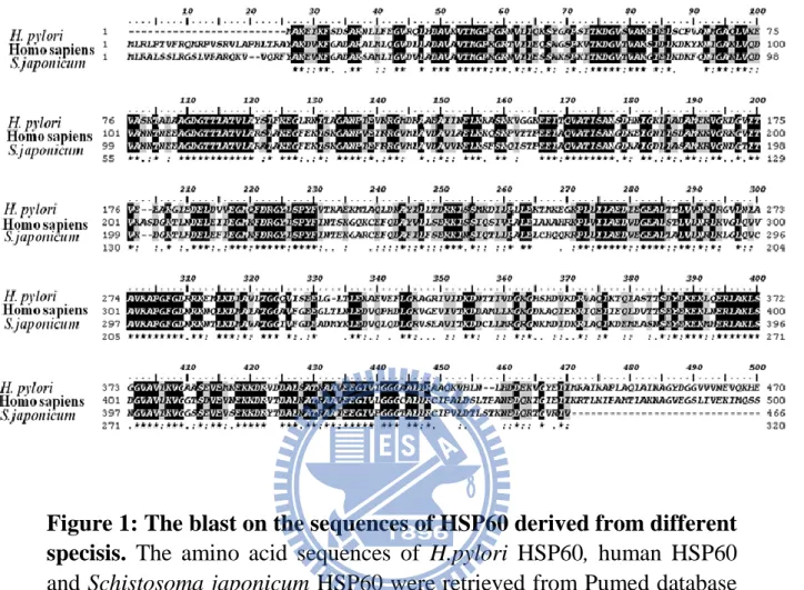
3.1 The blast on the sequences of HSP60 derived from different specisis…...…19
3.2 The expressionss of different HpHSP60 fragments………...…19
3.3 The effects of HpHSP60 on PBMCs on proliferation……….……...…20
3.4 Production of cytokines in PBMCs...…21
3.5 The effects of rHpHSP60 fragments on THP-1 proliferation...…21
3.6 Production of cytokines in THP-1 cells...…22
3.7 The research for the effective epitop for proliferative inhibition in HpHSP60..22
Chapter 4 Discussion………..24
Figure and Legend………...………..29
x
Figure and Legend
Figure 1 The blast on the sequences of HSP60 derived from different specisis.…….29 Figure 2 The expressionss of different HpHSP60 fragments...…………...………….30 Figure 3 Treatment with whole and different fragment HpHSP60 proteins to PBMCs decrease the proliferation in the present of anti-CD3 antibody………..…..31 Figure 4 Production of cytokines in PBMCs………...………...….32 Figure 5 Treatment with whole and different fragment HpHSP60 proteins to THP-1 cells decrease the proliferation.………...………...33 Figure 6 Production of cytokines in THP-1 cells…..………...………...….34 Figure 7 The characterizations and application of monoclonal Ab………....35
1
Chapter 1 Introduction
Helicobacter pylori (H. pylori), is well-known gastric-parasitical pathogen which has been declared by the World Health Organization and International Agency for Research on Cancer consensus group as a carcinogen (Organization and Humans 1994). The H. pylori living in the gastric mucosal can cause chronic infection, which result in chronic gastritis, peptic ulcer (Cello 1995), gastric cancer (Uemura et al. 2001, Peek Jr and Blaser 2002) or mucosa-associated lymphoid tissue (MALT) lymphoma (Parsonnet et al. 1994). Variety of virulence factors that derived from H. pylori has been proven to associate with immune evasion, including CagA (Peek Jr and Blaser 2002, Hocker and Hohenberger 2003, Huang et al. 2003), VacA (Peek Jr and Blaser 2002, Hocker and Hohenberger 2003), Heat Shock Protein 60 (HpHSP60), LPS, and arginase . CagA can upregulation of gastric epithelial IL-8 secretion (Crabtree et al. 1995). VacA produces proinflammatory cytokines (TNF-α, IL1-β, IL-6, IL-10 and IL-13) by induced bone marrow-derived mast cells or macrophage (Supajatura et al. 2002). HpHSP60 and LPS of H. pylori can result in gastric inflammation. HpHSP60 positively modulates mononuclear cells (Gobert et al. 2004) and LPS positively
2
modulates epithelial cells (Su et al. 2003) to secret inflammatory cytokines via Toll-like receptor-4 (TLR4) signal transduction pathway, respectively. HpHSP60 has been first identified as an adhesion molecule that can bind to host gastric epithelial cells and mucin(Huesca et al. 1996). Recently, for gastric tumor progression, HpHSP60 promote the migration ability of cancer cells, increase in the angiogenic activity of endothelial cells, induction of pro-inflammatory cytokine in both gastric epithelial cells and monocytes (Lin et al. 2010). These literatures suggest that HpHSP60 is an important factor that induces human excessive inflammation by multiple mechanisms.
Different species of Heat shock protein 60 (HSP60) play the different roles in the human body. For example, HSP60 of Chlamydia can promote lung inflammation (Cappello et al. 2009), but Schistosoma japonicum’s (S.japonicum) HSP60 induces regulatory T cell (Treg) generation and suppresses immunity (Wang et al. 2009) or human’s HSP60 activates native mouse B cell to proliferate and secrete IL-10 cytokines (Cohen-Sfady et al. 2009). On the other hand, HSP60s from different species are highly similar for their protein sequences (Ellis 1992), the alignment of amino acid sequence between H. pylori,
3
S.japonicum and homo sapiens HSP60 is up to 52% of similarity. In addition, S.japonicum and homo sapiens also play the similar role in immune suppression. After treated with S.japonicum HSP60, T cells releases of IL-10 and TGF-β. It play a role for TLR2 in mediated CD4+
CD25+ Treg induction (Wang et al. 2009). Human HSP60 and its
peptide amino acids 437-460 has been shown to change proinflammatory cytokines (Delaleu et al. 2008). In 2006, Dr. Zanin-Zhorov, A. showed human HSP60 treatment can enhance the IL-10 and TGF-β secretions of Treg via TLR2 to inhibit the expressions of IFN-γ and TNF- of CD4+CD25- T cells (Zanin-Zhorov et al. 2006b). Recent studies have
shown that H. pylori infection leads to CD4+CD25+Foxp3 regulatory T
cells increase (Lundgren et al. 2005, Kandulski et al. 2008). In our unpublished paper have shown that H. pylori HSP60 can decrease human peripheral blood mononuclear cells (PBMCs) proliferation rate, cytokine secretion, and induce cell cycle arrest. HpHSP60 also show its potential to elevate the secretion of anti-inflammatory cytokine IL-10 and TGF- n human monocytes (Lin et al. 2009). As well-known, IL-10 and TGF- play important roles for involved in the immunosuppressive function and increase of Treg cell (Dao Nguyen and Robinson 2004,
4
Zheng et al. 2004). Therefore, these findings indicate that HpHSP60 may have opposite function in regulating immune response. The domains of HpHSP60 inducing inflammation have been explored and confirmed. However, the domains which are capable to induce immune suppression still remain unknown. Thus, the aim of this study is to investigate which domains of HpHSP60 function for immune suppression.
To investigate the domains of immune suppression associated with HpHSP60, we construct whole HSP60 (sequence: a.a.1-547) and five partial fragments (sequence: a.a.1-200, 101-200, 1-250, 200-300, 300-547). Through the identification of the fragments, it may not only find out the inhibitory domain for T-cell proliferation but also get the effective sequence for the Treg cell increasing. In the study, we are aim at finding the immune regutation effects of rHpHSP60, HpHSP60101-200,
HpHSP601-200, HpHSP601-250, and HpHSP60300-547 on immune cell.
Monitoring the proliferation and cytokine expression confirm the immune suppression ability. One monoclonal antibodies (mAb) were also prepared by immunizing BALB/c mice with rHpHSP60. Human PBMCs the proliferation rate and immune suppress cytokine expression decreased was inhibited by one of the mAb against HpHSP60. According to the
5
results, we suggested the proliferative inhibition is on T cell populations mainly through IL-10 expression and the sequexcnce from 101 to 200 of H. pylori HSP60 may be closely associated with immune suppression.
6
Chapter 2 Materials and methods 2.1 Materials
2.1.1 Reagents
The following reagents and chemicals were obtained as indicated: RPMI 1640, Fetal Bovine Serum (FBS). Penicillin/ streptomycin/ Amphotericin (PSA) from Biological industries (Beithaemek, Israel). DNA agarose, Tryptone, Kanamycin and Tris from MdBio Inc. (Rockville, MD, USA). Restriction enzymes were obtained from Promega Inc. (WI, USA). Isopropyl-beta-D-thiogalactopyranoside (IPTG), NaCl, yeast extract, agar, Tris-HCl, Triton X-100, TEMED and imidazole from Amresco Inc. (Solon, OH, USA). Recombinant TGF-β1 was from Peprotech Inc. (Rocky Hill, NJ). RNase A, Thiazolyl blue tetrazdium bromide (MTT), Ammonium persulfate (APS), Sosium dodecyl sulfate (SDS), 3,3,5,5-tetramethylbenzidine (TMB), and dimethyl sulfoxide (DMSO) from SIGMA-ALDRICH (Steinheim, Germany). EDTA and chloroform from TEDIA (Fairfield, OH, USA). H3PO4, KH2PO4, Na2HPO4, Tween 20, MgCl2, and KHCO3 from SHOWA (Tokyo, Japan). Coomassie Plus reagent (Thermo Scientific,
7
Rockford, IL, USA), HAT and HT medium (Hybri-Max; Sigma Chemical Co., St. Louis, Mo), Nitrocellulose membrane (Hybond-ECL extra; Amersham, Buckingham, UK) .
2.1.2 Antibodies
The following antibodies were obtained as indicated: Mouse anti-human CD3 (OKT3) was kindly provided from Dr. Steve R. Roffler (Institute of BioMedical, Sciences ACADEMIA SINCA), goat anti-mouse (IgG, IgA, IgM)-HRP antibody was from Millipore Co. (Billerica, MA, USA), mouse anti-human CD4-FITC (RPA-T4) from Biolegend (Sandiego, CA, USA), mouse anti-human FoxP3-PE (259D/C7) from BD Biosciences (Bedford, MA, USA).
2.1.3 Kit
Human IL-8, IL-10, TNF-α, TGF-β ELISA kit was obtained from R&D systems (Minneapolis, MN, USA), Coomasie PlusTM Protein Assay
8 Pierce (Rockford, IL, USA).
2.1.4 Instruments
HisTrapTMHP column was from GE healthcare (Uppsala, Sweeden).
FASCan floecytometry from Becton, Dickinson and Company (Franklin Lakes, NJ, USA). ABI prism 7000 from Applied Biosystems (Drive Foster City, CA, USA).
2.1.5 Cell lines
The human monocytic cell line (THP-1) and mouse myeloma cell line (FO) were obtained from the Bioresourece Collection and Research Center (Hsinchu, Taiwan).
2.1.6 Animal
Balb/c mice were obtained from National Science Council (NSC) of Taiwan.
9 2.1.7 Others
Escherichia coli (BL-21 and DH5α) were obtained from Teastern Biotech Co. Peripherak blood mononuclear cells (PBMCs) from human whole blood.
2.2 Methods
2.2.1 Cell culture condition
THP-1 were cultured in RPMI 1640 medium supplemented with 0.05 mM 2-mercantoethanol, 2 g/L sodium bicarbonate, 50 μg/ml of PSA, and 10% heat-inactivated fetal bovine serum (FBS). FO were cultured in DMEM medium supplemented with 1.5 g/L sodium biucarbonate, 10 % heat-inactivated FBS, and 50 μg/ml of PSA. PBMCs were cultured in RPMI medium supplemented with 1.5 g/L sodium biucarbonate, 10 % heat-inactivated FBS, and 50 μg/ml of PSA. Hybridoma were culture in DMEM medium supplemented with 1.5 g/L sodium biucarbonate, 20 % heat-inactivated FBS, and 50 μg/ml of PSA.
10
2.2.2 Expression and purification of rHpHSP60 proteins Transformation of E.coli
E. coli [BL21(DE3)] (Yeastern Biotech, Taipei, Taiwan) were mixed
gently with 1 ng DNA then incubated on ice for 30 min. After incubation, cells were heat shock at 42℃ for 90 seconds followed by incubating on ice for 2 min. Next, placed the competent cells to 250 μl LB and incubated on 37C for 1 hour. Cells were plated on LB plate containing kanamycin (30 μg/ml) at 37C for 16 hours.
Midi plasmid DNA preparation
Pick up a single colony was picked and inoculated in 100 ml LB medium containing kanamycin (30 μg/ml). After 16 hours incubation at 37C, the bacteria were recovered by centrifugation at 8000 rpm for 15 mins. Remove the supernatant as dry as possible, and then DNA was preparated by QIAamp DNA Midi kit according to the manifacture’s protocols. The precipitated DNA was washed by adding 1ml of 70% ethanol, and then dissolved in DDW. Measure the absorbance at 260 and
11
280 nm and assay by using restriction enzyme digestion for checking the DNA quantity and quality.
Expression and purification of rHpHSP60 protein
E. coli [BL21(DE3)] (Yeastern Biotech, Taipei, Taiwan) were transformed with 1ng of DNA and grew on LB plate containing kanamycin (30 μg/ml) at 37C for 16 hours. Then single colony was picked and inoculated in 100 ml LB medium containing kanamycin (30μg/ml)). After 16 hours incubation at 37C, the bacteria in the LB broth were refreshed in 900 ml LB with vigorous shaking. Assayed the OD value until OD600 reaches 0.6~0.8, then protein induction was performed by adding 1.25 ml of IPTG (800mM) and continually incubated for 4 hours. After 4 hours incubation, harvested the bacteria by centrifugation at 5000 rpm for 15 min at 4 C. Discarded the supernatant and resuspended the pellet with 30 ml binding buffer containing urea (20 mM Na2HPO4, 0.5 M NaCl, 40 mM imidazole, 8 M urea, pH 7.4). Total
lysates were sonicated for 15 min and then centrifuged at 12,000 rpm for 30 min to collect the supernatant which containing rHpHSP60.
12
HisTrapHP column (General Electric, NY, USA) (1cm) was used to purify rHpHSP60. All the buffers and protein samples needed to be filtered with 0.45µm syringe filter. After protein sample loading into the column, washed the column with 30X volume of binding buffer to remove the unwanted proteins and then eluted the rHpHSP60 with 10X volume of elution buffer (20 mM Na2HPO4, 0.5 M NaCl, 500 mM
imidazole, pH 7.4). Eluted rHpHSP60 were collected and loaded into G-25 column (General Electric, NY, USA) to remove the unnecessary salt and replace the buffer with PBS (Phosphate Buffer Saline, 140 mM NaCl, 2.7 mM KCl, 10 mM Na2HPO4, 1.8 mM KHPO4, pH 7.4). Protein
concentration was quantified by Coomassie Plus reagent (BSA was used as the standard). SDS-PAGE and western blotting with anti-His conjugated HRP (ROCKLAND, PA, USA) were used to confirm the purity of rHpHSP60.
2.2.3 Peripheral blood mononuclear cells (PBMCs) isolation
PBMCs were isolated from human whole blood by using Ficoll-PaquePlus. Dilute human whole blood with equal volume of PBS.
13
Added 6 ml Ficoll-PaquePlus into the 15 ml centrifuge tube and loaded 8 ml of the diluted blood sample on Ficoll-Paque Plus reagent carefully. Then centrifuged the tubes at 400g for 40 min at 18C. After centrifugation, removed the plasma layer and collect the PBMCs carefully between the plasma layer and Ficoll-PaquePlus solution. Washed the cell with 2X volume of PBS, centrifuged at 1,500 rpm for 15 min. Discarded the supernatant and added 10 ml of ACK lysis buffer (0.15M NH4Cl, 10 mM KHCO3, 0.1 mM Na2EDTA) to lyse the red blood
cells. Centrifuged at 1,500 rpm for 15 min to remove the supernatant and then washed the cell with 10ml PBS again. Counted the cell number and seeded the PBMCs into 96-well culture plate with RPMI1640 culture medium.
2.2.4 PBMC proliferation assay
To mimic the action occur when the immune cells encounter antigen, anti-CD3 antibody (kindly provided from Dr. Steve R. Roffler) (OKT3, an antibody that specifically binds to CD3 surface marker and transduces signals continually to activate T lymphocytes) was used to selectively
14
activate T lymphocyte. Briefly, PBMCs were isolated from healthy donors then seeded into anti-CD3 mAb (0.03µg/well) pre-coated 96-well plates (2*105/well, triplicate for each treatment) and treated with different
proteins (200 ng/ml) of HpHSP60, HpHSP60101-200, HpHSP601-200,
HpHSP601-250, HpHSP60200-300, HpHSP60300-547 for four days incubation.
Cell proliferation was verified by MTT assay. In briefly, 96-well was centrifuged at 1,500 rpm for 15 min to remove the supernatant. Added 100 µl MTT solution into each well and incubated the plate at 37C for four hours. Then centrifuged again, removed the supernatant and dissolved the purple crystal with 100 µl DMSO each well. Shook the plate for 10 min then measured the OD value at 595nm. The proliferation index was calculated with following equation: proliferation = (OD value of protein-treated PBMC) (OD value of PBMC treated without HpHSP60)*100%.
2.2.5 IL-8, IL-10, TNF-α, TGF-β secretion from PBMCc cells
PBMCs cells grown on 96 well plates were stimulated with 200 ng human HSP60, 200 ng HpHSP60, HpHSP60101-200, HpHSP601-200,
15
HpHSP601-250, HpHSP60200-300, HpHSP60300-547 at 37℃ in RPMI
medium for 2 days. Supernatants and cells were separated by centrifugation at 4000rpm for 5 minutes. Supernatants were assayed for IL-8, IL-10, TNF-α, TGF-β production by ELISA kit. (R&D system, MN, USA).
2.2.6 IL-8, IL-10, TNF-α, TGF-β secretion from THP-1 cells
THP-1 cells grown on 96 well plates were stimulated with 200 ng human HSP60, 200 ng HpHSP60, HpHSP60101-200, HpHSP601-200,
HpHSP601-250, HpHSP60200-300, HpHSP60300-547 at 37℃ in THP-1 growth
medium for 24 hours. Supernatants and cells were separated by centrifugation at 4000 rpm for 5 minutes. Supernatants were assayed for IL-8, IL-10, TNF-α, TGF-β production, and IL-8, IL-10, TNF-α, TGF-β was measured by IL-8, IL-10, TNF-α, TGF-β ELISA development kit. (R&D system, MN, USA).
2.2.7 Production of monoclony antibody Animal care and use
16
The monoclony antibody productions were utilized Balb/c mice with 5-7 weeks of age. The mice were fed in animal room at Chiao Tung University during thr period of immunization. Feed and water were available daily.
Preparation of mouse polyclony antibodies against HpHSP60
Female Balb/c mice, aged 5-7 weeks, were used for immunization. HpHSP60 in sterilized phosphate buffered saline (PBS), containing 0.12M NaCl, 0.02 M phosphate, pH 7.4, was mixed and homogenized with an equal volume of complete Freund’s adjuvant by a three-way
stopcock. Each mouse was initially given a total emulsion of 0.3 ml containing 100μg of protein with 6 subcutaneous injections onto the back and an intraperitoneal injection. At day 7, an identical dose with incomplete was given intraperitoneally followed by two intramuscular injections without adjuvant at day 14. Seven days following a final booster, blood was collected in 0.1 % (wt/vol) EDTA and plasma was obtained. This plasma was used as a souerce for conventional polyclonal
17
antibody against HpHSP60. Additional booster injections were given when necessary.
Preparation of mouse polyclony antibodies against HpHSP60
The spleen obtained was used for preparing hybridoma fusion after immunization. In brief, myeloma cell line (FO) was fused with spleen cells from immunized Balb/c mice at a ration of 1:5. Fusion was carried out within 2 min at 37℃ using 1 ml of 50% (wt/vol) polyethylene glycol containing 10 % (vol/vol) DMSO. Cell mixture was then washed and resuspended in HAT medium containing approximately 1×10⁵ FO cells
per ml. The suspended cells were distributed as 100 μl per well in 96-well plates and incubared at 37℃ in incubator followed by an addition of 100μl of fresh HAT medium after 7 days. Subsequently, culture medium was assayed for the production of specific antibodyes by ELISA. After primary screening, desired hybridomas were selected and recloned.
18
The results are presented as mean ± SD. Significant is calculated by
19
Chapter 3 Results
3.1 The blast on the sequences of HSP60 derived from different specisis.
The sequences of H. pylori, homo sapiens and S. japonicum HSP60 proteins were retrieved from the NCBI Protein database. The protein secquence alignment showed that the identity is 52% (Fig. 1) to reveal that their amino acids are similar. There are two highly similar regions at “80 to 200” and “360 to 420 “.
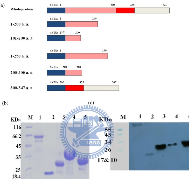
3.2 The expressionss of different HpHSP60 fragments.
As our previous study, HpHSP60 can inhibit the proliferation of
human PBMC. Thus, we have interesting in whether the different fragments of HpHSP60 have different activities on the PBMC proliferation. To further investigate the proliferative activities of the different fragments of Helicobacter pylori heat shock protein 60, we constructed different transgenes to encode different HpHSP60 fragments. Figure 2a showed the structures of these transgenes which were inserted
20
into pET100/D-TOPO. All recombinant proteins could be expressed and purified, and results showed their molecular weights were about 14.8 K.D., 25.35 K.D., 30.99 K.D., 15.08 K.D., and 30.28 K.D., respectively (Fig. 2b and 2c).
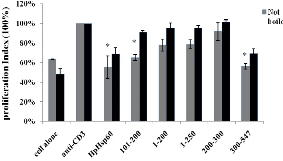
3.3 The effects of HpHSP60 on PBMCs on proliferation
Firstly, we examined the proliferative effects of different fragments of rHpHSP60s on the proliferation of human PBMC. In this system, PMBCs were activated by anti-CD3 monoclonal antibodies to induce the T-lymphocytes proliferation. Figure 3 showed that rHpHSP60, HpHSP60101-200, HpHSP601-200, HpHSP601-250, and HpHSP60300-547 could
inhibit the proliferations of PBMCs. However, treatments with different boiled rHpHSP60 fragments have different effects. Figure 3 showed that boiled HpHSP60101-200, HpHSP601-200 and HpHSP601-250 would not result
in the phenomenon of proliferation inhibition, whereas the boiled process had no effect on the proliferative inhibition of PBMCs for rHpHSP60 and HpHSP60300-547. Based on the above reaults, the results revealed the
21
but HpHSP60300-547 may inhibit the PBMC proliferation by its linear
sequence.
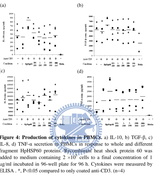
3.4 Production of cytokines in PBMCs
Several publications revealed that Helicobacter pylori heat shock protein 60 would stimulate IL-10, TGF-β, IL-8 or IL-6 secretion in human monocytic cells. Thus, the fragments of HpHSP60 were examined their effects on these cytokine expressions in PBMC activation system. The results showed that HpHSP60300-547 could induce expressions of
IL-10, IL-8 and TNF-α and HpHSP60101-200 could increase IL-10 cytokine
expression (Fig. 4). Except of the two fragments, all fragments of HpHSP60 lost the abilities to induce thses four cytokine expressions (Fig. 4).
3.5 The effects of rHpHSP60 fragments on THP-1 proliferation
To determine whether the effects of rHpHSP60 fragments could affect the growth of monocyte, different dosages of rHpHSP60 fragments
22
(50-300 μg/ml) were used to treat THP-1 cells. The results showed rHpHSP60 and rHpHSP60 fragments including HpHSP60101-200,
HpHSP601-200, HpHSP601-250 could inhibit the THP-1 cells proliferations
but HpHSP60200-300 and HpHSP60300-547 didn’t (Fig. 5).
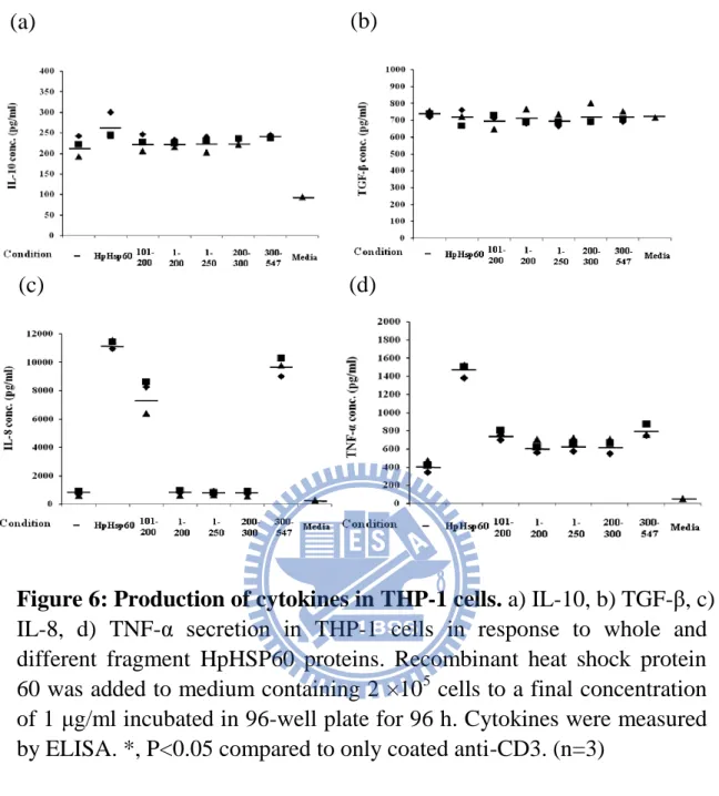
3.6 Production of cytokines in THP-1 cells
Several publications revealed that Helicobacter pylori heat shock protein 60 would stimulate IL-10, TGF-β, IL-8 or IL-6 secretion in human monocytic cells. Thus, the fragments of HpHSP60 were examined their effects on these cytokine expressions in THP-1 cells system. The results showed that HpHSP60300-547 and HpHSP60101-200 could induce expressions
of IL-8 and TNF-α cytokine expression (Fig. 6). Except of the two fragments, all fragments of HpHSP60 lost the abilities to induce thses four cytokine expressions (Fig. 6).
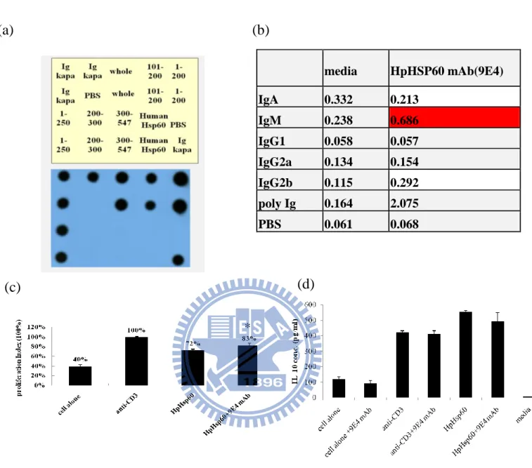
3.7 The research for the effective epitop for proliferative inhibition in HpHSP60
23
The previous results showed that treatments of HpHSP60101-200 and
HpHSP60300-547 could inhibit the proliferation of PBMCs activated by
anti-CD3 antibodies. To research which epitop(s) involved in the proliferative inhibition, anti-HpHSP60 monoclonal antibodies were prepared. The Fig 7a showed that 9E4 monoclonal Ab recognized the fragments including HpHSP60 1-200, HpHSP60101-200 and HpHSP601-250
recombinant proteins, which indicated that 9E4 mAb should recognize the epitop in HpHSP60101-200. The isotype of 9E4 mAb was identified as
IgM (Fig 7b.). Next, we examined whether the 9E4 mAb has the ability to block the proliferative inhibition of PBMCs. Figure 7c and Figure 7d revealed that 9E4 mAb decreasd the proliferative inhibition and IL-10 cytokine expression of PBMCs by rHpHSP60 in the present of anti-CD3 antibody (Fig 7c. and Figure 7d). In future, several anti-HpHSP60 mAbs will be prepared to further examine their functions on the proliferative inhibition of PBMCs.
24
Chapter 4 Discussion
We investigated the immune suppression domain of HpHSP60 involved in the inhibitory activity to anti-CD3 antibody-induced T-cell proliferation. The mechanisms might be the same homological between HpHSP60 and human HSP60. Many literatures reptorted human HSP60 can act as a ligand bind both innate immune cells (macrophages and dendritic cells (Chen et al. 1999, Habich et al. 2002)) and be adaptive immune cells (T cells and B cells (Zanin-Zhorov et al. 2003, Cohen-Sfady et al. 2005)) TLR receptors. When human HSP60 binding with immune cells can secrete IL-10 and enhance regulator T cells function (Zanin-Zhorov et al. 2006a). The sequences alignment of H. pylori, homo sapiens, S. japonicum HSP60 proteins showed that the identity is 52% (Fig. 1), HpHSP60 can interact with immune cell by instead of human HSP60 to achieve inhibit PBMCs proliferation (Fig. 3).
The effects of HpHSP60 on PBMCs were not only on cell growth but also on cytokine secretion. Since Treg cells inhibit proliferation of CD4+
T cells either via IL-10 (Quinn et al. 2000) and TGF-β (Yoshimura et al. 2010), we demonstrated which domains of HpHSP60 function for
25
immune suppression by monitoring the two cytokines. Fig 4 indicated different fragments of rHpHSP60 induced different cytokine expression, PBMCs treatment HpHSP60101-200 and HpHSP60300-547 could increase
IL-10 cytokine expression. According to Song-Nan et al report, the region HpHSP60300-435 was associated with induction of IL-8 in
monocytic cells, we truncated recombinant proteins HpHSP60300-547
confirmed the system workimg that PBMCs tremented with HpHSP60300-547 could expression IL-8 (Fig. 4).
Interestingly, the fragment of HpHSP60300-547 might have two
functions including the inflammation and immune suppression. In other words, this fragment included immune suppression and inflammation domain. HpHSP60300-435 have been exploed that treated with this
recombint proteins could induce dramatically IL-8 releases (Lin et al. 2005), immune suppression domains of HpHSP60300-435 was located in
HpHSP60435-547. In 2008, Delaleu et al. reported human HSP60 a.a.
437-460 led to alter inflammatory chemotaxis and down-regulate Th1 and Th2 effector responses (Zanin-Zhorov et al. 2006a). The human HSP60437-460 (amino acid sequence: vlgggcallrcipaldsltpaned) compared
26
after treated HpHSP60300-547 induced both IL-8 and IL-10 expression. On
the other hand, HpHSP60101-200 also could inhibit T cells’ proliferation as
HpHSP60 (Fig. 3) and induce dramatically IL-10 cytokine releases, but it would abolish this activity without (Fig. 4).
To exclude the possibility for the LPS contamination would cause the same inhibitory phenomenon, and investigate which amino secquences or structure of HpHSP60 fragments for lead to immune suppression, we boiled HpHSP60 to denature proteins but not LPS. In the result, the effect of HpHSP60101-200, HpHSP601-200, HpHSP601-250 on proliferation was
completely inhibited by boiling, which indicated the inhibitory effect was not likely due to the LPS contamination (data not show). However, the proliferative inhibition of PBMCs didn’t completely revise by treatments of rHpHSP60 and HpHSP60300-547. Based on the above reaults, we knew
HpHSP60101-200 and HpHSP60300-547 may be involved the immune
suppression, and HpHSP60101-200 inhibited the proliferations of PBMCs
by its structure, but HpHSP60300-547 inhibited the proliferations by amino
acids sequence.
PBMCs were mainly composed of monocytes and lymphocytes that were involved in both adaptive and innate immunity, and many literatures
27
also reported HpHSP60 was correlation with monocytes (Gobert et al. 2004, Lin et al. 2005). Fig. 5 showed rHpHSP60 fragments could decrease THP-1 proliferation, and different fragments of rHpHSP60 induced different cytokine expression. According to our lab’s unpublished paper have shown that the generation of regulatory T cell by HpHSP60 engageing with Smad-independent TGF-β signaling pathway. Thus, we presumed the HpHSP60 could effect to both monocytes and lymphocytes. In nature state, PBMCs were increase IL-10 and TGF-β cytokine expression after treated with HpHSP60. IL-10 is an anti-inflammatory cytokine (Quinn et al. 2000) and TGF-β can enhance the production of regulatory T cell (Yoshimura et al. 2010). The mechanism of proliferative inhibition was on T cell populations mainly and caused by HpHSP60-induced Treg.
This was confirmed using one mAb prepared by immunizing mice with HpHSP60101-200 fragments. The monoclonal Ab can specific reacted
with HpHSP60101-200 fragments; even human HSP60 has high homology;
yet, it still not reacted. HpHSP60101-200 mAb reacted with rHpHSP60, and
datas showed anti-HSP60101-200 antibodies inhibit the proliferation
28
This result confirms that HpHSP60101-200 maybe is immune suppression
domain.
In conclusion, this study showed that Helicobacter pylori-derived HSP60 might have two functions, included the inflammation and immune suppression. The proliferative inhibition is on T cell populations mainly through IL-10 expression or TGF-β could enhance the production of regulatory T cell. Furthermore, the monoclonal Abs against rHpHSP60101-200 could block the immune suppression ability of HpHSP60.
These results propose that the sequence from 101 to 200 of H. pylori HSP60 may be closely associated with immune suppression. However, it was still not clear about rHpHSP60300-547 for immune suppression. In the
future, we will continued to prepare the monoclonal Abs against rHSP60 (sequence: a. a. 300-547) to prove that rHSP60 (sequence: a. a. 300-547) could down-regulate the immune responses. On the other hand, we will synthetized peptide of rHpHSP60101-200 and rHpHSP60300-547 for
demonstrating the immune suppression domain located on above fragments. This finding may be useful to further study for the chronic infection of H. pylori.
29
Figure and Legend
Figure 1: The blast on the sequences of HSP60 derived from different specisis. The amino acid sequences of H.pylori HSP60, human HSP60
and Schistosoma japonicum HSP60 were retrieved from Pumed database and analyzed by BioEdit.
30
Figure 2: The expressionss of different HpHSP60 fragments. a) The figure simply showed that the different sequence of HpHSP60. b) SDS-PAGE and c) Western blotting analyses of recombinant HpHSP60. Commassie blue staining of different fragments run on a 10%
SDS-PAGE. Lane M means the molecular marker and different fragments (lanes 1: whole protein; lanes 2: 101-200; lanes 3: 1-200; lanes 4: 1-250; lanes 5: 300-547).
(a)
31
Figure 3: Treatment with whole and different fragment HpHSP60 proteins to PBMCs decrease the proliferation in the present of anti-CD3 antibody. PBMCs were isolated from healthy donors and
seeded in 96-well, which was coated with anti-CD3 antibody (OKT3, 1ug/ml). Different fragments of recombinant proteins were co-culture with PBMCs. MTT assay was used to detect the proliferation of PBMCs. *, P<0.05 compared to only coated anti-CD3. (n=4)
* *
32
Figure 4: Production of cytokines in PBMCs. a) IL-10, b) TGF-β, c)
IL-8, d) TNF-α secretion in PBMCs in response to whole and different fragment HpHSP60 proteins. Recombinant heat shock protein 60 was added to medium containing 2 ×10⁵ cells to a final concentration of 1 μg/ml incubated in 96-well plate for 96 h. Cytokines were measured by ELISA . *, P<0.05 compared to only coated anti-CD3. (n=4)
(a)
(b) (a)
(d) (c)
33
Figure 5: Treatment with whole and different fragment HpHSP60 proteins to THP-1 cells decrease the proliferation. Different dosages of
whole and different fragment HpHSP60 proteins were co-culture with THP-1 cells. MTT assay was used to detect the proliferation of THP-1 cells. *, P<0.05 compared to without HpHSP60 treatment. (n=3)
*
*
*
* *
34
Figure 6: Production of cytokines in THP-1 cells. a) IL-10, b) TGF-β, c)
IL-8, d) TNF-α secretion in THP-1 cells in response to whole and different fragment HpHSP60 proteins. Recombinant heat shock protein 60 was added to medium containing 2 ×10⁵ cells to a final concentration of 1 μg/ml incubated in 96-well plate for 96 h. Cytokines were measured by ELISA. *, P<0.05 compared to only coated anti-CD3. (n=3)
(a) (b)
(c)
(d) (c)
35
Figure 7: The characterizations and application of monoclonal Ab. a)
Dot blotting for investigating the HpHsp60 monoclonal to recognize which fragments. 50 ng of different fragments HpHsp60 were spotted on the nitrocellulose membrane and probed with HpHsp60 mAb (1:10000) followed by goat anti-mouse IgG Abs conjugated with HRP (1:10000). b) Determine of the isotype of HpHSP60 mAb by ELISA. c) Anti-HSP60101-200 antibodies inhibit the proliferation decreasing of
PBMCs in the present of anti-CD3 antibody. d) Anti-HSP60101-200
antibodies inhibit the IL-10 cytokines expression of PBMCs in the present of anti-CD3 antibody.
*, P<0.05 compared to with HpHsp60 treatment and with coated anti-CD3. (n=3) media HpHSP60 mAb(9E4) IgA 0.332 0.213 IgM 0.238 0.686 IgG1 0.058 0.057 IgG2a 0.134 0.154 IgG2b 0.115 0.292 poly Ig 0.164 2.075 PBS 0.061 0.068 (a) (b) (c) (d) *
36
References
Cappello, F., Conway De Macario, E., Di Felice, V., Zummo, G. & Macario, A., 2009. Chlamydia trachomatis infection and anti-hsp60 immunity: The two sides of the coin. PLoS Pathog, 5 (8), e1000552.
Cello, J.P., 1995. Helicobacter pylori and peptic ulcer disease. American Journal of Roentgenology, 164 (2), 283.
Chen, W., Syldath, U., Bellmann, K., Burkart, V. & Kolb, H., 1999. Human 60-kda heat-shock protein: A danger signal to the innate immune system. The Journal of Immunology, 162 (6), 3212.
Cohen-Sfady, M., Nussbaum, G., Pevsner-Fischer, M., Mor, F., Carmi, P., Zanin-Zhorov, A., Lider, O. & Cohen, I.R., 2005. Heat shock protein 60 activates b cells via the tlr4-myd88 pathway. The Journal of Immunology, 175 (6), 3594.
Cohen-Sfady, M., Pevsner-Fischer, M., Margalit, R. & Cohen, I.R., 2009. Heat shock protein 60, via myd88 innate signaling, protects b cells from apoptosis, spontaneous and induced. The Journal of Immunology, 183 (2), 890.
37
Crabtree, J., Covacci, A., Farmery, S., Xiang, Z., Tompkins, D., Perry, S., Lindley, I. & Rappuoli, R., 1995. Helicobacter pylori induced interleukin-8 expression in gastric epithelial cells is associated with caga positive phenotype. Journal of clinical pathology, 48 (1), 41. Dao Nguyen, X. & Robinson, D.S., 2004. Fluticasone propionate
increases cd4+ cd25+ t regulatory cell suppression of
allergen-stimulated cd4+ cd25- t cells by an il-10-dependent
mechanism. Journal of allergy and clinical immunology, 114 (2), 296-301.
Delaleu, N., Madureira, A.C., Immervoll, H. & Jonsson, R., 2008. Inhibition of experimental sjogren's syndrome through immunization with hsp60 and its peptide amino acids 437¡v460. Arthritis & Rheumatism, 58 (8), 2318-2328.
Ellis, J., 1992. Cytosolic chaperonin confirmed. NATURE, 358, 191-192. Gobert, A.P., Bambou, J.C., Werts, C., Balloy, V., Chignard, M., Moran,
A.P. & Ferrero, R.L., 2004. Helicobacter pylori heat shock protein 60 mediates interleukin-6 production by macrophages via a toll-like receptor (tlr)-2-, tlr-4-, and myeloid differentiation factor 88-independent mechanism. Journal of Biological Chemistry, 279
38 (1), 245.
Habich, C., Baumgart, K., Kolb, H. & Burkart, V., 2002. The receptor for heat shock protein 60 on macrophages is saturable, specific, and distinct from receptors for other heat shock proteins. The Journal of Immunology, 168 (2), 569.
Hocker, M. & Hohenberger, P., 2003. Helicobacter pylori virulence factors--one part of a big picture. Lancet, 362 (9391), 1231.
Huang, J.Q., Zheng, G.F., Sumanac, K., Irvine, E.J. & Hunt, R.H., 2003. Meta-analysis of the relationship between caga seropositivity and gastric cancer. Gastroenterology, 125 (6), 1636-1644.
Huesca, M., Borgia, S., Hoffman, P. & Lingwood, C.A., 1996. Acidic ph changes receptor binding specificity of helicobacter pylori: A binary adhesion model in which surface heat shock (stress) proteins mediate sulfatide recognition in gastric colonization. Infection and immunity, 64 (7), 2643.
Kandulski, A., Wex, T., Kuester, D., Peitz, U., Gebert, I., Roessner, A. & Malfertheiner, P., 2008. Naturally occurring regulatory t cells (cd4+,
cd25high, foxp3+) in the antrum and cardia are associated with
39 β]1. Helicobacter, 13 (4), 295-303.
Lin, C.S., He, P.J., Tsai, N.M., Li, C.H., Yang, S.C., Hsu, W.T., Wu, M.S., Wu, C.J., Cheng, T.L. & Liao, K.W., 2010. A potential role for helicobacter pylori heat shock protein 60 in gastric tumorigenesis. Biochemical and biophysical research communications, 392 (2), 183-189.
Lin, C.Y., Huang, Y.S., Li, C.H., Hsieh, Y.T., Tsai, N.M., He, P.J., Hsu, W.T., Yeh, Y.C., Chiang, F.H., Wu, M.S., Chang, C.C. & Liao, K.W., 2009. Characterizing the polymeric status of helicobacter pylori heat shock protein 60. Biochem Biophys Res Commun, 388 (2), 283-9.
Lin, S.N., Ayada, K., Zhao, Y., Yokota, K., Takenaka, R., Okada, H., Kan, R., Hayashi, S., Mizuno, M. & Hirai, Y., 2005. Helicobacter pylori heat-shock protein 60 induces production of the pro-inflammatory cytokine il8 in monocytic cells. Journal of medical microbiology, 54 (3), 225.
Lundgren, A., Stromberg, E., Sjoling, A., Lindholm, C., Enarsson, K., Edebo, A., Johnsson, E., Suri-Payer, E., Larsson, P. & Rudin, A., 2005. Mucosal foxp3-expressing cd4+ cd25high regulatory t cells in
40
helicobacter pylori-infected patients. Infection and immunity, 73 (1), 523.
Organization, W.H. & Humans, I.a.F.R.O.C.W.G.O.T.E.O.C.R.T., 1994. Iarc monographs on the evaluation of carcinogenic risks to humans: Schistosomes, liver flukes and helicobacter pylori: International Agency for Research on Cancer.
Parsonnet, J., Hansen, S., Rodriguez, L., Gelb, A.B., Warnke, R.A., Jellum, E., Orentreich, N., Vogelman, J.H. & Friedman, G.D., 1994. Helicobacter pylori infection and gastric lymphoma. New England Journal of Medicine, 330 (18), 1267-1271.
Peek Jr, R.M. & Blaser, M.J., 2002. Helicobacter pyloriand gastrointestinal tract adenocarcinomas. NATURE REVIEWS| CANCER, 2, 29.
Quinn, T.J., Taylor, S., Wohlford-Lenane, C.L. & Schwartz, D.A., 2000. Il-10 reduces grain dust-induced airway inflammation and airway hyperreactivity. J Appl Physiol, 88 (1), 173-9.
Su, B., Ceponis, P.J.M., Lebel, S., Huynh, H. & Sherman, P.M., 2003. Helicobacter pylori activates toll-like receptor 4 expression in gastrointestinal epithelial cells. Infection and immunity, 71 (6),
41 3496.
Supajatura, V., Ushio, H., Wada, A., Yahiro, K., Okumura, K., Ogawa, H., Hirayama, T. & Ra, C., 2002. Cutting edge: Vaca, a vacuolating cytotoxin of helicobacter pylori, directly activates mast cells for migration and production of proinflammatory cytokines. The Journal of Immunology, 168 (6), 2603.
Uemura, N., Okamoto, S., Yamamoto, S., Matsumura, N., Yamaguchi, S., Yamakido, M., Taniyama, K., Sasaki, N. & Schlemper, R.J., 2001. Helicobacter pylori infection and the development of gastric cancer. New England Journal of Medicine, 345 (11), 784-789.
Wang, X., Zhou, S., Chi, Y., Wen, X., Hoellwarth, J., He, L., Liu, F., Wu, C., Dhesi, S. & Zhao, J., 2009. Cd4+ cd25+ treg induction by an hsp60 derived peptide sjmhe1 from schistosoma japonicum is tlr2 dependent. European journal of immunology, 39 (11), 3052-3065. Yoshimura, A., Wakabayashi, A. & Mori, T., 2010. Cellular and
molecular basis for the regulation of inflammation by tgf-beta. J. Biochem., 147, 781-792.
Zanin-Zhorov, A., Cahalon, L., Tal, G., Margalit, R., Lider, O. & Cohen, I.R., 2006a. Heat shock protein 60 enhances cd4 cd25 regulatory t
42
cell function via innate tlr2 signaling. J Clin Invest, 116 (7), 2022-2032.
Zanin-Zhorov, A., Cahalon, L., Tal, G., Margalit, R., Lider, O. & Cohen, I.R., 2006b. Heat shock protein 60 enhances cd4+cd25+ regulatory t cell function via innate tlr2 signaling. The Journal of Clinical Investigation, 116, 2022–2032.
Zanin-Zhorov, A., Nussbaum, G., Franitza, S., Cohen, I.R. & Lider, O., 2003. T cells respond to heat shock protein 60 via tlr2: Activation of adhesion and inhibition of chemokine receptors. The FASEB journal, 17 (11), 1567.
Zheng, S.G., Wang, J.H., Gray, J.D., Soucier, H. & Horwitz, D.A., 2004. Natural and induced cd4+ cd25+ cells educate cd4+ cd25- cells to
develop suppressive activity: The role of il-2, tgf-β], and il-10. The Journal of Immunology, 172 (9), 5213.