Anti-oxidative and Anti-inflammatory Protection from Carnosine in
1Striatum of MPTP-treated Mice
23 4
SHIH-JEI TSAI#,§, WEI-WEN KUOø, WEN-HU LIU‡, MEI-CHIN YIN*, 5
6
#
School of Medicine, Chung Shan Medical University, Taichung, Taiwan 7
§
Department of Neurology, Chung Shan Medical University Hospital, Taichung, Taiwan 8
ø
Department of Biological Science & Technology, China Medical University, Taichung City, 9
Taiwan 10
‡Radiation Safety Office, Chung Shan Medical University, Taichung, Taiwan 11
*
Department of Nutrition, China Medical University, Taichung City, Taiwan 12
Department of Health and Nutrition Biotechnology, Asia University, Taichung County, 13
Taiwan 14
15 16
running title: neuroprotective effects of carnosine 17
To whom correspondence should be addressed: Dr. Mei-chin Yin, Professor, Department 18
of Nutrition, China Medical University, 16th Floor, 91, Hsueh-shih Rd., Taichung City, 19 Taiwan, ROC 20 TEL: 886-4-22053366 ext. 7510 21 FAX: 886-4-22062891 22 Email: [email protected] 23
ABSTRACT
1
Mice treated with 1-methyl-4-phenyl-1,2,3,6-tetrahydropyridine (MPTP) were used to 2
examine the neuroprotective effects of carnosine. Carnosine at 0.5, 1 and 2 g/L was 3
directly added to the drinking water for 4 wk. MPTP treatment significantly depleted 4
striatal glutathione content, reduced the activity of glutathione peroxidase (GPX), 5
superoxide dismutase (SOD) and catalase, increased malondialdehyde and reactive oxygen 6
species levels, and elevated interleukin-6, nitrite and tumor necrosis factor-production as 7
well as enhanced inducible nitric oxide synthase (iNOS) activity in striatum (P<0.05). 8
The pre-intake of carnosine significantly attenuated MPTP-induced glutathione loss, 9
retained the activity of GPX and SOD, diminished oxidative stress, and lowered 10
inflammatory cytokines and nitrite levels as well as suppressed iNOS activity (P<0.05). 11
MPTP treatment significantly suppressed GPX mRNA expression and enhanced iNOS 12
mRNA expression (P<0.05). Carnosine pre-intake significantly elevated GPX mRNA 13
expression and declined iNOS mRNA expression (P<0.05). Pre-intake of carnosine also 14
significantly improved MPTP-induced dopamine depletion and maintained 15
3,4-dihydroxyphenylacetic acid and homovanillic acid levels (P<0.05). These results 16
suggest that carnosine could provide anti-oxidative and anti-inflammatory protection for the 17
striatum againstthedevelopmentofParkinson’sdisease. 18
19
KEYWORDS: carnosine; Parkinson’sdisease; oxidative stress; iNOS activity; mRNA 20
expression 21
Abbreviations
1
DA, dopamine; DAT, dopamine transporter; DOPAC, 3,4-dihydroxyphenylacetic acid; 2
GPX, glutathione peroxidase; GSH, glutathione; HVA, homovanillic acid; IL-1, 3
interleukin-1; iNOS, inducible nitric oxide synthase; MDA, malonyldialdehyde; MPTP, 4
1-methyl-4-phenyl-1,2,3,6-tetrahydropyridine; NO, nitric oxide; PD, Parkinson’sdisease; 5
ROS, reactive oxygen species; RT-PCR, reverse transcription-polymerase chain reaction; 6
SOD, superoxide dismutase; TNF-, tumor necrosis factor-; 7
INTRODUCTION
1
Parkinson’sdisease (PD)isoneofthe majorneurodegenerativediseasesin theworld. 2
It is characterized by massive degeneration of nigrostriatal dopamine (DA) neurons in the 3
substantia nigra pars compacta and the resultant deficiency in the neurotransmitter DA at 4
the nerve terminals in the striatum (1, 2). The biochemical and cellular changes that occur 5
after administration of 1-methyl-4-phenyl-1,2,3,6-tetrahydropyridine (MPTP) in animals 6
are remarkably similar to those seen in idiopathic PD; thus, MPTP-induced PD has been 7
widely used as a model for investigating pathogenic mechanisms of PD (3, 4). Oxidative 8
stress and neuroinflammatory processes have been implicated as important mechanisms 9
responsible for neuronal death in PD because reactive oxygen species (ROS), oxidized DA 10
metabolites, nitric oxide (NO) and inflammatory cytokines are toxic to nigral neurons (5, 6). 11
Thus, there is an increasing interest to examine the use of appropriate agent(s) to prevent or 12
attenuate oxidative and inflammatory damage in PD (7, 8). 13
Carnosine (beta-alanyl-l-histidne) is endogenously synthesized peptide present in brain, 14
skeletal muscle and liver (9). It has been reported that carnosine concentration in rat 15
tissues could be increased by dietary supplementation (10). Several studies have indicated 16
that this compound could provide both anti-oxidative and anti-inflammatory protection 17
against diabetic deterioration and ethanol-induced chronic liver injury in mice (11, 12). 18
Shen et al. (13) and Fu et al. (14) reported that carnosine could attenuate 19
N-methyl-D-aspartate- and Abeta42-induced neurotoxicity in differentiated rat PC12 cells 20
through carnosine-histidine-histamine pathway and/or inhibiting glutamate release. Those 21
previous studies support that carnosine is a potent neuroprotective agent against oxidative 22
and inflammatory progression in neurodegenerative diseases; however, further animal study 23
In this study, MPTP was used to induce neurotoxicity in mice. Both anti-oxidative 1
and anti-inflammatory activities of carnosine were examined in the mouse striatum, in 2
which the impact of this agent at various doses on striatal content of glutathione (GSH), 3
ROS and nitrite, activity of glutathione peroxidase (GPX) and inducible nitric oxide 4
synthase (iNOS), and level of tumor necrosis factor (TNF)-and interleukin (IL)-6 was 5
determined. Furthermore, the effect of this agent on striatal level of DA metabolites such 6
as 3,4-dihydroxyphenylacetic acid (DOPAC) and mRNA expression of DA transporter 7
(DAT) was also evaluated. 8
9
MATERIALS AND METHODS
10
Animals and Diets. Three- to four-week-old male C57BL/6 mice were obtained
11
from National Laboratory Animal Center (National Science Council, Taipei City, Taiwan). 12
Mice were housed on a 12-h light-12-h dark schedule, and fed with water and mouse 13
standard diet for one week acclimation. Use of the mice was reviewed and approved by 14
both Chung Shan Medical University and China Medical University animal care 15
committees. 16
Experimental Design. Carnosine (98%), purchased from Sigma Chemical Co. (St.
17
Louis, MO, USA), at 0.5, 1 or 2 g/L, was directly added to the drinking water. After 18
4-wk supplementation, mice were treated by daily subcutaneous injection of vehicle 19
saline or MPTP (24 mg/kg body weight) for 6 consecutive days. Mice were sacrificed 20
by decapitation. Brain was quickly removed and the striatum was collected. The 21
striatum at 0.15 g was homogenized on ice in 2 mL of phosphate buffer (pH 7.2) and the 22
filtrate was collected. Protein concentration of striatal filtrate was determined by a 23
commercial assay kit (Pierce Biotechnology Inc., Rockford, IL, USA) with bovine serum 24
albumin used as standard. In all experiments, the sample was diluted to a final 1
concentration of 1 mg protein/mL. 2
Determination of MPP+ Level. The MPP+ level in striatum was determined 3
according to HPLC method of Richardson et al. (15). Briefly, striata were sonicated in 4
5% trichloroacetic acid and centrifuged for 10 min at 14,000 g, and the supernatants were 5
collected for analysis. HPLC was equipped with a reverse-phase C18 column (Alltech 6
Associates Inc., Deerfield, IL, USA), and the ultraviolet detector was set at 290 nm. 7
MPP+was identified by comparing retention time with a known standard and concentration 8
was calculated from a standard curve. 9
Measurement of DA, DOPAC and Homovanillic Acid (HVA). The levels of DA,
10
DOPAC and HVA were determined by HPLC methods (15). Briefly, the striatum was 11
homogenized in 0.1 mol/L of perchloric acid containing 0.1 mM 12
ethylene-diaminetetraacetic acid. After centrifuging at 12,000 g for 60 min at 4°C, the 13
supernatant was collected for analysis. HPLC equipped with a coulometric electrode 14
array detector was used to quantify. 15
Determination of Lipid Oxidation and ROS. Malonyldialdehyde (MDA), an index
16
of lipid peroxidation, was measured by using a commercial assay kit (OxisResearch, 17
Portland, OR, USA). The method described in Gupta et al. (16) was used to measure 18
ROS level. Briefly, 10 mg tissue was homogenized in 1 mL of ice cold 40 mM Tris–HCl 19
buffer (pH 7.4), and further diluted to 0.25% with the same buffer. Then, samples were 20
divided into two equalfractions. In one fraction,40 μL 1.25 mM 2’, 7’-dichlorofluorescin 21
diacetatein methanolwasadded forROS estimation. Anotherfraction,in which 40 μL 22
methanol was added, served as a control for auto fluorescence. After incubating for 23
using a fluorescence plate reader. 1
Analyses for Carnosine, GSH and Total Antioxidant Capacity. Carnosine
2
concentration was quantified according to the method described in Kamal et al. (17) by 3
HPLC equipped with a 5-m Waters Symmetry C18 column (250 x 4.6 mm). GSH 4
concentration in striatal filtrate was determined by a commercial colorimetric GSH assay 5
kit according to the manufacturer’s instruction (OxisResearch, Portland, OR, USA). 6
Reduced GSH was determined in this study. Total antioxidant capacity was measured via 7
monitoring the change in absorbance at 593 nm by the method of Benzie and Strain (18), in 8
which ferric tripyridyltriazine complex could be reduced by nonenzymatic antioxidants 9
such as ascorbic acid presented in the sample. 10
Catalase, superoxide dismutase (SOD) and GPX Activity Assay. The activities of
11
catalase, SOD, and GPX in the striatum were determined by catalase, SOD, and GPX assay 12
kits (Calbiochem, EMD Biosciences, Inc., San Diego, CA, USA). The enzyme activity 13
was expressed in U/mg protein. 14
Cytokine Measurements. Striatal levels of IL-1, IL-6, TNF- and monocyte
15
chemoattractant protein (MCP)-1 were measured by ELISA methods using cytoscreener 16
immunoassay kits (Bio-Source International, Camarillo, CA, USA). The sensitivities of 17
assay with the detection limit were 5 pg/mL for IL-1and IL-6, and 10 pg/mL for TNF- 18
and MCP-1. 19
Nitrite Assay and NOS Activity. The production of nitric oxide was determined by
20
measuring the formation of nitrite. Briefly, 100L of supernatant was treated with nitrate 21
reductase, NADPH and FAD, and incubated for 1 h at 37 ºC in the dark. After 22
centrifuging at 6,000 g, the supernatant was mixed with Griess reagent for color 23
development. The absorbance at 540 nm was measured and compared with a sodium 1
nitrite standard curve. The methods described in Sutherland et al. (19) were used to 2
measure total NOS and iNOS activities. Total NOS activity was determined via 3
incubating 30 L of homogenate with 10 mM -nicotinamide adenine dinucleotide 4
phosphate, 10 mM L-valine, 3000 U/mL calmodulin, 5 mM tetrahydrobiopterin, 10 mM 5
CaCl2, and a mixture of 100M L-arginine containing L-[3H]arginine. iNOS activity was
6
measured excluding CaCl2 and adding 10 mM ethylene glycol tetraacetic acid. Then,
7
reaction was stopped by 1 mL of 20 mM HEPES buffer (pH 5.5). A dowex column was 8
used to separate L-[3H]arginine and L-[3H]citrulline. The amount of L-[3H]citrulline was 9
assessed by liquid scintillation counter (Beckman Coultier, LS6500, Fullerton, CA, USA). 10
Reverse Transcription Polymerase Chain Reaction (RT-PCR) for mRNA
11
Expression. Part of the striatum was homogenized in guanidinethiocyanate, and total RNA
12
was extracted. Two micrograms of total RNA was used to generate cDNA, which was 13
amplified using Taq DNA polymerase. PCR was carried out in 50 mL of reaction mixture 14
containing Taq DNA polymerase buffer (20 mM Tris-HCl, pH 8.4, 50 mM KCl, 200 mM 15
dNTP, 2.5 mM MgCl2, 0.5 mM of each primer) and 2.5 U Taq DNA polymerase. The
16
specific oligonucleotide primers for DAT, GPX, iNOS and glyceraldehyde-3-phosphate 17
dehydrogenase (GAPDH, the housekeeping gene) are as follows (8, 20), DAT: forward 18
5’-ATC AAC CCA CCG CAG ACA CCA GT-3’,reverse,5’-GGC ATC CCG GCA ATA 19
ACC AT-3’;GPX:forward,5’-CCT CAA GTA CGT CCG GCC TG-3’,reverse,5’-CAA 20
CAT CGT TGC GAC ACA CC-3’;iNOS;forward,5’-ATG ACC AGT ATA AGG CAA 21
GC-3’,reverse,5’-GCT CTG GAT GAG CCT ATA TTG-3’; GAPDH:forward,5’-TGA 22
TGA CAT CAA GAA GGT GGT GAA G-3’,reverse,5’-CCT TGG AGG CCA TGT 23
AGG CCA T-3’. The cDNA was amplified under the following reaction conditions: 94 ºC 1
for 1 min, 57 ºC for 1 min, and 72 ºC for 1 min. 28 cycles were performed for GAPDH 2
and 32 cycles were performed for DAT, GPX and iNOS. A 10-L aliquot of each PCR 3
product was analyzed on a 2% agarose gel containing 0.5 g/mL of ethidium bromide. 4
Quantitative analysis for PCR products was performed by a BAS 2000 BIO-image analyzer 5
(Fuji Photo Film Co., Tokyo, Japan), in which PCR products were illuminated by 6
computerized densitometric scanning of the images. mRNA level was calculated as 7
percentage of the control group. 8
Statistical Analysis. The effect of each treatment was analyzed from 10 mice (n = 10)
9
in each group. All data were expressed as mean ± standard deviation (SD). Statistical 10
analysis was done using one-way analysis of variance (ANOVA), and post-hoc 11
comparisons were carried out using Dunnet's t-test. P values <0.05 were considered as
12 significant. 13 14 RESULTS 15
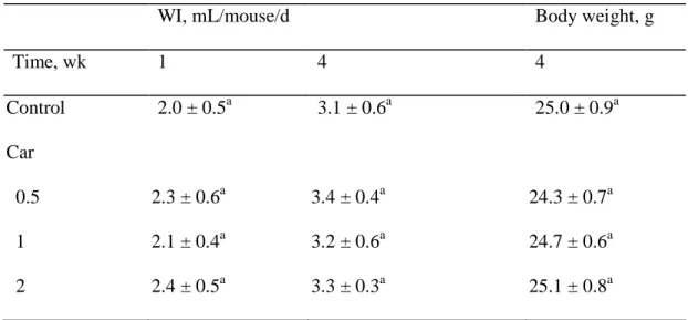
As shown in Table 1, the intake of carnosine did not affect daily water intake and final 16
body weight (P>0.05). The treatment of carnosine and/or MPTP did not significantly 17
affect the weight of whole brain and striatum (data not shown). Compared with MPTP 18
treatment alone, the pre-intake of carnosine did not significantly affect striatal MPP+level 19
(P>0.05, data not shown). 20
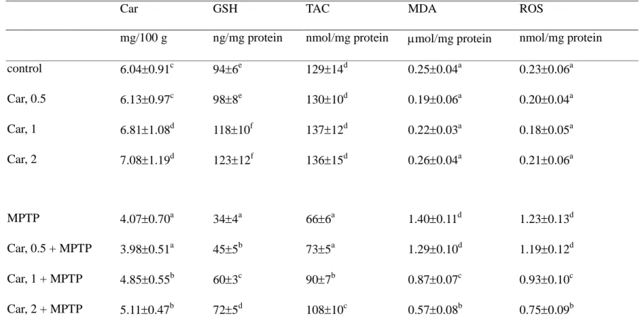
The effects of carnosine intake and/or MPTP treatment on striatal levels of carnosine, 21
GSH, total antioxidant capacity, MDA and ROS are presented in Table 2. The intake of 22
carnosine at 1 and 2 g/L significantly increased carnosine and GSH content in striatum 23
(P<0.05). MPTP treatment significantly decreased levels of carnosine, GSH and total 1
antioxidant capacity, and increased the production of MDA and ROS (P<0.05). However, 2
the pre-intake of carnosine dose-dependently diminished MPTP-induced GSH loss 3
(P<0.05). Furthermore, carnosine pre-treatments at 1 and 2 g/L significantly retained 4
carnosine and total antioxidant capacity levels, as well as decreased production of MDA 5
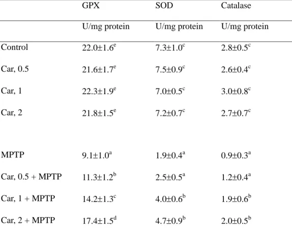
and ROS (P<0.05). The effect of carnosine and/or MPTP treatment on the activity of 6
GPX, SOD and catalase is presented in Table 3. Carnosine pre-treatments alone did not 7
affect the activity of these enzymes (P>0.05). MPTP treatment significantly reduced the 8
activity of three test enzymes (P<0.05); however, the pre-intake of carnosine 9
dose-dependently attenuated MPTP-induced GPX activity loss; but only at 1 and 2 g/L 10
significantly retained the activity of SOD and catalase (P<0.05). 11
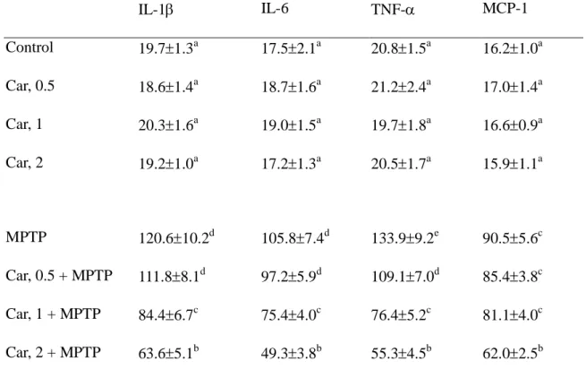
The effect of carnosine and/or MPTP treatment on the level of IL-1, IL-6, TNF- 12
and MCP-1 is presented in Table 4. Carnosine pre-treatments alone did not affect these 13
cytokines (P>0.05); however, MPTP treatment significantly increased the release of four 14
test cytokines (P<0.05). The pre-intake of carnosine dose-dependently decreased TNF- 15
production; but this agent significantly lowered IL-1and IL-6 levels at 1 and 2 g/L 16
(P<0.05). Carnosine pre-intake only at 2 g/L significantly reduced MPTP-caused MCP-1 17
release (P<0.05). 18
As shown in Figure 1, MPTP treatment significantly increased nitrite production and 19
elevated total NOS and iNOS activities (P<0.05). The pre-intake of carnosine 20
dose-dependently decreased nitrite production and iNOS activity (P<0.05); but this agent at 21
1 and 2 g/L significantly lowered total NOS activity (P<0.05). MPTP treatment 22
significantly down-regulated DAT and GPX mRNA expression, and up-regulated iNOS 23
mRNA expression (Figure 2, P<0.05). The pre-intake of carnosine dose-dependently 1
enhanced GPX expression and suppressed iNOS expression (P<0.05). Carnosine 2
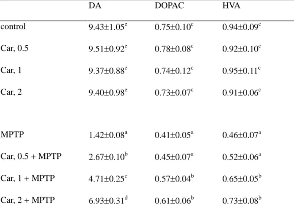
pre-treatments failed to affect DAT expression (P>0.05). As shown in Table 5, MPTP 3
treatment significantly decreased the striatal content of DA, DOPAC and HVA (P<0.05). 4
The pre-intake of carnosine dose-dependently attenuated MPTP-induced DA loss; but only 5
at 1 and 2 g/L significantly retained DOPAC and HVA content (P<0.05). 6
7
DISCUSSION
8
In our present study, carnosine pre-intake markedly attenuated MPTP-caused 9
oxidative and inflammatory stress by lowering ROS, NO and inflammatory cytokines 10
production, as well as mediating activity and mRNA expression of GPX and iNOS in 11
striatum. Because carnosine pre-intake did not affect the striatal MPP+ level; thus, the 12
observed antioxidant protective action from this agent was not due to its scavenging 13
activity on MPP+. Therefore, our results support that carnosine is an effective 14
anti-oxidative and anti-inflammatory agent against the development of neurodegenerative 15
diseases such as PD. Since carnosine could mediate the mRNA expression of GPX and 16
iNOS, this agent might exert its functions at the level of transcription. 17
The increased carnosine content in brain via dietary intake as we observed implied that 18
this compound was able to penetrate the blood-brain barrier. It is reported that carnosine 19
could scavenge free radicals and chelate divalent metal ions (21). Thus, partial 20
anti-oxidative protection for MPTP-treated mice from this compound should be ascribed to 21
its free radical scavenging action. Postmortem study indicated that GSH content in 22
substantia nigra of PD patients was decreased; and GSH depletion has been proposed as the 23
brain may delay PD progression. In our present study, carnosine intake elevated GSH 1
content in brain from mice without MPTP treatment. This finding suggested that 2
carnosine might be able to spare GSH and favor GSH homeostasis, which definitely 3
contributed to enhance anti-oxidative protection for brain. In addition, we notified that 4
carnosine pre-intake effectively attenuated MPTP-caused decline in striatal GSH and total 5
antioxidant capacity. These results implied that carnosine participated in the 6
anti-oxidative defense to protect brain of MPTP-treated mice via sparing other antioxidant 7
agents or elevating the overall reducing power of this tissue. On the other hand, we found 8
that carnosine intake markedly alleviated subsequent MPTP-caused activity decrease in 9
GPX, SOD and catalase, and dose-dependently up-regulated mRNA expression of GPX, 10
which further diminished oxidative damage in this tissue. Therefore, the results of our 11
present study support that carnosine could mitigate oxidative injury in brain of 12
MPTP-treated mice via both non-enzymatic and enzymatic antioxidant protective actions. 13
Increased level of proinflammatory cytokines such as TNF- and IL-6 in the 14
nigrostriatal region of postmortem brains from patients with sporadic PD is reported (23). 15
The inhibition of TNF-response has been considered as a promising target for developing 16
anti-parkinsonian drugs for inflammatory treatment in PD (24). Our present study found 17
that carnosine dose-dependently decreased MPTP-induced TNF-production, and also 18
effectively lowered IL-1and IL-6 release at 1 and 2 g/L. Thus, carnosine could alleviate 19
inflammatory damage via diminishing inflammatory cytokines production. On the other 20
hand, iNOS is one of three NOS forms in the central nerves system. Overexpressed iNOS 21
and elevated NO production are the most important neurotoxic effectors contributed to the 22
loss of dopaminergic neurons and inflammatory deterioration of PD (25). Furthermore, 23
marked up-regulation of iNOS in the nigrostriatal region of postmortem brains from PD 1
patients has been observed (26). Thus, inflammatory response in PD could be also 2
improved via suppressing iNOS activity and lowering NO level. In our present study, 3
carnosine pre-intake dose-dependently suppressed iNOS mRNA expression and inhibited 4
activity of total NOS and iNOS, which consequently lowered NO production. It is 5
reported that TNF--mediated activation of NF-kappaB is responsible for iNOS 6
upregulation (27). However, Eberhardt et al. (28) indicated that NO is required for the 7
expression of proinflammatory cytokines in macrophages. Obviously, there is a closed 8
link in inflammatory regulation between NO/iNOS and proinflammatory cytokines such as 9
TNF-. Thus, carnosine seems a more efficient anti-inflammatory agent because it could 10
suppress both production and activity of NO, iNOS and proinflammatory cytokines. 11
DAT is involved in DA homeostasis and sensitivity to dopaminergic neurotoxicants 12
(29). As reported by others, MPTP depleted the striatal DA level (1) and suppressed gene 13
expression of DAT (30). The results of our present study agreed those previous studies. 14
However, we found the pre-intake of carnosine substantially locked MPTP-induced DA 15
depletion in the striatum without alleviating MPTP-induced DAT depletion. Apparently, 16
the increased DA level from this compound was not associated with DAT expression. It 17
is highly possible that carnosine by its anti-oxidative and anti-inflammatory actions directly 18
protected nigrostriatal dopaminergic neurons and ameliorated DA degeneration in the 19
substantia nigra pars compacta. Since DA depletion was improved, the increased levels of 20
DOPAC and HVA, metabolites of DA, could be explained. Carnosine is a naturally 21
occurring dipeptide. Thus, the supplement of this compound might be safe. The major 22
food source of carnosine is muscle foods such as chicken and beef (31). These muscle 23
foods also contain considerable fat. Based on healthy consideration, it may not be 1
practical to increase muscle foods consumption in order to obtain carnosine. 2
In conclusion, the pre-intake of carnosine effectively alleviated MPTP-induced 3
oxidative stress, inflammatory damage and DA loss. This agent exhibited anti-oxidative 4
and anti-inflammatory activities by increasing GSH and carnosine content, elevating the 5
activity of GPX and SOD, decreasing IL-6 and TNF-levels, suppressing NO production 6
and iNOS activity, as well as regulating mRNA expression of GPX and iNOS in striatum, 7
which consequently retained levels of neurotransmitters such as DA, DOPAC and HVA. 8
These results suggest that carnosine is a potent neuroprotective agent against the 9
development of PD. 10
11 12
LITERATURE CITED
1
1. Schapira, A.H.Presentand future drug treatmentforParkinson’sdisease.J. Neurol.
2
Neurosurg. Psychiatry 2005, 76, 1472-1478.
3
2. Fuxe, K.; Manger, P.; Genedani, S.; Agnati, L. The nigrostriatal DA pathway and 4
Parkinson's disease. J. Neural. Transm. Suppl. 2006, 70, 71-83. 5
3. Fabre, E.; Monserrat, J.; Herrero, A.; Barja, G.; Leret, M.L. Effect of MPTP on brain 6
mitochondrial H2O2 and ATP production and on dopamine and DOPAC in the 7
striatum. J. Physiol. Biochem. 1999, 55, 325-331. 8
4. Bezard, E.; Dovero, S.; Prunier, C.; Ravenscroft, P.; Chalon, S.; Guilloteau, D.; 9
Crossman, A.R.; Bioulac, B.; Brotchie, J.M.; Gross, C.E. Relationship between the 10
appearance of symptoms and the level of nigrostriatal degeneration in a progressive 11
1-methyl-4-phenyl-1,2,3,6-tetrahydropyridine-lesioned macaquemodelofParkinson’s 12
disease. J. Neurosci. 2001, 21, 6853-6861. 13
5. Beal, M.F.Mitochondria,oxidativedamage,and inflammation in Parkinson’sdisease. 14
Ann. N. Y. Acad. Sci. 2003, 991, 120-131.
15
6. Miller, R.L.; James-Kracke, M.; Sun, G.Y.; Sun, A.Y. Oxidative and inflammatory 16
pathwaysin Parkinson’sdisease.Neurochem. Res. 2009, 34, 55-65.
17
7. Suganuma, H.; Hirano, T.; Arimoto, Y.; Inakuma, T. Effect of tomato intake on 18
striatalmonoaminelevelin amousemodelofexperimentalParkinson’sdisease.J.
19
Nutr. Sci. Vitaminol. 2002, 48, 251-254.
20
8. Chen, C.M.; Yin, M.C.; Hsu, C.C.; Liu, T.C. Anti-oxidative and anti-inflammatory 21
effects of four cysteine-containing agents in striatum of MPTP-treated mice. Nutrition 22
2007, 23, 589–597. 23
9. Bonfanti, L.; Peretto, P.; De Marchis, S.; Fasolo, A. Carnosine-related dipeptides in the 1
mammalian brain. Prog. Neurobiol. 1999, 59, 333-353. 2
10. Maynard, L.M.; Boissonneault, G.A.; Chow, L.K.; Bruckner, G.G. High levels of 3
dietary carnosine are associated with increased concentrations of carnosine and 4
histidine in rat soleus muscle. J. Nutr. 2001, 131, 287-290. 5
11. Liu, W.H.; Liu, T.C.; Yin, M.C. Beneficial effects of histidine and carnosine on 6
ethanol-induced chronic liver injury. Food Chem. Toxicol. 2008, 46, 1503-1509. 7
12. Hipkiss, A.R. Carnosine,diabetesand Alzheimer’sdisease.Expert. Rev. Neurother.
8
2009, 9, 583-585.
9
13. Shen, Y.; Hu, W.W.; Fan, Y.Y.; Dai, H.B.; Fu, Q.L.; Wei, E.Q.; Luo, J.H.; Chen, Z. 10
Carnosine protects against NMDA-induced neurotoxicity in differentiated rat PC12 11
cells through carnosine-histidine-histamine pathway and H(1)/H(3) receptors. Biochem. 12
Pharmacol. 2007, 73, 709-717.
13
14. Fu, Q.; Dai, H.; Hu, W.; Fan, Y.; Shen, Y.; Zhang, W.; Chen, Z. Carnosine protects 14
against Abeta42-induced neurotoxicity in differentiated rat PC12 cells. Cell Mol. 15
Neurobiol. 2008, 28, 307-316.
16
15. Richardson, J.R.; Caudle, W.M.; Wang, M.; Dean, E.D.; Pennell, K.D.; Miller, G.W. 17
Developmental exposure to the pesticide dieldrin alters the dopamine system and 18
increasesneurotoxicity in an animalmodelofParkinson’sdisease.FASEB J. 2006, 20,
19
1695-1697. 20
16. Gupta, R.; Dubey, D.K.; Kannan, G.M.; Flora, S.J.S. Concomitant administration of 21
Moringa oleifera seed powder in the remediation of arsenic-induced oxidative stress in 22
mouse. Cell Biol. Int. 2007, 31, 44-56. 23
17. Kamal, M.A.; Jiang, H.; Hu, Y.; Keep, R.F.; Smith, D.E. Influence of genetic knockout 1
of Pept2 on the in vivo disposition of endogenous and exogenous carnosine in 2
wild-type and Pept2 null mice. Am. J. Physiol. Regul. Integr. Comp. Physiol. 2009, 3
296, R986-991.
4
18. Benzie, I.F.; Strain, J.J. The ferric reducing ability of plasma (FRAP) as a measure of 5
“antioxidantpower”:theFRAP assay.Anal. Chem. 1996, 239, 70-76.
6
19. Sutherland, B.A.; Shaw, O.M.; Clarkson, A.N.; Jackson, D.N.; Sammut, I.A.; 7
Appleton, I. Neuroprotective effects of (-)-epigallocatechin gallate following 8
hypoxia-ischemia-induced brain damage: novel mechanisms of action. FASEB J. 2005, 9
19, 258-260.
10
20. Xu, Z.; Cawthon, D.; McCastlain, K.A.; Slikker, W. Jr.; Ali, S.F. Selective alterations 11
of gene expression in mice induced by MPTP. Synapse. 2005, 55, 45-51. 12
21. Lee, J.W.; Miyawaki, H.; Bobst, E.V.; Hester, J.D.; Ashraf, M.; Bobst, A.M. 13
Improved functional recovery of ischemic rat hearts due to singlet oxygen scavengers 14
histidine and carnosine. J. Mol. Cell Cardiol. 1999, 31, 113-121. 15
22. Bharath, S.; Hsu, M.; Kaur, D.; Rajagopalan, S.; Andersen, J.K. Glutathione, iron and 16
Parkinson’sdisease.Biochem. Pharmacol. 2002, 64, 1037-1048.
17
23. Nagatsu, T.; Mogi, M.; Ichinose, H.; Togari, A. Changes in cytokines and 18
neurotrophinsin Parkinson’sdisease.J. Neural. Transm. 2000, 60, 277-290.
19
24. Ferger, B.; Leng, A.; Mura, A.; Hengerer, B.; Feldon, J. Genetic ablation of tumor 20
necrosis factor-alpha (TNF-alpha) and pharmacological inhibition of TNF-synthesis 21
attenuates MPTP toxicity in mouse striatum. J. Neurochem. 2004, 89, 822–833. 22
25. Okuno, T.; Nakatsuji, Y.; Kumanogoh, A.; Moriya, M.; Ichinose, H.; Sumi, H.; 23
of inducible nitric oxide synthase and cyclooxygenase-2 via CD 40: relevance to 1
Parkinson's disease. J. Neurosci. Res. 2005, 81, 874-882. 2
26. Knott, C.;Stern, G.; Wilkin, G.P. Inflammatory regulatorsin Parkinson’sdisease: 3
iNOS, lipocortin-1, and cyclooxygenase-1 and -2. Mol. Cell Neurosci. 2000, 16, 4
724-739. 5
27. Madrigal, J.L.; Hurtado, O.; Moro, M.A.; Lizasoain, I.; Lorenzo, P.; Castrillo, A.; 6
Boscá, L.; Leza, J.C. The increase in TNF-alpha levels is implicated in NF-kappaB 7
activation and inducible nitric oxide synthase expression in brain cortex after 8
immnbilization stress. Neuropsychopharmacology 2002, 26, 155-163. 9
28. Eberhardt, W.; Beeg, T.; Beck, K.F.; Walpen, S.; Gauer, S.; Böhles, H.; Pfeilschifter, J. 10
Nitric oxide modulates expression of matrix metalloproteinase-9 in rat mesangial cells. 11
Kidney Int. 2000, 57, 59-69.
12
29. Miller, G.W.; Gainetdinov, R.R.; Levey, A.I.; Caron, M.G. Dopamine transporters and 13
neuronal injury. Trends Pharmacol. Sci. 1999, 20, 424–429. 14
30. Kurosaki, R.; Muramatsu, Y.; Watanabe, H.; Michimata, M.; Matsubara, M.; Imai, Y.; 15
Araki, T. Role of dopamine transporter against MPTP
16
(1-methyl-4-phenyl-1,2,3,6-tetrahydropyridine) neurotoxicity in mice. Metab. Brain 17
Dis. 2003, 18, 139–146. 18
31. Park, Y.J.; Volpe, S.L.; Decker, E.A. Quantitation of carnosine in humans plasma after 19
dietary consumption of beef. J. Agric. Food Chem. 2005, 53, 4736-4739. 20
Table 1. Water intake (WI) and body weight of mice consumed 0.5, 1 or 2 g/L carnosine
1
(Car) at 1 and/or 4 week. Data are mean ± SD (n=10). 2
WI, mL/mouse/d Body weight, g
Time, wk 1 4 4 Control 2.0 ± 0.5a 3.1 ± 0.6a 25.0 ± 0.9a Car 0.5 2.3 ± 0.6a 3.4 ± 0.4a 24.3 ± 0.7a 1 2.1 ± 0.4a 3.2 ± 0.6a 24.7 ± 0.6a 2 2.4 ± 0.5a 3.3 ± 0.3a 25.1 ± 0.8a a
Means in a column without a common letter differ, P<0.05. 3
Table 2. Effect of carnosine (Car) alone or plus MPTP treatment on content of Car, GSH, total antioxidant capacity (TAC), MDA
and ROS in the striatum. Values are meanSD, n=10.
Car GSH TAC MDA ROS
mg/100 g ng/mg protein nmol/mg protein mol/mg protein nmol/mg protein
control 6.040.91c 946e 12914d 0.250.04a 0.230.06a Car, 0.5 6.130.97c 988e 13010d 0.190.06a 0.200.04a Car, 1 6.811.08d 11810f 13712d 0.220.03a 0.180.05a Car, 2 7.081.19d 12312f 13615d 0.260.04a 0.210.06a MPTP 4.070.70a 344a 666a 1.400.11d 1.230.13d Car, 0.5 + MPTP 3.980.51a 455b 735a 1.290.10d 1.190.12d Car, 1 + MPTP 4.850.55b 603c 907b 0.870.07c 0.930.10c Car, 2 + MPTP 5.110.47b 725d 10810c 0.570.08b 0.750.09b
Table 3. Effect of carnosine (Car) alone or plus MPTP treatment on activity of GPX, SOD
and catalase in the striatum. Values are meanSD, n=10.
GPX SOD Catalase
U/mg protein U/mg protein U/mg protein
Control 22.01.6e 7.31.0c 2.80.5c Car, 0.5 21.61.7e 7.50.9c 2.60.4c Car, 1 22.31.9e 7.00.5c 3.00.8c Car, 2 21.81.5e 7.20.7c 2.70.7c MPTP 9.11.0a 1.90.4a 0.90.3a Car, 0.5 + MPTP 11.31.2b 2.50.5a 1.20.4a Car, 1 + MPTP 14.21.3c 4.00.6b 1.90.6b Car, 2 + MPTP 17.41.5d 4.70.9b 2.00.5b
Table 4. Effect of carnosine (Car) alone or plus MPTP treatment on level (pg/mL) of
IL-1,IL-6, TNF-and MCP-1 in the striatum. Values are mean SD, n=10.
IL-1 IL-6 TNF- MCP-1 Control 19.71.3a 17.52.1a 20.81.5a 16.21.0a Car, 0.5 18.61.4a 18.71.6a 21.22.4a 17.01.4a Car, 1 20.31.6a 19.01.5a 19.71.8a 16.60.9a Car, 2 19.21.0a 17.21.3a 20.51.7a 15.91.1a MPTP 120.610.2d 105.87.4d 133.99.2e 90.55.6c Car, 0.5 + MPTP 111.88.1d 97.25.9d 109.17.0d 85.43.8c Car, 1 + MPTP 84.46.7c 75.44.0c 76.45.2c 81.14.0c Car, 2 + MPTP 63.65.1b 49.33.8b 55.34.5b 62.02.5b Means in a column without a common superscript letter differ, P< 0.05.
Figure 1. Effect of carnosine (Car) alone or plus MPTP treatment on nitrite level, total NOS and iNOS activity in the striatum. Data are meanSD (n=10). a-eMeans among bars without a common letter differ, P<0.05.
Nitrite e d c b a a 0 5 10 15 20 25 30 35
control Car, 2 MPTP Car, 0.5+MPTP Car, 1+MPTP Car, 2+MPTP
u M /m g p ro te in
total NOS activity d d c b a a 0 10 20 30 40 50 60
control Car, 2 MPTP Car, 0.5+MPTP Car, 1+MPTP Car, 2+MPTP
p m o l/ m in /m g p ro te in
iNOS activity e d c b a a 0 2 4 6 8 10 12 14 16 18 20
control Car, 2 MPTP Car, 0.5+MPTP Car, 1+MPTP Car, 2+MPTP
p m o l/ m in /m g p ro te in
Figure 2. mRNA expression of DAT, GPX and iNOS in striatum from mice treated with
carnosine (Car) alone or plus MPTP. Data are mean SD (n=10). a-eMeans among bars without a common letter differ, P<0.05.
DAT b b a a a a 0 20 40 60 80 100 120
control Car, 2 MPTP Car, 0.5+MPTP Car, 1+MPTP Car, 2+MPTP
re la ti v e m R N A ex p re ss io n (% )
a e e a e a d b c c b d 0 50 100 150 200 GPX iNOS re la ti v e m R N A ex p re ss io n (% )
DAT
GPX
iNOS
GAPDH
1 2 3 4 5 6
Number 1 to 6 (from left to right) represents control, Car2, MPTP, Car0.5+MPTP, Car1+MPTP, Car2+MPTP, respectively.
Table 5. Effect of carnosine (Car) alone or plus MPTP treatment on content (ng/mg) of
DA, DOPAC and HVA in the striatum. Values are meanSD, n=10.
DA DOPAC HVA control 9.431.05e 0.750.10c 0.940.09c Car, 0.5 9.510.92e 0.780.08c 0.920.10c Car, 1 9.370.88e 0.740.12c 0.950.11c Car, 2 9.400.98e 0.730.07c 0.910.06c MPTP 1.420.08a 0.410.05a 0.460.07a Car, 0.5 + MPTP 2.670.10b 0.450.07a 0.520.06a Car, 1 + MPTP 4.710.25c 0.570.04b 0.650.05b Car, 2 + MPTP 6.930.31d 0.610.06b 0.730.08b