廣東山葡萄之藥理學研究— 對鎮痛、抗炎、乙醯轉移酵素及人類血癌細胞株方面之研究; Pharmacological studies of Ampelopsis cantoniensis An approach on antinociceptor, anti-inflammation, N-acetyltransferase and HL-60 Cell Line
159
0
0
全文
(2) 2. 急性毒性實驗·····································································································38 3. 廣東山葡萄抽取物對於鎮痛及抗發炎上之影響·············································39 4. 廣東山葡萄抽取物對於大鼠肝臟組織以及血液中芳香胺類化學物質乙醯轉 移酵素 N-acetyltransferase (NAT)之影響·························································40 5. 廣東山葡萄抽取物對於人類血癌細胞株(Human leukemia cell line; HL-60)上 之影響·················································································································43 第五章 討論··············································································································115 第六章 結論··············································································································127 參考文獻····················································································································129 英文摘要····················································································································142. 2.
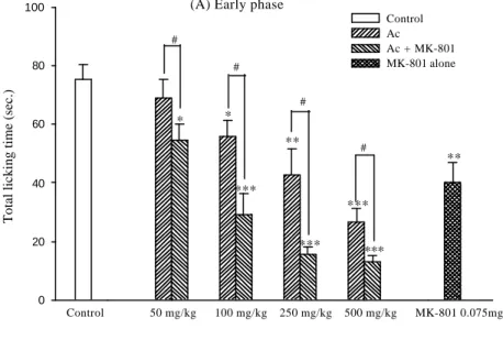
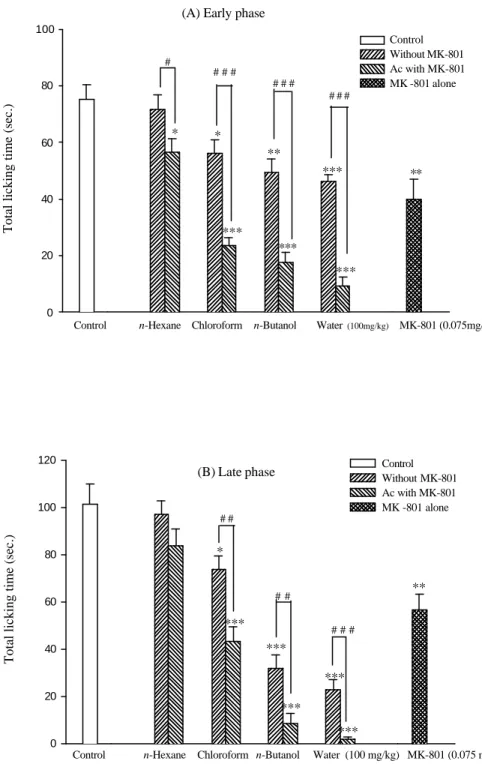
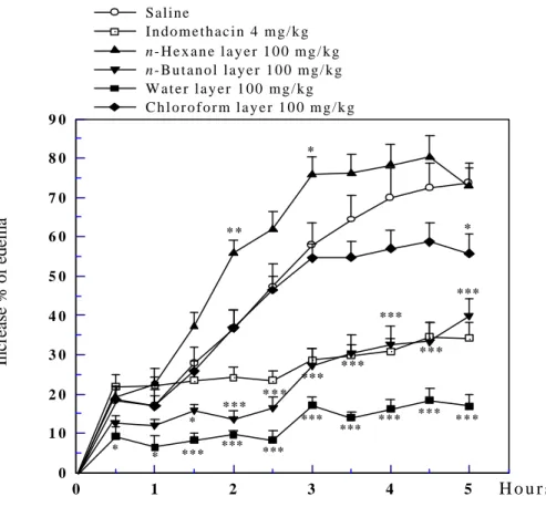
(3) 圖. 次. Fig. 1. Original plant and slices of Ampelopsis cantoniensis. ··································36 Fig. 2. Fractionation scheme of methanol extract of Ampelopsis cantoniensis. ······37 Fig. 3. The LD50 of crude extract of Ampelopsis cantoniensis in mice orally.········ 38 Fig. 4. The effects of crude extract of Ampelopsis cantoniensis or combination with MK-801 in formalin test.···············································································46 Fig. 5. The effects of various extract layers of Ampelopsis cantoniensis or combination with MK-801 in formalin test.··················································47 Fig. 6. The effects of crude extract of Ampelopsis cantoniensis on the carrageenininduced hind-paw edema. ·············································································48 Fig. 7.. The effects of various extract layers of Ampelopsis cantoniensis on the carrageenin-induced hind-paw edema. ·························································49. Fig. 8. The profile from HPLC indicated that the effects of crude extract of Ampelopsis cantoniensis on the N-acetylation of 2-AF in rat’s liver tissue cytosols.·········································································································50 Fig. 9.. The effects of crude extract of Ampelopsis cantoniensis on the N-acetylation of 2-AF in rat’s liver tissue cytosols.····························································51. Fig. 10. The effects of crude extract of Ampelopsis cantoniensis on the N-acetylation of PABA in rat’s liver tissue cytosols.··························································51 Fig. 11. The effects of crude extract of Ampelopsis cantoniensis on the N-acetyla tion of 2-AF in male rat’s liver tissue cytosols.····················································52 Fig. 12. The effects of crude extract of Ampelopsis cantoniensis on the N-acetylation of 2-AF in female rat’s liver tissue cytosols. ···············································52 Fig. 13. The effects of crude extract of Ampelopsis cantoniensis on the N-acetylation of 2-AF in rat’s blood cytosols.·····································································53 Fig. 14. The effects of crude extract of Ampelopsis cantoniensis on the N-acetylation of PABA in rat’s blood cytosols···································································53 Fig. 15. The effects of various extracted of Ampelopsis cantoniensis on the N3.
(4) acetylation of 2-AF in male rat’s liver tissue cytosols.·································54 Fig. 16. The effects of various extracted of Ampelopsis cantoniensis on the Nacetylation of 2-AF in female rat’s liver tissue cytosols·······························54 Fig. 17. The effects of various extracted of Ampelopsis cantoniensis on the Nacetylation of PABA in male rat’s liver tissue cytosols.·······························55 Fig. 18. The effects of various extracted of Ampelopsis cantoniensis on the Nacetylation of PABA in female rat’s liver tissue cytosols.····························55 Fig. 19. The effects of crude and various extracted layers of Ampelopsis cantoniensis administered 24 hrs before 2-AF on the N-acetylation of 2-AF in rat’s liver tissue cytosols. ······························································································57 Fig. 20. The effects of crude and various extracted layers of Ampelopsis cantoniensis administered simultaneously with 2-AF on the N-acetylation of 2-AF in rat’s liver tissue cytosols.······················································································57 Fig. 21. The effects of crude and various extracted layers of Ampelopsis cantoniensis administered 24 hrs before 2-AF on the N-acetylation of 2-AF in rat’s blood cytosols.·········································································································58 Fig. 22. The effects of crude and various extracted layers of Ampelopsis cantoniensis administered simultaneously with 2-AF on the N-acetylation of 2-AF in rat’s blood cytosols.·······························································································58 Fig. 23. The effects of crude and various extracted layers of Ampelopsis cantoniensis administered 24 hrs before 2-AF on the N-acetylation of 2-AF in rat’s kidney tissue cytosols. ··················································································59 Fig. 24. The effects of crude and various extracted layers of Ampelopsis cantoniensis administered simultaneously with 2-AF on the N-acetylation of 2-AF in rat’s kidney tissue cytosols.···················································································59 Fig. 25. The effects of crude and various extracted layers of Ampelopsis cantoniensis administered 24 hrs before 2-AF on the N-acetylation of 2-AF in rat’s urine.··············································································································60. 4.
(5) Fig. 26. The effects of crude and various extracted layers of Ampelopsis cantoniensis administered simultaneously with 2-AF on the N-acetylation of 2-AF in rat’s urine.··············································································································60 Fig. 27. Viability of HL-60 cells after treatme nt with crude extract of Ampelopsis cantoniensis for 24 hours. ············································································61 Fig. 28. Viability of HL-60 cells after treatment with various extracted layers of Ampelopsis cantoniensis for 24 hours. ·························································62 Fig. 29. The effects of crude extract of Ampelopsis cantoniensis on the morphological appearance in HL-60 cells.···········································································63 Fig. 30. The effects of n-hexane layer of Ampelopsis cantoniensis on the morphological appearance in HL-60 cells. ··················································64 Fig. 31. The effects of chloroform layer of Ampelopsis cantoniensis on the morphological appearance in HL-60 cells. ··················································64 Fig. 32. The effects of n-butanol layer of Ampelopsis cantoniensis on the morphological appearance in HL-60 cells. ··················································65 Fig. 33. The effects of water layer of Ampelopsis cantoniensis on the morphological appearance in HL-60 cells. ··········································································65 Fig. 34. The effects of crude extract of Ampelopsis cantoniensis on the DNA fragmentation in HL-60 cells. ······································································66 Fig. 35. The effects of various extract layers of Ampelopsis cantoniensis on the DNA fragmentation in HL-60 cells for 24 hours. ·················································67 Fig. 36. The effects of crude extract of Ampelopsis cantoniensis on the apoptosis in HL-60 cells. ··································································································68 Fig. 37. The effects of n-hexane layer of Ampelopsis cantoniensis on the apoptosis in HL-60 cells. ··································································································69 Fig. 38. The effects of chloroform layer of Ampelopsis cantoniensis on the apoptosis in HL-60 cells. ·····························································································70 Fig. 39. The effects of n-butanol layer of Ampelopsis cantoniensis on the apoptosis in HL-60 cells. ··································································································71 5.
(6) Fig. 40. The effects of water layer of Ampelopsis cantoniensis on the apoptosis in HL-60 cells. ··································································································72 Fig. 41. Crude extract of Ampelopsis cantoniensis induced apoptosis of HL-60 cells which were examined by flow cytometry. ··········· ·······································73 Fig. 42. n-Hexane layer extract of Ampelopsis cantoniensis induced apoptosis of HL-60 cells which were examined by flow cytometry.································74 Fig. 43. Chloroform layer extract of Ampelopsis cantoniensis induced apoptosis of HL-60 cells which were examined by flow cytometry.································75 Fig. 44. n-Butanol layer extract of Ampelopsis cantoniensis induced apoptosis of HL-60 cells which were examined by flow cytometry.································76 Fig. 45. Water layer extract of Ampelopsis cantoniensis induced apoptosis of HL-60 cells which were examined by flow cytometry.············································77 Fig. 46. The effects of crude extract of Ampelopsis cantoniensis on the cell cycle in HL-60 cells. The HL-60 cells were incubated with various concentrations of crude extract of Ampelopsis cantoniensis for 6 hrs, and they were examined by flow cytometry. ·······················································································78 Fig. 47. The effects of crude extract of Ampelopsis cantoniensis on the cell cycle in HL-60 cells. The HL-60 cells were incubated with various concentrations of crude extract of Ampelopsis cantoniensis for 12 hrs, and they were examined by flow cytometry. ···························· ·········································79 Fig. 48. The effects of crude extract of Ampelopsis cantoniensis on the cell cycle in HL-60 cells. The HL-60 cells were incubated with various concentrations of crude extract of Ampelopsis cantoniensis for 24 hrs, and they were examined by flow cytometry. ·····································································80 Fig. 49. The effects of crude extract of Ampelopsis cantoniensis on the cell cycle in HL-60 cells. The HL-60 cells were incubated with various concentrations of crude extract of Ampelopsis cantoniensis for 36 hrs, and they were examined by flow cytometry. ······································································81 Fig. 50. The effects of crude extract of Ampelopsis cantoniensis on the cell cycle in 6.
(7) HL-60 cells. The HL-60 cells were incubated with various concentrations of crude extract of Ampelopsis cantoniensis for 48 hrs, and they were examined by flow cytometry. ··································································82 Fig. 51. The effects of n- hexane layer extract of Ampelopsis cantoniensis on the cell cycle in HL-60 cells. The HL-60 cells were incubated with various concentrations of n-hexane layer extract of Ampelopsis cantoniensis for 24 hrs, and they were examined by flow cytometry. ········································83 Fig. 52. The effects of chloroform layer extract of Ampelopsis cantoniensis on the cell cycle in HL-60 cells. The HL-60 cells were incubated with various concentrations of chloroform layer extract of Ampelopsis cantoniensis for 24 hrs, and they were examined by flow cytometry. ········································84 Fig. 53. The effects of n-butanol layer extract of Ampelopsis cantoniensis on the cell cycle in HL-60 cells. The HL-60 cells were incubated with various concentrations of n-butanol layer extract of Ampelopsis cantoniensis for 24 hrs, and they were examined by flow cytometry. ········································85 Fig. 54. The effects of water layer extract of Ampelopsis cantoniensis on the cell cycle in HL-60 cells. The HL-60 cells were incubated with various concentrations of water layer extract of Ampelopsis cantoniensis for 24 hrs, and they were examined by flow cytometry. ···············································86 Fig. 55. The effects of crude extract of Ampelopsis cantoniensis on the intracellular cyclins distribution in HL-60 cells which were examined by flow cytometry. ····································································································87 Fig. 56. The effects of n- hexane layer extract of Ampelopsis cantoniensis on the intracellular cyclins distribution in HL-60 cells which were examined by flow cytometry.·····························································································88 Fig. 57. The effects of chloroform layer extract of Ampelopsis cantoniensis on the intracellular cyclins distribution in HL-60 cells which were examined by flow cytometry.·····························································································89 Fig. 58. The effects of n-butanol layer extract of Ampelopsis cantoniensis on the 7.
(8) intracellular cyclins distribution in HL-60 cells which were examined by flow cytometry.·····························································································90 Fig. 59. The effects of water layer extract of Ampelopsis cantoniensis on the intracellular cyclins distribution in HL-60 cells which were examined by flow cytometry.·····························································································91 Fig. 60. RT-PCR analysis for intracellular cyclins distribution of HL-60 cells repressing in the presence of different dose of crude extract of Ampelopsis cantoniensis. ·································································································92 Fig. 61. RT-PCR analysis of CDK 2 and beta-actin in the HL-60 cells for treatment with different dose of crude extract of Ampelopsis cantoniensis. ···············93 Fig. 62. RT-PCR analysis of cyclin E and beta-actin in the HL-60 cells for treatment with different dose of crude extract of Ampelopsis cantoniensis. ···············94 Fig. 63. RT-PCR analysis of cyclin A and beta-actin in the HL-60 cells for treatment with different dose of crude extract of Ampelopsis cantoniensis.················95 Fig. 64. RT-PCR analysis of cyclin B and beta-actin in the HL-60 cells for treatment with different dose of crude extract of Ampelopsis cantoniensis. ···············96 Fig. 65. RT-PCR analysis of NAT 1 and beta-actin in the HL-60 cells for treatment with different dose of crude extract of Ampelopsis cantoniensis. ···············97 Fig. 66. RT-PCR analysis of NAT 2 and beta-actin in the HL-60 cells for treatment with different dose of crude extract of Ampelopsis cantoniensis. ···············98 Fig. 67. RT-PCR analysis of cyclin D2 and beta-actin in the HL-60 cells for treatment with different dose of crude extract of Ampelopsis cantoniensis. ···············99 Fig. 68. RT-PCR analysis of cyclin D3 and beta-actin in the HL-60 cells for treatment with different dose of crude extract of Ampelopsis cantoniensis.··············100 Fig. 69. RT-PCR analysis for intracellular cyclins distribution of HL-60 cells repressing in the presence of different dose of water layer of Ampelopsis cantoniensis. ·······························································································101 Fig. 70. RT-PCR analysis of CDK 2 and beta-actin in the HL-60 cells for treatment with different dose of water layer of Ampelopsis cantoniensis. ················102 8.
(9) Fig. 71. RT-PCR analysis of cyclin E and beta-actin in the HL-60 cells for treatment with different dose of water layer of Ampelopsis cantoniensis.·················103 Fig. 72. RT-PCR analysis of cyclin A and beta-actin in the HL-60 cells for treatment with different dose of water layer of Ampelopsis cantoniensis. ················104 Fig. 73. RT-PCR analysis of cyclin B and beta-actin in the HL-60 cells for treatment with different dose of water layer of Ampelopsis cantoniensis. ················105 Fig. 74. RT-PCR analysis of NAT 1 and beta-actin in the HL-60 cells for treatment with different dose of water layer of Ampelopsis cantoniensis. ················106 Fig. 75. RT-PCR analysis of NAT 2 and beta-actin in the HL-60 cells for treatment with different dose of water layer of Ampelopsis cantoniensis. ················107 Fig. 76. RT-PCR analysis of cyclin D2 and beta-actin in the HL-60 cells for treatment with different dose of water layer of Ampelopsis cantoniensis. ················108 Fig. 77. RT-PCR analysis of cyclin D3 and beta-actin in the HL-60 cells for treatment with different dose of water layer of Ampelopsis cantoniensis. ················109 Fig. 78. RT-PCR analysis of caspase-3 in the HL-60 cells for treatment with different dose of crude extract of Ampelopsis cantoniensis. ····································110 Fig. 79. Ampelopsis cantoniensis affects the caspase 3 and caspase 9 gene expression on the HL-60 cells by cDNA microarrays. ················································110 Fig. 80. Ampelopsis cantoniensis affects the caspase5 and caspase 8 gene expression on theHL-60 cells by cDNA microarrays. ·················································111 Fig. 81. Ampelopsis cantoniensis affects the cytochrome C and cytochrome C1 gene expression on the HL-60 cells by cDNA microarrays. ······························111 Fig. 82. Ampelopsis cantoniensis affects the cyclin E and cyclin E2 gene expression on the HL-60 cells by cDNA microarrays. ················································112 Fig. 83. Ampelopsis cantoniensis affects the cyclin D1 and cyclin D2 gene expression on the HL-60 cells by cDNA microarrays. ················································112 Fig. 84. Ampelopsis cantoniensis affects the cyclin D3 and CDK2 gene expression on the HL-60 cells by cDNA microarrays. ·····················································113 Fig. 85. Ampelopsis cantoniensis affects the cyclin A and Cyclin A1 gene expression 9.
(10) on the HL-60 cells by cDNA microarrays. ················································113 Fig. 86. Ampelopsis cantoniensis affects the cyclin B1 and Cyclin B2 gene expression on theHL-60 cells by cDNA microarrays. ·················································114 Fig. 87. Ampelopsis cantoniensis affects the E2F transcription factor gene expression on the HL-60 cells by cDNA microarrays. ·················································114. 10.
(11) 表. 次. Tab. 1. The inhibition (%) of various extracted layers of Ampelopsis cantoniensis on N-acetylation of 2-AF in male rat’s blood cytosols. ·······································56 Tab. 2. The inhibition (%) of various extracted layers of Ampelopsis cantoniensis on N-acetylation of 2-AF in female rat’s blood cytosols. ····································56 Tab. 3. The inhibition (%) of various extracted layers of Ampelopsis cantoniensis on N-acetylation of PABA in male rat’s blood cytosols. ·····································56 Tab. 4. The inhibition (%) of various extracted layers of Ampelopsis cantoniensis on N-acetylation of PABA in female rat’s blood cytosols.···································56. 11.
(12) 略. 語. 表. Ac: Ampelopsis cantoniensis 2-AF: 2-Aminofluorene AAF: Acetyl-aminofluorene Ac-PABA: Acetyl-para-aminobenzoic acid CDK: Cyclin-dependent kinase DNA: Deoxyribonucleic acid DTT: Dithiothreitol EDTA: Ethylenediaminetetraacetate FACS: Flow cytometry FBS: Fetal bovine serum HL-60: Human leukemia cell line HPLC: High performance liquid chromatography MPF: Maturation promoting factor NAT: N-acetyltransferase NMDA: N-methyl- D-aspartate PABA: Para-aminobenzoic acid PAF: Platelet-activating factor PBS: Phosphate buffered saline PGs: Prostaglandins PI: Propidium iodine PMSF: Phenylmethylsulfonyl fluoride RNA: Ribonucleic acid RT-PCR: Reverse transcriptase polymerase chain reaction. 12.
數據

+7


相關文件
The multi-task learning problem comes from our biological application: Drosophila gene expression pattern analysis (funded by NSF and
An Empirical Study of the Influences of Emotional Intelligence, Sex Discrimination, Change Leadership and LMX on Female Leadership Effectiveness.. 研 究
Laser Capture Microdissection Microdissection of Fluorescently of Fluorescently Labeled Embryonic Cranial Neural Crest Cells Labeled Embryonic Cranial Neural Crest Cells..
The prepared nanostructured titania were applied for the photoanodes of dye-sensitized solar cell.. The photoanodes were prepared by the doctor blade technique and the area
Monopolies in synchronous distributed systems (Peleg 1998; Peleg
Corollary 13.3. For, if C is simple and lies in D, the function f is analytic at each point interior to and on C; so we apply the Cauchy-Goursat theorem directly. On the other hand,
Corollary 13.3. For, if C is simple and lies in D, the function f is analytic at each point interior to and on C; so we apply the Cauchy-Goursat theorem directly. On the other hand,
“The Connectivity Map: using gene-expression signatures to connect small molecules, genes, and disease.” Science 313(5795):..
