E L S E V I E R Journal of Chromatography A, 759 (1997) 193-201
JOURNAL OF
CHROMATOGRAPHY A
Determination of saikosaponins by micellar electrokinetic capillary
chromatography
Y o u - Z u n g H s i e h * , H s i - Y a H u a n g
Department of Applied Chemistry, National Chiao Tung University, 1001 Ta-Hsueh Road, Hsinchu 30050, Taiwan Received 12 June 1996; revised 13 August 1996; accepted 9 September 1996
Abstract
In this study, we examine the feasibility of determining saikosaponins a, bl, b2, c and d, i.e., the bioactive components in Bupleuri Radix, using the micellar electrokinetic capillary chromatography technique. Results obtained from using different anion surfactants, bile salt or sodium dodecyl sulfate (SDS), are also compared. By using bile salt as a pseudostationary phase, the baseline separation of saikosaponins is observed; however, the detection limit is unfavorable. Whereas the baseline resolution of saikosaponins is achieved after adding -,/-cyclodextrin in the SDS running buffer. In addition, another separating method is developed with the mixed micellar system of SDS and Brij 35 containing ~/-cyclodextrin. The contents of saikosaponins in traditional Chinese medicinal prescriptions, such as Sheau Chair Hwu Tang, Bupleuri Radix plant and Chair Hwu San are analyzed by one of the above methods. The entire separation can be completed within 9 min by these methods. The R.S.D. values of the migration times are all below 1%. Moreover, the detection limits of saikosaponins vary from 7 to 33 txg/ml.
Keywords: Buffer composition; Pharmaceutical analysis; Saikosaponins; Saponins
1. I n t r o d u c t i o n
Bupleuri Radix, referred to as Chair-Hwu in China, is a popular traditional Chinese drug. It has been used in medical treatment for over 2000 years and has been primarily used to treat cold fever, chill and fever in turn, the feeling of oppression and fullness in the chest and hypochondria [ 1 ]. Saponins contained in Bupleuri Radix are called saikosaponins and have been extensively studied in pharmacology and immunology [2,3]. Saikosaponins were verified as bioactive compounds with anti-inflammatory and anti-hepatotoxic activity [4].
Several studies on the characteristics of
*Corresponding author.
saikosaponins have addressed areas such as dis- tributions of saikosaponins in various kinds of tissues and the root part [5], change in saikosaponin content in Bupleuri Radix with different seasons 16], and saikosaponin content in crude drugs [7-9]. In those studies, saikosaponins were analyzed by thin-layer chromatography and high-performance liquid chro- matography.
Capillary electrophoresis (CE) is a highly effec- tive analytical tool, which has been extensively applied to separate various compounds, such as ionic and neutral compounds [10-14]. CE offers the advantages of high resolution, high separation ef- ficiency and the need for only a small amount of sample. Micellar electrokinetic capillary chromatog- raphy (MEKC) is another CE technique. Terabe et
0021-9673/97/$17.00 Copyright © 1997 Elsevier Science B.V. All rights reserved PII S0021-9673(96)00768-6
194 Y.-Z. Hsieh, H.-Y. Huang / J. Chromatogr. A 759 (1997) 193-201
al. showed that this technique may provide highly efficient separation of neutral and hydrophobic com- pounds by forming micelles in the buffer [15,16]. The underlying separation mechanism for MEKC is that analytes have different partition coefficients between an aqueous buffer and a micellar pseudo- stationary phase [ 15,17]. Previous investigators have proposed CE techniques to analyze Chinese medici- nal preparations [18,19]. Their results show that CE van be used for the qualitative and quantitative analysis of those medicinal preparations.
In the present study, we use MEKC to analyze saikosaponins in various medicinal samples. The bioactive saikosaponins under study are saikosaponins a, b l, b2, c and d. The factors affecting the separation efficiency are examined by combining different micelles and modifiers. Various separation conditions are used to minimize the interference from actual medicinal samples.
2. Experimental
2.2. Chemicals
Sodium dodecyl sulfate (SDS), sodium deoxy- cholate (SDC) and sodium cholate (SC), borax, boric acid, a-cyclodextrin and 13-cyclodextrin were pur- chased from Sigma (St. Louis, MO, USA). Saikosaponins a and c and ~/-cyclodextrin were obtained from Nacalai Tesque (Kyoto, Japan). Saikosaponin d was purchased from Toray (Siga, Japan). Saikosaponins b l and b2 were purchased from ALPS (Gifu, Japan). Polyoxyethylene (23) dodecanol (Brij 35) were obtained from Merck (Darmstadt, Germany). Bupleuri Radix plant and two Chinese medical samples containing Bupleuri Radix - Sheau Chair Hwu Tang and Chair Hwu San - were purchased in Taiwan. Medicine's major component is Bupleuri Radix. Manufacturing of the commercial product includes boiling in water, drying and powdering. All other chemicals were analytical grade and purchased from Merck. Water was purified in a Milli-Q water system (Millipore, Bedford, MA, USA).
2.3. Procedure 2.1. Apparatus
A Beckman P/ACE 5500 CE system (Beckman Instruments, Palo Alto, CA, USA) was used, equipped with a diode array detector, capable of scanning the full range of wavelengths from 190 to 800 nm. The detector could simultaneously record two wavelength signals through two detection chan- nels. Channel A was set at 194 nm and channel B was set at 252 nm for searching the optimum experimental condition. After the search, the de- tection wavelength was programmed to change dur- ing the separating process from 194 nm to 252 nm at the optimal condition. CE was performed in a 47 cm (40 cm to detector)X50 I.Lm I.D. fused-silica capil- lary tube (Polymicro Technologies, Phoenix, AZ, USA). The capillary column was assembled in the cartridge format (Beckman Instruments). The voltage of the electrophoresis separation was 20 kV. Pressure injection was the sample injection method. A person- al computer using System Gold software (Beckman Instruments) controlled the P/ACE instrument. Final- ly, data analysis was also performed with the System Gold software.
Stock solutions of the saikosaponins (5 mg/ml) were prepared in ethanol. Sample solutions with various concentrations were prepared by dilution with ethanol-water (2:3, v/v). Electrophoresis buf- fers were prepared by dissolving 0.1 M borax and 0.1 M sodium hydroxide (or 0.1 M boric acid) in water. A 1.5-g sample of each concentrated commer- cial medicinal preparation was accurately weighed. On the bases of the extraction methods employed in previous studies [5-8], a neutral or weakly alkaline solvent can be used to prevent acid-labile saikosaponins from forming diene saponins. Thus, each 1.5-g sample was mixed and extracted with 15 ml ethanol (99.8%) for 15 rain by using an ultrasonic bath. The mixed sample was then filtered through filter paper. This extracting and filtering procedure was repeated three times. A total of 45 ml sample extract was concentrated to a final volume of 3.0 ml, which was used for analysis.
3. Results and discussion
Y.-Z. Hsieh, H.-Y. Huang I J. Chromatogr. A 759 (1997) 193-201 195
saikosaponins a, b l , b2, c and d, were selected as analytes in this study. Fig. 1 displays their molecular structures. The five analytes are quite similar with respect to molecular structure. The molecular masses of saikosaponins a, b l , b2 and d are all 780 u and the molecular mass of saikosaponin c is 927 u. There are two pairs of isomers among these compounds. One pair is the saikosaponins a and d, and the other the saikosaponins b l and b2. The only difference in the structure between these two pairs is a functional group of the saikosaponins. In aqueous acidic con- ditions, saikosaponins a and d of the ether saponins can be converted to saikosaponins bl and b2 of the diene saponins.
The maximum absorbance wavelengths of saikosaponins a, c and d are around 194 nm. The other saikosaponins, bl and b2, have a maximum absorbance at 252 nm. There is no other intense absorbance for the five analytes between 194 nm and 252 nm wavelength. Since the two saikosaponin
groups have different maximum absorbance wave- lengths, two detection wavelengths were recorded in this study. After baseline resolution was obtained for the optimum separation conditions, the detection wavelength was programmed to change during the separation process. Thus, the signal peaks of analytes with different absorbance wavelengths could be displayed in one electropherogram.
Borate buffers with pH values ranging from pH 8.2 to 10.0 were initially used to separate the five analytes. The electropherograms are similar to one another in the sense that each electropherogram has only one peak, implying that all saikosaponin migra- tion velocities are identical with the electroosmotic flow velocities. These results suggest that all analytes do not dissociate in this pH range. Consequently, these analytes cannot be separated by their migration velocities in CE buffer without any modifiers. Thus, MEKC with various modifiers added in a micellar buffer was chosen in this study to separate the saikosaponins.
.,,,,'
Rl O ~ ~ R 3 R2
Rl R2 R3
saikosaponm a GIc~ 3 Fuc~ CH2OH 13-OH saikosaponin d GIc-+ 3 Fuc--+ CH2OH a-OH saikosaponin c GIc---~6GIe---~ CH3 13-OH
t4 Rha ~ C H 2 O H R i O ~ R3 R2 RI R2 R3
saikosaponi~ b l Gle--* 3 Fue -~ CH2OH t3-OH saikosaponin b2 Glc--* 3 Fuc --* CH2OH (z-OH Fig. 1. Molecular structures of saikosaponins.
3.1. MEKC with bile salt and cyclodextrin
Bile salt monomer is more polar than SDS mono- mer and the former has a hydroxyl-substituted steroid backbone. The saikosaponins have steroid- like structures similar to bile salt. Thus, bile salt was selected here as test surfactant in MEKC analysis. Fig. 2 shows the baseline separation of saikosaponins a, b2 and c with 20 mM sodium cholate (SC) added to the borate buffer. However, in this condition, saikosaponins bl and d have identical migration times. The successful separation of the three analytes by SC bile salt may be due to different strengths of the interaction between the saikosaponins and bile salt. Such interactions may be due to the similar steroid backbone structures of the saikosaponins and SC. The separation efficiency decreases with increas- ing SC concentration from 20 to 100 mM in the running buffer. Similar results are observed when another bile salt (SDC) was employed.
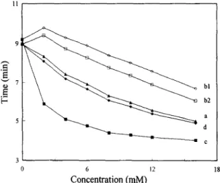
Because bile salt can only separate three saikosaponins, adding another modifier was consid- ered to increase the resolution. Fig. 3 shows the effect of the concentration of [3-cyclodextrin on the migration times of saikosaponins in a 30 mM SC bile salt buffer. Saikosaponins a, c and d migrate faster as the concentration of ~3-cyclodextrin increases. This
196 Y.-Z. Hsieh, H.-Y. Huang / J. Chrornatogr. A 759 (1997) 193-201
~
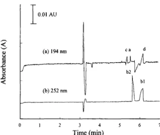
- 0.01 < ~ 2 F AU (a) 194 nm (b) 252 nm ca d i r i i 1 2 3 4 5 6 7 Time (min)Fig. 2. Separation of five saikosaponins at (a) 194 nm and (b) 252 nm. Peaks: a=saikosaponin a; bl=saikosaponin bl; b2= saikosaponin b2; c=saikosaponin c; d=saikosaponin d. Condi- tions: separation solution, 0.038 M borate buffer containing 20 mM SC, pH 10.0; capillary, 47 cm (40 cm to the detector)x50 p.m I.D.; applied voltage, 20 kV; detection wavelength, 194 and 252 nm; concentration of each saikosaponin, 0.50 mg/ml.
indicates that the ratio of the saikosaponin-cyclo- dextrin complex to saikosaponin increases when more 13-cyclodextrin is added to the buffer. However, the migration velocities of the other two saikosaponins, bl and b2, hardly change with the
concentration of [3-cyclodextrin. The above results suggest that saikosaponins a, c and d have much stronger interactions with 13-cyclodextrin than saikosaponins bl and b2. The molecular structure of cyclodextrin consists of a truncated cone with a relatively hydrophobic interior and a hydrophilic exterior. Since saikosaponins a, c and d are more hydrophobic than saikosaponins bl and b2, the different migration behaviors of the analytes with 13-cyclodextrin are possibly due to the analytes' hydrophobic property.
The migration velocities of the five analytes decrease as SC concentration increases from 30 to 100 mM in the buffer in which the concentration of [3-cyclodextrin remains at 10 mM. Because more negatively charged SC micelles exist as SC con- centration increases in the buffer, more analytes interact with the micelles. Consequently, the analysis time of saikosaponins increases. However, the res- olution of separation does not improve.
Baseline separation of the five saikosaponins (Fig. 4) is obtained by adding 5 mM 13-cyclodextrin to the borate buffer containing 30 mM SC. As indicated in Fig. 4, the analysis is completed within 7 min. Therefore, the above separation condition seems sufficient for a quantitative analysis of saiko- saponins. However, because bile salt has a large
6 • ~ bl '--" b2 [-- 5 d a c 4 h i i i 4 6 8 10 12 Concentration (raM)
Fig. 3. Effect of ~-cyclodextrin concentration on the migration time of saikosaponins. Conditions: separation solution, i3-cyclo- de×trin in 0.038 M borate buffer containing 30 mM SC. Other conditions as in Fig. 2. < ¢ . ) < I 0 . 0 1 A U b2 .__ b ~ I 2 3 4 5 0 7 Time (rain)
Fig. 4. Separation of five saikosaponins. Conditions: separation solution, 0.038 M borate buffer containing 30 mM SC and 5 mM [3-cyclodextrin, pH 10.0; detection wavelength, 194 nm (before 5.9 min) and 252 nm (after 5.9 min). Other conditions as in Fig. 2.
11
background absorbance near 190 nm, the detection limit would be insufficient in saikosaponins analysis. Consequently, other MEKC separation conditions must be sought to analyze actual saikosaponin samples.
3.2. M E K C with SDS and cyclodextrin
A broadened peak of saikosaponins appears at 9 min when 30 mM SDS is added in a pH 10.0 borate buffer. If the MEKC buffer pH value is changed to pH 9.0, the broadened peak elutes at 15 min. The migration time is even longer if the buffer's pH value is lower than 9.0. Compared with borate only buffers, the SDS buffers do not enhance the res- olution but, instead, increase the migration time. The fact that good resolution could not be obtained for the five saikosaponins is due to the strong solubiliza- tion effect of the SDS micelles. All the analytes have marked interactions with the SDS micelles; there- fore, the differences in the interactions are rather small. Thus, to obtain an enhanced resolution for these analytes, the influence of adding neutral ma- terial in the SDS buffer is discussed.
The separation of saikosaponins is improved by adding either e~-cyclodextrin or [3-cyclodextrin to the SDS buffer. However, baseline resolution of saikosaponins is still not achieved. Altering the concentration of ~/-cyclodextrin from 2 to 16 mM in the SDS buffer markedly affects the migration times of saikosaponins as indicated in Fig. 5. The five analytes can be sufficiently separated by only adding 2 mM "y-cyclodextrin to 10 mM ~-cyclodextrin in the running buffer. According to our results, several different combinations of ~/-cyclodextrin and SDS can be used to separate saikosaponins. The sepa- ration time is shortened from 10 to 8 min when 10 mM ~/-cyclodextrin is added. Experimental results demonstrate that the interactions between saikosaponins and ~-cyclodextrin are much stronger than those between saikosaponins and o~-cyclodextrin or [3-cyclodextrin. Also, the strong interactions be- tween the saikosaponins and the SDS micelles seem to cause the interactions between saikosaponins and ~x-cyclodextrin or [3-cyclodextrin to be not signifi- cant. The structure of ~/-cyclodextrin is similar to that of a-cyclodextrin and 13-cyclodextrin except for the size of the interior cavity. Thus, the ~t-cyclodextrin
b l b 2 a d c .9 E " - " 7 E-
Y.-Z. Hsieh, H.-Y, Huang / J. Chromatogr. A 759 (1997) 193-201 197
0 6 12 18
Concentration (raM)
Fig. 5. Effect of 7-cyclodextrin concentration on the migration time of saikosaponins. Conditions: separation solution, ~/-cyclo- dextrin in 0.038 M borate buffer containing 30 mM SDS. Other conditions as in Fig. 2.
cavity size plays an essential role in saikosaponin- cyclodextrin complex formation. The size of the ~-cyclodextrin cavity is sufficiently large to form a stable complex with the analyte. Therefore, the formation of more stable complexes in the buffer results in a shorter migration time.
Here, the effect of SDS concentration on sepa-
E 6 E- b l b 2 a d c 2 i i i 20 40 60 Concentration (raM)
Fig. 6. Effect of SDS concentration on the migration time of saikosaponins. Conditions: separation solution, SDS in 0.038 M borate buffer containing 10 mM ~/-cyclodextrin. Other conditions as in Fig. 2.
198 Y.-Z. Hsieh, H.-Y. Huang / J. Chromatogr. A 759 (1997) 193-201 0.005 AU c d a b2 h i i 0 2 4 6 8 10 Time (min)
Fig. 7. Separation of five saikosaponins. Conditions: separation solution, 0.038 M borate buffer containing 30 mM SDS and 10 mM ~-cyclodextrin, pH 10.0; detection wavelength, 194 nm (before 7 min) and 252 nm (after 7 min); concentration of saikosaponins a, c and d, 0.40 mg/ml; concentration of saikosaponins bl and b2, 0.10 mg/ml. Other conditions as in Fig. 2.
ration efficiency was examined by using several electrolyte buffers containing different SDS con- centrations. Fig. 6 presents the effect of SDS con- centration on the migration times of saikosaponins in borate buffer containing 10 mM ~/-cyclodextrin. As revealed in Fig. 6, all analytes are adequately separated at 30 mM SDS. At lower SDS concen- trations, several peaks overlap. At higher SDS concentrations, the migration velocities decrease. However, the separation time increases with no improvement in resolution. Thus, 30 mM SDS
together with 10 mM ~/-cyclodextrin was chosen for subsequent analysis. Fig. 7 depicts a baseline sepa- ration of saikosaponins. The analysis is completed within 9 min.
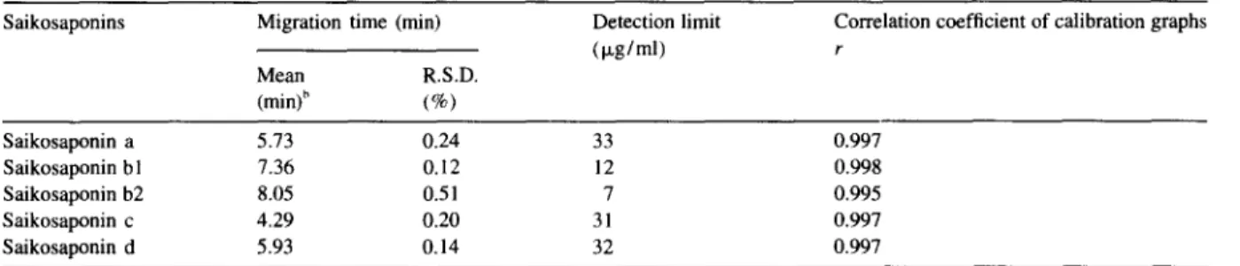
Table 1 lists the average migration times, the detection limits and the correlation coefficients of calibration graphs of the separation shown in Fig. 7. Calibration curves (peak area vs. concentration in Ixg/ml) are constructed in the range from 50 to 1000 Ixg/ml for the five saikosaponins. Linear relation- ships are found with the correlation coefficients exceeding 0.995. The R.S.D. of the migration time of each peak is below 0.51%. The detection limits of saikosaponins vary from 7 to 33 p~g/ml.
In order to identify saikosaponins in actual sam- ples, each peak was identified by comparing both the migration times of standard samples and the UV-Vis spectra of standard samples acquired by a UV-Vis diode array detector. With respect to the diode array UV spectra, the criterion for the correlation coeffi- cient of identification was set at 0.98. Moreover, a standard addition method was employed to further confirm the saikosaponins in the actual samples.
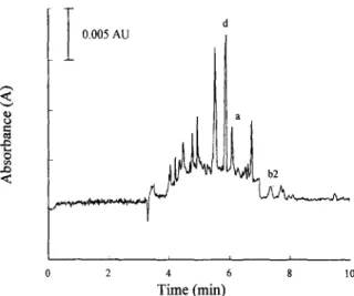
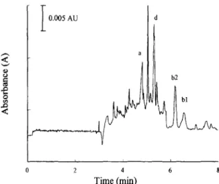
Bupleuri Radix, which has not been boiled and powdered, is usually used as an ingredient of a traditional Chinese drug. The plant of dried Bupleuri Radix was extracted by the experimental procedure mentioned above. Fig. 8_displays the Bupleuri Radix's extract analyzed by MEKC. More than ten peaks appear in this figure. However, saikosaponins a, b2 and d are clearly identified in the Bupleuri Radix extract. The average contents of saikosaponins a, b2 and d based on four extractions are 0.97, 0.83 and 12.1 mg/g, respectively. Several other unknown
Table 1
Migration times and detection limits of separation of saikosaponins with MEKC a Saikosaponins Migration time (min) Detection limit
(p,g/ml) Mean R.S.D.
(min)" (%)
Correlation coefficient of calibration graphs r Saikosaponin a 5.73 0.24 33 0.997 Saikosaponin b I 7.36 0.12 12 0.998 Saikosaponin b2 8.05 0.51 7 0.995 Saikosaponin c 4.29 0.20 31 0.997 Saikosaponin d 5.93 0.14 32 0.997
The separation buffer was a 38 mM borax buffer containing 30 mM SDS and 10 mM ~/-cyclodextrin. b n ~ 5 ,
Y.-Z. Hsieh, H.-Y. Huang / J. Chromatogr. A 759 (1997) 193-201 199 < e - < I 0.005 AU a ; i i i 2 4 6 8 10 Time (min)
Fig. 8. Separation of saikosaponin extract from a Bupleuri Radix plant. Other conditions as in Fig. 7.
peaks appear in the electropherogram. However, the saikosaponin peaks are clearly differentiated from those of unknown compounds in the extract.
Fig. 9 presents the separation results of Sheau Chair Hwu Tang, which is a commercial Chinese medicinal preparation. This medicine contains Bu- pleuri Radix, Scutellariae Radix, Ginseng Radix, Glycyrrhizae Radix, Pinelliae Tuber, Zingiberis Rhizoma, etc. A m o n g those, Bupleuri Radix is the major material in this medicine. Saikosaponins a and
< ¢ 3 O .g < I 0.005 AU da 2 4 6 8 10 Time (min)
Fig. 9. Separation of saikosaponin extract from a Chinese medici- nal preparation - Sheau Chair Hwu Tang. Other conditions as in Fig. 7.
d are observed in the electropherogram. The quan- tities of saikosaponins a and d in the preparation measured from four extractions are 2.2 and 4.7 m g / g with R.S.D. 1.1% and 2.1%, respectively.
3.3. Mixed micellar MEKC with SDS and Brij 35
Although a 30 mM SDS together with 10 mM 3,-cyclodextrin buffer offers an adequate resolution, the unknown components in the extracts occasionally interfere with the signals of the target analytes. An insufficient separation is observed for the extract of another medicinal sample, Chair Hwu San. Conse- quently, adding another modifier in the buffer to minimize interference is necessary for MEKC to efficiently separate this extract. ,~
Before the new method is tested on the medicinal sample, Chair Hwu San, satisfactory analysis needs to be established on the five saikosaponins. A neutral surfactant, Brij 35, was chosen to reduce the interfer- ence. The migration time markedly decreases after addition of Brij 35 to the buffer. Thus, a pH 9.0 buffer with 60 mM SDS was used to maintain the elution range. As shown in Fig. 10, the separation of saikosaponins is sufficient by adding 5 mM ~/-cyclo- dextrin in the borate buffer containing 60 mM SDS
< ,.Q < I 0,005 AU b2 bl c 0 2 4 6 8 Time (min)
Fig. 10. Separation of five saikosaponins. Conditions: separation solution, 0.065 M borate buffer containing 60 mM SDS, 5 mM Brij 35, and 5 mM ~-cyclodextrin, pH 9.0; detection wavelength, 194 nm (before 5.8 rain) and 252 nm (after 5.8 min); con- centration of each saikosaponin, 0.40 mg/ml. Other conditions as in Fig. 2.
200 Y.-Z. Hsieh, H.-Y. Huang / J. Chromatogr. A 759 (1997) 193-201 I 0.005 AU < 0 o
I
h i i 2 4 6 8 Time (rain)Fig. 11. Separation of saikosaponin extract from a Chinese medicinal preparation - Chair Hwu San. Other conditions as in Fig. 10.
and 5 m M Brij 35. All saikosaponins are baseline resolved and the total analysis time is less than 7 min. Moreover, once the ratio o f Brij 35 to "y- cyclodextrin increases, the resolution o f saikosaponins a and d improves. Experimental re- sults such as the reproducibility, the detection limits and the correlation coefficients o f calibration graphs from the buffer containing Brij 35 are similar to those from the 30 m M SDS with 10 m M ~/-cyclo- dextrin buffer, except that the analysis time is shortened from 9 to 7 min.
Another commercial product o f Bupleuri Radix,
Chair Hwu San, was analyzed by this modified method (Fig. 11). More than ten peaks appear in this figure. According to the experimental results, the medicine contains saikosaponins a, b 1, b2 and d. The quantities o f saikosaponins a, b l, b2 and d in the medicine are 4.2, 0.41, 0.74 and 4.7 m g / g , respec- tively. Table 2 summarizes the analysis results for the actual samples - Bupleuri Radix plant, Sheau Chair Hwu Tang and Chair Hwu San - analyzed by two different methods.
Results in this study demonstrate that the contents of saikosaponins in Bupleuri Radix plant, Sheau Chair Hwu Tang and Chair Hwu San are different. The Bupleuri Radix plant is not pretreated by boiling in water, drying and powdering as the other two commercial medicines. Thus, the discrepancies be- tween contents of saikosaponins among these sam- ples may be due either to different sources o f the material (Bupleuri Radix) or to different pretreatment processes by the manufacturer.
It should also be noted that traditional Chinese medicinal preparations usually have more than five materials in one medicine, thereby making the medicine itself quite complex. Interference from other components in the medicine may reduce the resolution o f saikosaponins contained in Bupleuri Radix. Therefore, depending on the contents o f the medicine, a minor modification of the analysis method must be made for different routine analyses. Our experimental results offer some guidelines for selecting various combinations o f y-cyclodextrin and SDS to separate saikosaponins in actual samples.
Table 2
Contents of five saikosaponins in Chinese medicinal preparations
Saikosaponins Bupleuri Radix ~ Sheau Chair Hwu Tang a Chair Hwu San b
Mean R.S.D. Mean R.S.D. Mean R.S.D.
(mg/g) c (%) (mg/g)" (%) (mg/g) ~ (%) Saikosaponin a 0.97 1.7 2.2 1.1 4.2 2.9 Saikosaponin b 1 nd ~ - nd - 0.41 2.3 Saikosaponin b2 0.83 1.0 nd - 0.74 1.9 Saikosaponin c nd - nd - nd - Saikosaponin d 12.1 2.5 4.7 2.1 4.7 2.6
The separation buffer was a 38 mM borax buffer containing 30 mM SDS and 10 mM ~,-cyclodextrin.
"The separation buffer was a 65 mM borax buffer containing 30 mM SDS, 5 mM Bfij 35 and 5 mM "V-cyclodextrin. c n=4.
Y.-Z. Hsieh, H.-Y. Huang / J. Chromatogr. A 759 (1997) 193-201 201
Adding Brij 35 can provide a better selection for preparation of the buffer used to determine saikosaponins.
4. Conclusion
In the present study, separation methods with good resolution are successfully demonstrated for the bioactive components of Bupleuri Radix, including saikosaponins a, b l, b2, c and d. A satisfactory resolution can be achieved by adding ~/-cyclodextrin in either the SDS micellar buffer or the mixed micellar buffer of SDS and Brij 35. The MEKC technique can rapidly determine the saikosaponins in commercial and traditional Chinese medicine. Thus, MEKC potentially offers a rapid and reliable quality assurance method to analyze the contents of major components in traditional Chinese medicine. Further applications are under study.
Acknowledgments
This research was supported by the National Science Council of Taiwan (NSC 85-2113-M-009-
007).
References
[2] M.H. Yen, C.C. Lin and C.M. Yen, Phytotherapy Res., 9 (1995) 351.
[3] M. Ohtsuka, K. Fukuda, H. Yano and M. Kojiro, Japanese J. Cancer Res., 86 (1995) 1131.
[4] L. Pistelli, A. Cammilli, A. Manunta, A. Marsili and I. Morelli, Phytochemistry, 33 (1993) 1197.
[5] T. Tani, T. Katsuki, M. Kubo and S. Arichi, J. Chromatogr., 360 (1986) 407.
[6] M. Minami, M. Sugino, M. Sadaoka, K. Ashida and K. Ogaki, Yakugaku Zasshi, 115 (1995) 145.
[7] H. Kanazawa, Y. Nagata, Y. Matsushima, M. Tomoda and N. Takai, J. Chromatogr., 507 (1990) 327.
[8] H. Kanazawa, Y. Nagata, Y. Matsushima, M. Tomoda and N. Takai, J. Chromatogr., 630 (1993) 408.
[9] N. Ebata, K. Nakajima, K. Hayashi, M. Okada and M. Maruno, Phytochemistry, 41 (1996) 895.
[10] F.E.P. Mikkers, F.M. Everaerts and T.P.E.M. Verheggen, J. Chromatogr., 169 (1979) 11.
[11] J.W. Jorgenson and K.D. Lukacs, Science, 222 (1983) 266. [12] F. Foret, L. Kriv and P. Bocek, in B.J. Radola (Editor), Capillary Zone Electrophoresis, Cambridge University Press, New York, 1993.
[13] S.F.Y. Li, Capillary Electrophoresis, Elsevier, Amsterdam, 1993.
[14] M. Novotny, H. Soini and M. Stefansson, Anal. Chem., 66 (1994) 646A.
[15] S. Terabe, K. Otsuka and T. Ando, Anal. Chem., 57 (1985) 834.
[16] H. Nishi, T. Fukuyama, M. Matsuo and S. Terabe, J. Chromatogr., 513 (1990) 279.
[17] R.O. Cole, M.J. Sepaniak, W.L. Hinze, J. Gorse and K. Oldiges, J. Chromatogr., 557 (1991) 113.
[18] H-R. Chen and S-J. Sheu, J. Chromatogr. A, 704 (1995) 141. [19] C.L. Flurer, L.A. Lin, R.D. Satzger and K.A. Wolnik, J.
Chromatogr. B, 669 (1995) 133. [1] S-Q. Luo, L-Z. Lin and G. A. Cordell, Phytochemistry, 33