Fabrication of a miniature CMOS-based optical biosensor
Wei-Jen Ho
a, Jung-Sheng Chen
b, Ming-Dou Ker
b, Tung-Kung Wu
a,
Chung-Yu Wu
b, Yuh-Shyong Yang
a, Yaw-Kuen Li
c, Chiun-Jye Yuan
a,∗ aDepartment of Biological Science and Technology, National Chiao Tung University, 75 Bo-Ai Street, Hsinchu, Taiwan 300, ROCbDepartment of Electronics Engineering, National Chiao Tung University, Hsinchu, Taiwan 300, ROC cDepartment of Applied Chemistry, National Chiao Tung University, Hsinchu, Taiwan 300, ROC Received 6 August 2006; received in revised form 19 December 2006; accepted 20 December 2006
Available online 7 January 2007
Abstract
This work presents a novel, miniature optical biosensor by immobilizing horseradish peroxidase (HRP) or the HRP/glucose oxidase (GOx) coupled enzyme pair on a CMOS photosensing chip with a detection area of 0.5 mm× 0.5 mm. A highly transparent TEOS/PDMS Ormosil is used to encapsulate and immobilize enzymes on the surface of the photosensor. Interestingly, HRP-catalyzed luminol luminescence can be detected in real time on optical H2O2and glucose biosensors. The minimum reaction volume of the developed optical biosensors is 10L. Both optical H2O2
and glucose biosensors have an optimal operation temperature and pH of 20–25◦C and pH 8.4, respectively. The linear dynamic range of optical H2O2and glucose biosensors is 0.05–20 mM H2O2and 0.5–20 mM glucose, respectively. The miniature optical glucose biosensor also exhibits
good reproducibility with a relative standard deviation of 4.3%. Additionally, ascorbic acid and uric acid, two major interfering substances in the serum during electrochemical analysis, cause only slight interference with the fabricated optical glucose biosensor. In conclusion, the CMOS-photodiode-based optical biosensors proposed herein have many advantages, such as a short detection time, a small sample volume requirement, high reproducibility and wide dynamic range.
© 2007 Published by Elsevier B.V.
Keywords: Sol–gel; CMOS; Optical biosensor; Ormosil
1. Introduction
Optical biosensors represent a recent trend in biosensor devel-opment owing to their high sensitivity, ease of operation, high accuracy and wide detection capacity (Blum and Gautier, 1991; Golden et al., 1994; Preuschoff et al., 1994; Chen et al., 1996; Marquette and Blum, 1999; Ruano et al., 2000; Tsafack et al., 2000; Ivanov et al., 2001; Askari et al., 2002; Lu et al., 2003, 2004; Huang et al., 2004; Song et al., 2005). In particular, luminescence-based optical biosensors exhibit various advan-tages over conventional electrochemical biosensors, including insensitivity to electrical disturbance, a low detection limit and a wide linear working range (Blum and Gautier, 1991; Rubtsova et al., 1998; Marquette and Blum, 1999). Most optical biosensors are developed based on optical fiber technology, which is used as a waveguide medium to transmit light signals from the
immo-∗Corresponding author. Tel.: +886 3 573 1735; fax: +886 3 572 9288. E-mail address:[email protected](C.-J. Yuan).
bilized enzyme to the photomultiplier tube (Blum and Gautier, 1991; Golden et al., 1994; Chen et al., 1996; Marquette and Blum, 1999; Tsafack et al., 2000). The need for an expensive set-up, involving a photomultiplier tube and associated equip-ment, limits the application of the fiber-optic biosensors to the development of simple portable devices for fast screening and in-field/on-site monitoring.
An early attempt to fabricate a miniature optical biosensor was made by replacing the photomultiplier tube with a pho-todiode to detect luminol luminescence directly (Preuschoff et al., 1994). Although the achievable detection limit of hydrogen peroxide was low, the overall set-up of this optical biosensor was complex. An elaborate, powerful and miniature photodiode at low cost can be fabricated by employing the mature com-plementary metal oxide semiconductor (CMOS) technology. Many attempts have demonstrated that the CMOS photosen-sor is a powerful light detecting device in the bioluminescent reactions (Lu et al., 2003, 2004; Huang et al., 2004). However, the development of a real optical biosensor by immobilizing an enzyme on the surface of a CMOS photosensing chip has
0956-5663/$ – see front matter © 2007 Published by Elsevier B.V. doi:10.1016/j.bios.2006.12.031
not been demonstrated. It is postulated that the immobilization of the target enzyme on the surface of a CMOS-photosensor may allow a CMOS-based biosensor to meet the requirements of rapid response time, small reaction volume and large dynamic detection range.
Encapsulation of an enzyme using a sol–gel matrix has been shown to be an effective method of entrapping enzymes in a manner that maintains most of their activity (Wang et al., 2000; Diwan and Park, 2001; Zhou et al., 2002; Youan, 2003; Ho et al., 2006). Silica sol–gel has been used in the development of biosensors (Shtelzer and Braun, 1994; Wang et al., 1998; Yao and Takashima, 1998; Wolfbeis et al., 2000; Li et al., 2002; Park and Clark, 2002; Ho et al., 2006) because of its simplicity of preparation, optical transparency, chemical inertness, low-temperature processing and mechanical stability (Singh et al., 1999; Pandey et al., 2003; Tang et al., 2003). The high trans-parency makes silica sol–gel an appropriate material for the fabrication of the optical biosensors (Schulze-Nahrup et al., 2004).
This study demonstrates a novel, miniature optical biosensor that was developed by integrating the CMOS photosensor, the sol–gel technique and the chemilumminescence (CL) detection method. This miniature CMOS-based optical biosensor not only exhibits high detection capability, short detection time and high reproducibility, but also requires only a small sample. With these characteristics, the developed CMOS-based optical biosensor has great potential in the future development of a simple portable biosensing system for rapid screening and in-field/on-site mon-itoring.
2. Materials and methods 2.1. Reagents
-d-Glucose, horseradish peroxidase (HRP, 116 U/mg),
luminol, tetraethylorthosilane (TEOS), silanol-terminated
poly(dimethylsiloxane) (PDMS, molecular weight 1700)
and Tris Base were purchased from Sigma. H2O2 (30%)
was purchased from Riedel-deHa¨en. Glucose oxidase from
Aspergillus niger (GOx, 200 U/mg) was purchased from Fluka.
Chlorobutadiene was purchased from Kuo Sen Enterprise Co., Ltd. Other chemicals and reagents were of analytical grade and used without further purification.
2.2. Preparing solution of enzymes
HRP stock solution (15.5 mg/ml) was prepared in 100 mM Tris–HCl buffer, pH 8.6. Glucose oxidase stock solution (144 mg/ml) was prepared in 100 mM phosphate buffer, pH 7.0.
The stock enzyme solutions were stored at−20◦C. The HRP
and GOx solutions were melted on ice just before use and were diluted with pH 7.4 phosphate buffer saline (1× PBS). One unit of HRP corresponds to the formation of 1.0 mg of purpurogallin from pyrogallol in 20 s at pH 6.0 and 20◦C. One unit of GOx is defined as oxidizing 1mol glucose per minute at pH 7.0 and 25◦C.
Fig. 1. Experimental setup for fabricating a miniature CMOS-based optical biosensor. (a) The CMOS photodiode was fixed on the PCB board, which was connected to E3646 and 34401A. (b) An E3646A power supply (Agilent) was utilized to apply a revise bias to the CMOS photodiodes. (c) A 34401A mul-timeter (Agilent) was utilized to measure the luminescence generated current. (d) A PC connected to E3646 and 34401A via a GPIB constituted the automatic detection system.
2.3. Apparatus
Fig. 1displays the complete experimental setup of the minia-ture CMOS optical biosensing system. The instruments were an E3646A power supply (Agilent), a 34401A multimeter (Agi-lent), a General Purpose Interface Bus (GPIB) card (National Instruments) and a personal computer (PC). Each pixel of the CMOS photosensor was implemented with a P–N junction diode using a P-substrate and an N+ active diffusion layer, which
was fabricated with a standard 0.35m CMOS process by the
Vanguard International Semiconductor Corporation (VIS) at the Science-Based Industrial Park, Hsinchu, Taiwan. The sensing
area of the CMOS photosensor (0.5 mm× 0.5 mm) was
passi-vated with a thin layer of SiO2approximately 5m thick. After
fabrication, the testing chip is packaged with cypress package and fixed on a print circuit board (PCB) (part (a) ofFig. 1) to develop a CMOS-based optical biosensing system. The I/O pads with bond wires of the CMOS photosensor chip are sealed by using the rubber adhesive, chlorobutadiene. A bias resistance (Rb) was connected between the power supply (part (b) of the Fig. 1), which is set to 3 V, and the cathode (K) of the CMOS pho-tosensor to convert the current signal of the CMOS phopho-tosensor linearly to the voltage signal. The positive and negative probes of multi-meter are connected to the cathode (K) and the anode (A) nodes, respectively, of the CMOS photosensor to measure the voltage difference VOUT(output sensing voltage) from the
photosensor. The signal from the cathode was adopted as the
VOUT and connected to the positive probe of the multi-meter
(part (c) of the Fig. 1; Agilent 34401A). The ground of the bias voltage source was connected to the negative probe of a multi-meter. The voltage signals were digitized and transferred as digital data to a personal computer with GPIB communi-cation using a simple data collection program (part (d) of the
Fig. 1).
2.4. Enzyme immobilization
TEOS was used directly without further purification. The TEOS sol was prepared by mixing TEOS (6.6 ml), H2O (4.9 ml)
and 0.01 M HCl (0.75 ml) in a 15 ml plastic tube with vigor-ous vortexing for about 30 min or until a clear homogenevigor-ous solution was obtained. The TEOS/PDMS sol was prepared by
mixing TEOS sol and PDMS in a ratio of 3:1 (v/v) (Ho et
al., 2006). The enzyme stock solution (5l) was mixed with
15l TEOS/PDMS Ormosil sol and 10 l of the resulting
mix-ture was spread on the surface of a CMOS photodiode to prepare enzyme-encapsulated TEOS/PDMS. The HRP stock solution is 1.8 U/l, and the HRP/GOx stock solution comprised 1.8 U/l HRP and 28.8 U/l GOx. Enzyme-TEOS/PDMS com-posite was allowed to be polymerized at room temperature for about 30 min to form a film on the surface of the CMOS photosensor. The resulting enzyme-immobilized CMOS
pho-todiodes were further aged for another 24 h at 4◦C before
use.
2.5. Activity assay
The activity assays of HRP and GOx-HRP in Ormosil film
were initiated by adding 10l CL reagent (55 mM luminol in
100 mM Tris–HCl, pH 8.6), containing various concentrations of glucose and H2O2. The reactions were performed at 25◦C and
the output signal was monitored in real time using the Agilent 34401A multi-meter. The data were collected and analyzed using the PC.
3. Results and discussion
3.1. Fabrication of CMOS-based optical biosensor
Although silica sol–gel has many advantages in the immo-bilization of enzymes, brittleness is the main disadvantage that limits its application in industry. A TEOS/poly-dimethylsiloane (PDMS) hybrid material (TEOS/PDMS Ormosil) was recently developed and shown to have great potential to immobilize
enzymes (Ho et al., 2006). This organic–inorganic hybrid
material was demonstrated to overcome the drawbacks of con-ventional silica sol–gel by improving the stability of entrapped enzymes. Therefore, the TEOS/PDMS Ormosil was adopted to immobilize horseradish peroxidase (HRP) on the surface of the CMOS photodiode to form a miniature optical H2O2biosensor
(Fig. 2A). As presented inFig. 2B, the HRP-catalyzed, H2O2
-dependent luminol luminescence was detected in real time on the developed miniature optical H2O2biosensor. The luminescence
rapidly reached its peak in about 5 s, before rapidly declining, which result is consistent with the fact that HRP catalyzed a flash-type luminol luminescent reaction (Kamidate et al., 1994). Interestingly, only a small sample volume (∼10 l) was required for the developed optical biosensor. This result reveals that the fabricated optical H2O2biosensor is useful for monitoring the
real-time responses to HRP-catalyzed luminol luminescence in a small volume.
Directly coating enzyme-TESO/PDMS Ormosil composite onto the CMOS photodiode reduced the distance between the photodiode and the emitting source of luminol luminescence. The Inverse Square Law states that the illuminance (E) or light intensity per unit area of a target surface varies in inverse
pro-Fig. 2. Real-time measurement of HRP-catalyzed luminol luminescence on a miniature optical H2O2biosensor. (A) Simple diagram of CMOS-based optical biosensor. The P–N junction diode was implemented with N+ diffusion and a P-well and was manufactured using a standard 0.35m CMOS process. The enzyme–Ormosil composite was coated directly onto the photodiode. A mixture of analyte and reagent (10l) was added to the enzyme–Ormosil composite to start the reaction. Luminescence generated during the enzyme reaction was detected by the bottom CMOS photodiode, which transforms luminescence to photo-current. (B) Real-time detection curve of HRP-catalyzed luminescence on CMOS-based H2O2biosensor. The reaction was performed at 25◦C in 100 mM Tris–HCl buffer (pH 8.5) in the presence of 1 mM H2O2and 10 mM luminol. The total reaction volume was 10l.
portion to the square of the distance (d) (E = I/d2) (Morrison and Whiteside, 1984). In the previously developed luminescence sensing systems (Lu et al., 2003, 2004), the distance between the illuminance source and photodiode is around 2 mm; whereas it is approximately 5m in this experimental setup. Therefore, with same light source, the illuminance on the photodiode of this design is approximately five orders more sensitive than that of the previously developed luminescence system. Furthermore, direct immobilization of enzyme on the surface of CMOS photodiode eliminate the light scattering effect and allows a current to occur over the threshold of the CMOS photo-diode even with a volume of around 10l. Hence, the miniature optical H2O2 biosensor proposed herein has great potential
for the future development of a portable luminescence sensing system.
3.2. Characterization of CMOS photodiode-based optical H2O2biosensor
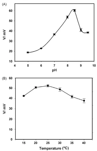
The effects of luminol concentration, temperature and pH on the activity of immobilized HRP and on the performance of the CMOS photodiode were further evaluated. The effect of the pH on the HRP-catalyzed luminol luminescence at pH values of between 5 and 9.5 was also studied. The activity of HRP was measured at 25◦C in buffers at various pH values in
the presence of 4 mM H2O2and 10 mM luminol. As shown in
Fig. 3A, the HRP-catalyzed luminescence detected on the opti-cal H2O2 biosensor increased from pH 5 to 8.5, and reached
a maximum at a pH of 8.5. The activity of HRP then declined from pH 8.5 to 9.5. The temperature is known to influence both the dark current of a CMOS photodiode and the activity of an immobilized enzyme on it. Thus, the effect of temperature on the HRP-catalyzed luminol luminescence was tested at tem-peratures of between 15 and 40◦C (Fig. 3B, solid line). With
Fig. 3. Effect of temperature and pH on the detection of HRP-catalyzed luminol luminescence. (A) Effect of pH on the detection of HRP-catalyzed luminol lumi-nescence. Measurements were made with 10 mM luminol as substrate and 4 mM H2O2at room temperature and the indicated pH value. Data were presented as mean± S.D. from three independent experiments. (B) Effect of temperature on the detection of HRP-catalyzed luminescence. Measurements were made in 100 mM Tris–HCl buffer (pH 8.5) that contained 10 mM luminol and 4 mM H2O2. Reagents were individually incubated at the desired temperature for 10 min. The total reaction volume was 10l. Each datum was the mean ± S.D. from three independent measurements.
10 mM luminol and 4 mM H2O2, the luminescence detected on
the developed optical H2O2 biosensor reached a maximum at
20–25◦C. The luminescent response outside this temperature range reduced approximately 20%. Interestingly, the dark cur-rent of the CMOS photodiode chip increased only 7.6% with the increase in temperature from 20 to 30◦C. At 20◦C, the volt-age caused by dark current is 113.23 mV, while it is 116.99 and 121.85 mV at 25 and 30◦C, respectively. These results reveal that the developed optical H2O2biosensor is appropriate for use
at room temperature. Additionally, the HRP-catalyzed lumines-cence in various concentrations of luminol (0.5–60 mM) and H2O2 (0.5–20 mM) was investigated on the developed optical
H2O2biosensor. As displayed inFig. S1, at a fixed concentration
of H2O2, the luminescence reached a plateau when the luminol
concentration was about double the concentration of H2O2. This
observation is consistent with the results of the work ofBerezin et al. (1975). Accordingly, the following real-time detection of HRP-catalyzed luminescence was performed in the presence of 55 mM luminol at pH 8.5 and 25◦C.
Fig. 4. Calibration graphs of the miniature optical H2O2and glucose biosensors. (A) Real-time detection of H2O2(0.05–20 mM) on the optical H2O2 biosen-sor. The reactions were performed in 100 mM Tris–HCl in the presence of 55 mM luminol at pH 8.5 and 25◦C. Calibration curve of miniature optical H2O2biosensor. Data are presented as mean± S.D. from at least three indepen-dent experiments. (B) Calibration curve of fabricated miniature optical glucose biosensor. Measurements were made with 55 mM luminol and various concen-trations of glucose (0.5–20 mM) in Tris–HCl buffer at pH 8.5 and 25◦C. The inset graph shows the photocurrents of glucose optical biosensor with glucose concentration between 75M and 1 mM. The total reaction volume was 10 l. Data are presented as mean± S.D. from at least three independent experiments.
The miniature optical H2O2biosensor can detect luminescent
responses of various concentrations of H2O2in real time. The
calibration curve of hydrogen peroxide was linear from 50M to 20 mM (Fig. 4A) with a correlation coefficient of 0.991. The detection limit of this system was estimated approximately 25M. These results indicate that a miniature optical H2O2
biosensor having a high sensitivity and wide detection range can be fabricated by simply integrating a CMOS photodiode with sol–gel technology.
3.3. Fabricating a GOx/HRP coupled enzyme-base optical glucose biosensor
A miniature optical glucose biosensor was further fabricated by integrating a GOx/HRP coupled enzyme pair and a CMOS photodiode. Like the optical H2O2biosensor, the developed
opti-cal glucose biosensor can also detect glucose in real time and has an optimal operational temperature of between 20 and 25◦C (data not shown). Outside this range, the detected luminescent response drops by about 20%. This result reveals that the CMOS-based optical glucose biosensor can also perform optimally at room temperature.
The current–time profile of various concentrations of glucose reveals that the fabricated optical glucose biosensor exhibited a linear dynamic range of 0.5–20 mM glucose with a corre-lation coefficient of 0.992 (Fig. 4B). The detection limit of
this biosensor was estimated approximately 75M glucose.
The concentration of glucose in normal sera is around 5 mM. The dynamic range of the developed optical glucose biosensor certainly enables the glucose level in blood samples to be deter-mined in a clinical laboratory. This work reveals that miniature optical biosensors for analyzing various substances can be fab-ricated by integrating different coupled redox enzyme systems and the CMOS photodiode.
3.4. Characterization of the miniature optical glucose biosensor
The reproducibility of the miniature optical glucose biosensor was investigated by repetitively measuring the
enzyme-Fig. 5. Reproducibility of the miniature optical glucose biosensor. The pho-tocurrent responses of optical glucose biosensor to 5 mM glucose were obtained at room temperature in 30 repeated experiments. Measurements were carried out in the presence of 55 mM luminol in Tris–HCl buffer at pH 8.5 and 25◦C.
catalyzed luminescence in the presence of 5.6 mM glucose and 55 mM luminol at pH 8.5 and 25◦C.Fig. 5reveals that the devel-oped optical glucose biosensor is highly reproducible with a mean photocurrent response of 64.8± 2.8 mV. The relative stan-dard deviation was determined to be 4.4% for 30 tests. Moreover, no leakage of the encapsulated enzymes in the TEOS/PDMS Ormosil was observed. This result shows that the fabricated opti-cal glucose biosensor is reliable and exhibits good operational stability and reproducibility in multiple-usage and continuous analysis.
The presence of naturally occurring potent electroactive species, such as ascorbic acid (AA), l-cysteine (l-Cys) and uric acid (UA), poses a great problem for electrochemical-type biosensors in clinical diagnosis. These electroactive species typ-ically cause severe interference during the measurement. At a working potential of over 0.4 V, marked Faradic responses can be produced by AA, l-Cys and UA (Mizutani et al., 1998; Matos et al., 2000). AA (0.1 mM), l-Cys (0.05 mM) and UA (0.5 mM) alone exhibited electrochemical responses of 0.22± 0.01 A, 0.09± 0.01 A and 0.14 ± 0.008 A, respectively, which were 8.4± 0.36%, 3.4 ± 0.46% and 5.4 ± 0.34% of the electrochem-ical response to 5.6 mM glucose (Table 1). In contrast, the
Table 1
Effect of natural interference substances on the amperometric and the miniature optical glucose biosensors during the detection of glucose
Substancee Amperometric biosensor CMOS biosensor
Iox(A)a %b V (mV)c %d 5.6 mM Glc 2.61± 0.03 100 68.10± 1.19 100 0.1 mM AA 0.22± 0.01 8.4± 0.36 0.0 0 0.05 mM l-Cys 0.09± 0.01 3.4± 0.46 0.0 0 0.5 mM UA 0.14± 0.01 5.4± 0.34 0.0 0 5.6 mM Glc + 0.1 mM AA 3.16± 0.04 121± 2.77 62.62± 1.36 92± 0.59 5.6 mM Glc + 0.05 mM l-Cys 2.95± 0.07 113± 3.62 65.78± 1.16 97± 1.44 5.6 mM Glc + 0.5 mM UA 2.79± 0.02 107± 1.59 65.37± 0.24 96± 2.04
aThe oxidative current (Iox) of indicated substances on the GOx immobilized screen-printing carbon electrode. The electrochemical reaction was performed in the 50 mM phosphate buffer, pH 7.4 under a working potential of 0.7 V.
b The percentage of oxidative current of indicated substance relative to the oxidative current of 5.6 mM glucose. cThe photocurrent response (V) of indicated substances on the miniature optical glucose biosensor.
d The percentage of photocurrent response of indicated substances relative to the photocurrent response of 5.6 mM glucose. eGlc: glucose; AA: ascorbic acid; l-Cys: l-cysteine; UA: uric acid.
developed optical glucose biosensor did not respond to these electroactive species. The measurement of 5.6 mM glucose in the presence of 0.1 mM AA, 0.05 mM l-Cys or 0.5 mM UA revealed increments of about 21%, 13% or 7% in the electrochemical responses on a GOx-immobilized screen-printed carbon paste
electrode (SPCE) (Table 1). AA (0.1 mM), l-Cys (0.05 mM)
and UA (0.5 mM) caused declines slightly in the photocurrent response of glucose on the developed optical glucose biosensor. These results indicate that these electrochemical active species cause only weak interference, even at double their physiolog-ical concentrations. Therefore, the fabricated optphysiolog-ical glucose biosensor has great potential for future applications in clinical diagnosis.
4. Conclusion
This work reveals that the miniature optical biosensors can be fabricated by integrating a CMOS-based photodiode, sol–gel technology and coupled redox enzyme pair. A highly transpar-ent organic–inorganic hybrid material (TEOS/PDMS Ormosil) was adopted to immobilize enzymes on the CMOS photodiode to form these miniature optical biosensors. These biosensing devices were demonstrated to be efficient at detecting enzyme-catalyzed luminescence in real time. Both single and coupled enzyme systems can be utilized in this design to fabricate minia-ture optical biosensors. Additionally, the developed miniaminia-ture optical biosensors exhibit a short response time, a small sample volume requirement, a large dynamic range and high sensitiv-ity in clinical diagnoses, food qualsensitiv-ity control and environmental
monitoring (DeBusschere and Kovacs, 2001; Simpson et al.,
2001; Golden and Ligler, 2002; Yang et al., 2002; Huang et al., 2004). The results of this study reveal that this design has a great potential to fabricate miniature optical biosensors for a wide range of biochemical and biological targets. Furthermore, a more sophisticated, powerful, miniature luminescence sensing system can easily be fabricated by combining both photodiodes and particular integrated circuits, for future biochip development (Millar, 1993; Jain, 2000; Fuchs et al., 2002).
Acknowledgements
The authors would like to thank the Center for Nano Science and Technology of the University System of Taiwan and the Ministry of Education of the Republic of China (Taiwan).
Appendix A. Supplementary data
Supplementary data associated with this article can be found, in the online version, atdoi:10.1016/j.bios.2006.12.031.
References
Askari, M.D., Miller, G.H., Vo-Dinh, T., 2002. Cancer Detect. Prevent. 26, 331–342.
Berezin, I.V., Ugarova, N.N., Dmitrieva-Getling, M.P., Kershengoltz, B.M., 1975. Biokhimiia 40, 475–483.
Blum, L.J., Gautier, S.M., 1991. L.J. Blum, P.R. Coulet (Eds.). Marcel Dekker, New York, pp. 213–247.
Chen, Z., Kaplan, D.L., Gao, H., Kumar, J., Marx, K.A., Tripathy, S.K., 1996. Mater. Sci. Eng. C 4, 155–159.
DeBusschere, B.D., Kovacs, G.T., 2001. Biosens. Bioelectron. 16, 543–556. Diwan, M., Park, T.G., 2001. J. Control. Release 73, 233–244.
Fuchs, B., Vogel, S., Schroeder, D., 2002. Med. Eng. Phys. 24, 695–701. Golden, J.P., Anderson, G.P., Rabbany, S.Y., Ligler, F.S., 1994. IEEE Trans.
Biomed. Eng. 41, 585–591.
Golden, J.P., Ligler, F.S., 2002. Biosens. Bioelectron. 17, 719–725. Ho, W.J., Yuan, C.J., Reiko, O., 2006. Anal. Chim. Acta 572, 248–252. Huang, S.H., Shih, Y.C., Wu, C.Y., Yuan, C.J., Yang, Y.S., Li, Y.K., Wu, T.K.,
2004. Biosens. Bioelectron. 19, 1627–1633.
Ivanov, Y.D., Kanaeva, I.P., Gnedenko, O.V., Pozdnev, V.F., Shumyantseva, V.V., Samenkova, N.F., Kuznetsova, G.P., Tereza, A.M., Schmid, R.D., Archakov, A.I., 2001. J. Mol. Recognit. 14, 185–196.
Jain, K.K., 2000. Pharmacogenomics 1, 289–307.
Kamidate, T., Katayama, A., Ichihashi, H., Watanabe, H., 1994. J. Biolumin. Chemilumin. 9, 279–286.
Li, C.I., Lin, Y.H., Shih, C.L., Tsaur, J.P., Chau, L.K., 2002. Biosens. Bioelectron. 17, 323–330.
Lu, U., Hu, B.C., Shih, Y.C., Wu, C.Y., Yang, Y.S., 2004. Biosens. Bioelectron. 19, 1185–1191.
Lu, U., Hu, B.C.P., Shih, Y.-C., Yang, Y.-S., Wu, C.-Y., Yuan, C.-J., Ker, M.-D., Wu, T.-K., Li, Y.-K., Hsieh, Y.-Z., Hsu, W., Lin, C.-T., 2003. IEEE Sens. J. 3, 310–316.
Marquette, C.A., Blum, L.J., 1999. Anal. Chim. Acta 381, 1–6.
Matos, R.C., Augelli, M.A., Lago, C.L., Angnes, L.L., 2000. Anal. Chim. Acta 404, 151–157.
Millar, N., 1993. Med. Device Technol. 4, 16–21.
Mizutani, F., Sato, Y., Sawaguchi, T., Yabuki, S., Iijima, S., 1998. Sens. Actuators B: Chem. 52, 23–29.
Morrison, J.D., Whiteside, T.C., 1984. Perception 13, 555–566.
Pandey, P.C., Upadhyay, S., Shukla, N.K., Sharma, S., 2003. Biosens. Bioelec-tron. 18, 1257–1268.
Park, C.B., Clark, D.S., 2002. Biotechnol. Bioeng. 78, 229–235.
Preuschoff, F., Spohn, U., Janasek, D., Weber, E., 1994. Biosens. Bioelectron. 9, 281–295.
Ruano, J.M., Benoit, V.V., Aitchison, J.S., Cooper, J.M., 2000. Anal. Chem. 72, 1093–1097.
Rubtsova, M., Kovba, G.V., Egorov, A.M., 1998. Biosens. Bioelectron. 13, 75–85.
Schulze-Nahrup, J., Gao, Z.M., Mark, J.E., Sakr, A., 2004. Int. J. Pharm. 270, 199–208.
Shtelzer, S., Braun, S., 1994. Biotechnol. Appl. Biochem. 19, 293–305. Simpson, M.L., Sayler, G.S., Patterson, G., Nivens, D.E., Bolton, E.K., Rochelle,
J.M., Arnott, J.C., Applegate, B.M., Ripp, S., Guillorn, M.A., 2001. Sens. Actuators B: Chem. 72, 134–140.
Singh, A.K., Flounders, A.W., Volponi, J.V., Ashley, C.S., Wally, K., Schoeniger, J.S., 1999. Biosens. Bioelectron. 14, 703–713.
Song, J.M., Culha, M., Kasili, P.M., Griffin, G.D., Vo-Dinh, T., 2005. Biosens. Bioelectron. 20, 2203–2209.
Tang, Y., Tehan, E.C., Tao, Z., Bright, F.V., 2003. Anal. Chem. 75, 2407–2413. Tsafack, V.C., Marquette, C.A., Pizzolato, F., Blum, L., 2000. Biosens.
Bioelec-tron. 15, 125–133.
Wang, B., Zhang, J., Dong, S., 2000. Biosens. Bioelectron. 15, 397–402. Wang, J., Pamidi, P.V., Rogers, K.R., 1998. Anal. Chem. 70, 1171–1175. Wolfbeis, O.S., Oehme, I., Papkovskaya, N., Klimant, I., 2000. Biosens.
Bio-electron. 15, 69–76.
Yang, J.M., Bell, J., Huang, Y., Tirado, M., Thomas, D., Forster, A.H., Haigis, R.W., Swanson, P.D., Wallace, R.B., Martinsons, B., Krihak, M., 2002. Biosens. Bioelectron. 17, 605–618.
Yao, T., Takashima, K., 1998. Biosens. Bioelectron. 13, 67–73. Youan, B.B., 2003. Drug Deliv. 10, 283–288.
Zhou, S., Deng, X., He, S., Li, X., Jia, W., Wei, D., Zhang, Z., Ma, J., 2002. J. Pharm. Pharmacol. 54, 1287–1292.