國 立 交 通 大 學
材 料 科 學 與 工 程 學 系
博 士 論 文
奈米顆粒及化學改質對藥物載體材料之物理性質與
生物醫學特性影響研究
Influence of nanoparticles and chemical modification on physical
properties and biomedical characteristics of drug carrier materials
研 究 生: 劉澤英
指導教授: 陳三元 博士
奈米顆粒及化學改質對藥物載體材料之物理性質與生物醫學特性影響研究
Influence of nanoparticles and chemical modification on physical properties and
biomedical characteristics of drug carrier materials
研 究 生:劉澤英 Student:Tse-Ying Liu
指導教授:陳三元 博士 Advisor:Dr. San-Yuan Chen
國 立 交 通 大 學
材料科學與工程系
博 士 論 文
A Dissertation
Submitted to Department of Materials Science and Engineering And Institute of Materials Science and Engineering
College of Engineering National Chiao Tung University in Partial Fulfillment of the Requirements
for the Degree of Doctor of Philosophy
in Materials Science and Engineering
June 2006
Hsinchu, Taiwan, Republic of China
奈米顆粒及化學改質對藥物載體材料之物理性質與生物醫學特性影響研究
研究生: 劉澤英 指導教授: 陳三元 博士
國立交通大學材料科學與工程學系 博士班
摘 要
本研究主要是研究缺鈣氫氧基磷灰石(Ca-deficient hydroxyapatite, CDHA) 奈米顆粒
對於PMMA 與幾丁聚醣高分子物理性質及藥物釋放行為的影響。首先,PMMA-CDHA
的流變行為與 CDHA 形貌與濃度的關聯性如下:增加 CDHA 濃度會使 shear-thinning 行為變得顯著,代表奈米顆粒間以凡得瓦力與機械接觸構成交互作用使流變行為偏離牛 頓流體,同時,複合材料中奈米顆粒的堆積效率與流變降服強度均與奈米顆粒長寬比有 高度相關。至於以 CDHA 奈米顆粒做為牛血清蛋白載體時,其攜藥奈米載體的合成方 式(原位製程與非原位製程)對牛血清蛋白攜帶量及後續蛋白質釋放行為的影響如下:對 這兩種製程而言,奈米載體之牛血清蛋白攜帶量均隨合成環境pH 值增加而減少,在 pH 值超過8 時,相對原位製程而言,透過非原位製程所合成的奈米載體具有較大的蛋白質 攜帶量,然而,其也明顯地對應一個突放(bursting release)的藥物釋放行為,這個突放行 為主要歸因於牛血清蛋白分子的脫附(desorption);相反地,透過原位製程所合成的奈米 載體則表現出較和緩的釋放行為,原因為透過原位製程使得牛血清蛋白分子與奈米載體 產生較強的交互作用,這樣的解釋可以透過高解析度穿透式電子顯微鏡(HR-TEM)來證 明﹔另一方面,在低pH 值環境下以原位製程所合成的奈米載體,則表現出兩階段釋放 行為,第一階段突放是來自於牛血清蛋白分子的脫附,第二階段的釋放是來自於奈米顆 粒c 軸(長軸)溶解時所釋放出的蛋白質分子。另外,CDHA 奈米顆粒對於攜藥幾丁聚醣
複合薄膜藥物釋放行為的影響如下: CDHA 奈米顆粒的添加量與複合材料合成方式均 會對擴散機制造成影響,以原位合成所製備的複合薄膜,其擴散機制由擴散控制機制朝 向膨潤控制機制變化。 為了進一步調控奈米粒子與幾丁聚醣高分子間之作用力,並擴展幾丁聚醣的生醫適 應性使其可包覆雙性藥物分子及DNA 等會在酸性環境中變性的生物分子,本研究成功 地合成一種無細胞毒性的雙性幾丁聚醣(NOCHC),其上之己醯基對於細胞存活率並無顯 著負面影響,本材料因有高度的親水性及部份疏水基所造成的靜電遮蔽效用,使得纖維 母細胞非常不易貼附,然而,己醯基的進一步增加似乎有助於纖維母細胞的貼附﹔另 外,己醯基的導入對於吸水率產生了非常顯著的影響,其在低溼度時會使水接合位置增 加,在飽和狀態時則會增加分子間的空間使得大量的自由水可以存在,這個現象,也造
成了NOCHC 的酸鹼敏感性大於 NOCC(N,O-carboxymethyl chitosan)﹔而且,疏水性的己
醯基會影響水分子的移動使材料的保水能力提升﹔另外,相較於 NOCC 與未改質的幾
丁聚醣,NOCHC 可以包覆較多的雙性藥物 Ibuprofen,然而, NOCHC 因膨潤度大所
以導致過於快速的藥物釋放,為了改善此特性,本研究使用疏水性的O-hexanoyl chitosan
(OHC)微球來分散於 NOCHC 基底中,可以達到程序式釋放的目的﹔另一方面,CDHA
奈米顆粒的混掺亦可調控此二者的藥物釋放行為,CDHA 奈米顆粒對於此二幾丁聚醣衍
生物藥物釋放行為的影響如下:CDHA 對於 NOCHC 有類似交聯的效應,可減緩釋放 速度並使擴散機制改變,然而,CDHA 則會破壞 OHC 的疏水結構使水份容易進入材料 內部,加速降解導致釋放速度增加。
Influence of nanoparticles and chemical modification on physical properties and
biomedical characteristics of drug carrier materials
Student: Tse-Ying Liu Advisor: Dr. San-Yuan Chen
Department of Materials Science and Engineering
National Chiao Tung University
ABSTRACT
Ca-deficient hydroxyapatite (CDHA) nanoparticles were incorporated with poly(methyl methacrylate) (PMMA), bovine serum albumin (BSA) and chitosan-based materials to explore the influence of filler-filler interaction, nanocarrier-protein interaction and filler-polymer interaction on the physical properties and drug release behaviors of the bioactive nanocomposites. Rheological behaviors of the PMMA-CDHA melting suspensions were systematically investigated in terms of solid loading and aspect ratio of the CDHA nanoparticles. It was found that an increase in solid contents causes pronounced shear-thinning behavior. This result suggests that a strong interaction, including van der Waals attraction and mechanical interlocking, between the CDHA nanoparticles makes the nanocomposite mixture more non-Newtonian. In addition, the packing efficiency and yield strength in the suspension were strongly influenced by the aspect ratio of nanoparticles. For the BSA-incorporated CDHA nanocarrier, the amount of BSA uptake by CDHA decreases with increasing pH. Besides, the release profile showed a bursting behavior for the nano-carrier prepared via the ex-situ process, which is probably due to the desorption of BSA molecules. In contrast, for the sample synthesized via the in-situ process at a higher pH level, a slower release profile without bursting behavior due to the dissolution of the BSA-incorporated CDHA crystal as seen from high solution TEM that indicates different extent of interaction between BSA and CDHA.
nanocomposite monolithic membrane was investigated. It demonstrates that the drug diffusion mechanism is altered by the CDHA-chitosan interaction which is strongly influenced by both the synthesis process and the concentration of the CDHA nanofiller in the membrane. In order to encapsulate the partially hydrophobic agents to prevent the agent from denature in acid environment, a novel carboxymethyl-hexanoyl (NOCHC) hydrogel with amphiphatic nature and pH-sensitivity was synthesized. The cell viability results indicate that carboxymethyl-hexanoyl chitosan did not show any sign of cytotoxicity in vitro. The degree of hexanoyl substitution did not affect the number of vital cells. In addition, the amount of fibroblast with spread out morphology increased with degree of the hexanoyl substitution. On the other hand, the hexanoyl substitution affected significantly the water-absorption ability by altering the number of water-binding sites and the state of water under low humidity and the fully-swollen state, respectively. Moreover, the presence of hydrophobic hexanoyl substitution retarded significantly water mobility during deswelling, causing better water-retention ability. Besides, compared with that of pristine chitosan and N,O-carboxymethyl chitosan (NOCC), the encapsulation efficiency of ibuprofen (IBU, partially hydrophobic agent) was significantly enhanced with the incorporation of the hexanoyl group. However, the bursting release of IBU was less prominent for the NOCHC samples with high degree of carboxymethyl substitution. Moreover, the pH-sensitivity was more pronounced for NOCHC than NOCC. In order to regulate the bursting release behavior of the NOCHC hydrogel in a highly swollen state, the release kinetics of microsphere-embedded hydrogel prepared by NOCHC and O-hexanoyl chitosan (OHC) was further investigated by incorporating various amounts of CDHA nanoparticles. It was found that the release duration of IBU from NOCHC was prolonged with the amounts of CDHA. On the contrary, the release duration of IBU from OHC (hydrophobic phase) was shortened through increasing the CDHA amount, which is due to the hydrophilic nature of the CDHA nanoparticles, destroying the intermolecular hydrophobic interaction and accelerating OHC degradation.
誌 謝
“昨夜西風凋碧樹,獨上高樓,望盡天涯路”道盡博士班研究生在多少長夜裡面對 研究挫折的徬徨與心境上的孤寂,如果沒有師長的指導與家人的支持,很難順利走完蕭 瑟的研究旅程! 首先要感謝指導教授陳三元博士與劉典謨博士在研究策略規劃上的導引及科學邏 輯的訓練,使我能夠順利從金屬材料背景順利切入高分子生醫材料的領域,繼續砌築知 識之磚;另外,衷心感謝國防醫學大學王順德教授、吳慶祥教授以及仁愛醫院夏清智主 任提供生物醫學實驗設施及寶貴的知識與建議,使得研究成果未來得以真正與生物醫學 領域相銜接;在高分子專業領域部份,感謝韋光華教授與林宏洲教授在授課過程中的指 引;對於中興大學徐善慧教授在細胞毒性實驗上的協助,在此也謹致感謝之意! 另外, 在電子顯微鏡學部份,非常感謝劉思謙博士的協助與指導!同時也謝謝同步輻射中心李 信義博士在 X-Ray 繞射儀使用上的協助! 感謝若豪及怡伶兩位學妹在協同研究上的貢獻,使得整體研究能夠順利進行;也謝 謝實驗室夥伴:晉慶、志成、繼聖、定宇、彥妤、昆和、翔詮、佳惠、冠廷、泓洲、尚 秀、虹蓓、書萍、白嘉、泰彥、彥谷、昀碩、茹蘭與琬琳的相伴,共享研究瓶頸之苦與 工四館烤肉之趣;同時也感謝國防醫學院學弟妹: 耀文、尚群、典妮與俊彥在生物實驗 上的協助;另外,所上其他同學及過去華通電腦老長官與伙伴在這四年中所給予的鼓勵 與協助在此一併致謝! 我要特別感謝妻子文如在過去四年獨立負擔家計與辛勤養育女兒翔瑜,她們二位讓 我有無比的勇氣面對所有的挫折與挑戰;另外,衷心地感激岳父岳母給予的支持與勉 勵,讓我無後顧之憂地從產業界轉戰學術界;最後,我要將最誠摯的謝意獻給我的父母, 感謝他們多年來的茹苦養育與默默的付出,謹以此論文獻給我摯愛的家人,感謝他們為 我所做的一切!!CONTENTS:
ABSTRACT (Chinese) i ABSTRACT iii ACKNOWLEDGEMENT v CONTENTS vi LIST OF TABLES x LIST OF FIGURES xi CHAPTER 1: Introduction 11.1. Introduction to bioactive nanocomposites 1
1.2. Motivation of this dissertation 2
1.3. Outline of this dissertation 3
CHAPTER 2: Literature Review
6
2.1. Inorganic nanomaterials for orthopedic application 6
2.1.1. Introduction to nanomaterials 6
2.1.2. Fundamental of calcium phosphate ceramics (CPCs) 6 2.1.3. Hydroxyapatite (HA) and Ca-deficient hydroxyapatite (CDHA) 7
2.2. Polymer-based orthopedic materials 9
2.2.1. Acrylic bone cement 9
2.2.2. PMMA-HAp nanocomposites 10
2.2.3. Chitosan and its derivatives 11
2.3. Drug release dynamics 14
2.3.1. Undegradable matrices 14
2.3.2. Biodegradable matrices 17
CHAPTER 3
Aspect Ratio of Bioactive Nanoparticles on Rheological Behavior of PMMA-Based Orthopedic Materials
26
3.1. Introduction 26
3.2. Experiment materials and methods 27
3.3. Results and discussion 29
3.4. Conclusion 35
CHAPTER 4
BSA-loaded Calcium-deficient Hydroxyapatite Nano-carriers for Controlled Drug Delivery
45
4.1. Introduction 45
4.2. Experiment materials and methods 46
4.3. Results and discussion 48
4.4. Conclusion 55
CHAPTER 5
Study on Drug Release Behaviour of CDHA/chitosan Nanocomposites-Effect of CDHA Nanoparticles
66
5.1. Introduction 66
5.3. Results and discussion 70
5.4. Conclusion 77
CHAPTER 6
Synthesis and Characterization of Amphiphilic Carboxymethyl-hexanoyl Chitosan Hydrogel: Water-retention Ability and Drug Encapsulation
87
6.1. Introduction 87
6.2. Experiment materials and methods 88
6.3. Results and discussion 92
6.4. Conclusion 99
CHAPTER 7
Characterization of Amphiphilic Carboxymethyl-hexanoyl Chitosan Hydrogel
108
7.1. Introduction 108
7.2. Experiment materials and methods 109
7.3. Results and discussion 112
7.4. Conclusion 116
CHAPTER 8
Sequential Rrelease Behaviors of Carboxymethyl-hexanoyl chitosan/O-hexanoyl chitosan Hydrogel: Role of Hydroxyapatite Nanoparticles.
123
8.1. Introduction 123
8.3. Results and discussion 127 8.4. Conclusion 131 CHAPTER 9 Conclusions 139 REFERENCES 142 PERSONAL INFORMATION 160 PUBLICATION 161
LIST OF TABLES
Table 2-1 Definitions of the terms used in nanostructured materials. 19
Table 2-2 Types of implant-tissue response. 20
Table 2-3 Present uses of bioceramics. 21
Table 2-4 Name, abbreviations and formula of the calcium phosphate. 22 Table 2-5 Source and composition of the commercialized bone cement. 23 Table 2-6 Diffusion exponent (n) of the power law and drug release mechanism
from polymeric controlled delivery system of different geometry.
23
Table 4-1 Synthetic conditions of the BSA-CDHA nano-carriers. 56 Table 5-1 Synthetic conditions of the CDHA/CS nanocomposites. 78 Table 6-1 Sample name and corresponding estimation of substitution degree by
1H-NMR.
100
Table 7-1 Sample name and corresponding estimation of substitution degree by 1H-NMR.
LIST OF FIGURES
Figure 2-1 Structural model of hydroxyapatite with the [0001] as projection axis. 24 Figure 2-2 Structures of cellulose, chitin and chitosan. 25 Figure 3-1 X-ray diffraction patterns of the calcium phosphate powders prepared
with various addition rates.
37
Figure 3-2 Shear rate-shear viscosity relationship for CDHA-PMMA mixtures with nano-needles of different aspect ratio.
38
Figure 3-3 Casson’s correlation for the mixtures with AR = 17. 39 Figure 3-4 Dependence of yield stress of the CDHA-PMMA mixtures on solid
concentration.
40
Figure 3-5 Dimensional configuration between two nano-needles with an interparticle distance of λ.
41
Figure 3-6 Maximum solid load (Φm) of the CDHA-PMMA mixtures under investigation can be determined via an extrapolation of the linear (1-η-1/2) - φ correlation at 1- η-1/2 = 1.
42
Figure 3-7 Dependence of maximum solid concentration on the aspect ratio of the CDHA particles.
43
Figure 3-8 The variation of the yield strength of the CDHA-PMMA mixtures as a function of particle aspect ratio at different solid loads.
44
Figure 4-1 FT-IR spectra of the nano-carriers synthesized in different conditions. 57 Figure 4-2 Dependence of Ca(CH3COO)2 concentrations on the CDHA
production yield rate. The corresponding ratio of Ca2+ to amino acid residue was calculated andshown in the second Y-axis.
Figure 4-3 XRD patterns of the BSA-loaded CDHA nano-carriers synthesized in different condition.
59
Figure 4-4 (a) HR-TEM photograph of the BSA-loaded CDHA nano-carriers prepared through in-situ processes at pH=9.5, (b) Corresponding color-enhanced image of (a).
60
Figure 4-5 DTG curves of the BSA-loaded CDHA nano-carriers synthesized in different conditions.
61
Figure 4-6 TEM micrographs of the BSA-loaded CDHA nano-carriers prepared through different processes, (a)-(c) showing the effect of processing variation on morphology and size of CDHA nano-crystals before release test, (d)-(f) showing a further reduction in both aspect ratio and size of the CDHA nano-crystals after release test compared to that before the test.
62
Figure 4-7 Dependence of the pH value of synthetic solution on the (a) aspect ratio of CDHA crystal, (b) amount of BSA uptake by CDHA nano-crystal.
63
Figure 4-8 BSA release profiles of the BSA-loaded CDHA nano-carriers synthesized in different conditions.
64
Figure 4-9 ELNES spectra near the Ca L2,3-edge of BSA-loaded CDHA nano-carriers prepared through in-situ processes at pH=9.5.
65
Figure 5-1 Selective SEM micrographs (cross-section) of CDHA/CS nanocomposite membranes.
79
Figure 5-2 Selective TEM micrographs of CDHA/CS nanocomposite membranes.
80
Figure 5-4 TGA curves of TPP cross-linked chitosan and CDHA/CS nanocomposite membranes prepared via in-situ process.
82
Figure 5-5 (a). Dependences of synthetic processes and amount of CDHA on the degree of cross-link and diffusion exponent of nanocomposite membranes. (b). Dependences of degree of cross-link on the diffusion exponents of nanocomposite membranes.
83
Figure. 5-6 DMA curves of chitosan and CDHA/CS nanocomposite membranes with CDHA content of10%.
84
Figure 5-7 Dependences of synthetic processes and CDHA content on the permeability (P) and partition coefficient (H) of CDHA/CS nanocomposite membranes.
85
Figure 5-8 Release profiles of CDHA/CS nanocomposite matrix membranes. 86
Figure 6-1 1H-NMR spectrum of sample NOCHC-2B. 101
Figure 6-2 Dependence of carboxymethyl substitution on water-absorption ability (Wc, solid line) and maximal non-freezable water amount (Wnf,max, dash line).
102
Figure 6-3 ATR-FTIR spectra of chitosan derivates. 103
Figure 6-4 Dependence of hexanoyl group on water-absorption ability (Wc, solid line) and maximal non-freezable water amount (Wnf,max, dash line).
104
Figure 6-5 Deswelling profiles of samples NOCC-3 and NOCHC-3A. 105
Figure 6-6 DSC curves of chitosan derivates (carboxymethyl substitution degree =0.5) measured at Wc = 200%. Dash lines represent the curve fitting by Lorentzian curve-fitting procedure.
Figure 6-7 Dependences of degrees of carboxymethyl and hexanoyl substitution on ibuprofen encapsulation efficiency.
107
Figure 7-1 Cytotoxicity of NOCHC samples with carboxymethyl substitution degree of 0.5.
118
Figure 7-2 Cell morphology on the 6-well plates (a), NOCHC-2A immobilized plates and (b) NOCHC-2B immobilized plates after cell seeding for 24 h.
119
Figure 7-3 Dependence of carboxymethyl and hexanoyl groups on pH values. 120
Figure 7-4 Dependence of the swellng ratio of NOCHC-3A and NOCPC samples on pH values.
121
Figure 7-5 Ibuprofen release profiles of NOCHC samples. 122 Figure 8-1 . Proton NMR spectra and molecular structures of (a) HNOCC and (b)
NOCC.
132
Figure 8-2 TEM images of (a) HNOCC-CDHA nano-composite membrane, (b) selected-area electron diffraction (SAD) pattern of CDHA, and (c) OHC-CDHA nano-composite membrane.
133
Figure 8-3 Influence of CDHA amount on the release profiles of OHC-CDHA monolithic membranes.
134
Figure 8-4 DMA curves of (a) OHC-CDHA and (b) HNOCC-CDHA monolithic membranes.
135
Figure 8-5 Influence of CDHA amount on the release profiles of HNOCC-CDHA monolithic membranes: (a) Linear scale; (b) logarithmic scale.
Figure 8-6 SEM images of HNOCC/OHC microsphere-embedded porous sponge: (a) 2500X; (b) 5000X.
137
Figure 8-7 Sequential release profile of HNOCC/OHC microsphere-embedded sponge.
CHAPTER 1
Introduction
1.1. Introduction to bioactive composite for drug carrier
Synthetic bioactive and biodegradable composite materials are becoming increasingly important as scaffolds for tissue engineering. Next-generation biomaterials should combine bioactive and biodegradable properties to activate in vivo mechanisms of tissue regeneration, stimulating the body to heal itself and leading to replacement of the scaffold by the regenerating tissue. The key successful point of the implantable devices for tissue regeneration is not only the suitable matrix with sufficient bioactivity and mechanical strength for progenitor cells, but also the sustained release of antibiotics and growth factors to eliminate infection and insure cell differentiation, respectively. Hence, drug-loaded implants with controlled release function were developed to optimize the therapeutic concentration of drug to the specific site of action, which eliminates the need for multiple intravenous injections and maintains the biological activity of the agents with short half-life such as platelet-derived growth factor and fibroblast growth factor.
Synthetic biocompatiable and biodegradable polymers are easily fabricated into complex structure via injection molding or solution casting method, yet insufficient bioactivity and mechanical strength are hard to meet the demands of surgery and in vivo physiologic environment. On the other hand, certain bioactive ceramics such as hydroxyapatite (HA) react with physiologic fluids to form tenacious bonds with hard tissue. However, these bioactive materials are relatively stiff, brittle, and difficult to be fabricated into complex shapes. Therefore, the tailored properties of bioactive composites such as rheological behavior, molecular relaxation kinetics, biologic response, mechanical strength and drug release profile as well as predictable degradation behavior can be manipulated through incorporating
bioactive inorganic phases into biomedical polymers. Moreover, the characteristics of polymers can be further manipulated through chemical modification by which both biological responses to implants and material responses to the physiological environment can be regulated.
1.2. Motivation of this dissertation
Poly(methymethacrylate) (PMMA), a self-curing cement, is the current standard for cement held prostheses, providing immediate structural support. However, the clinical use of this type of bone cement is typically accompanied by several complications. These complications are a result of the cement’s limited mechanical properties and poor adhesion with bone which may lead to implant failure and periprosthetic osteolysis. On the other hand, chitosan (CS) is abundant in nature and currently receiving a great deal of attention for medical and pharmaceutical applications due to its biodegradable nature and biocompatibility, allowing its use in various medical applications such as bone substitute and drug-loaded scaffolds for bone tissue engineering. Nevertheless, chitosan lacks sufficient mechanical strength, which restricts its uses for load-bearing applications, especially in orthopedics. On this base, many researchers proposed that the incorporation of hydroxyapatite particles (HAp) into PMMA and chitosan can enhance osteoconductivity and mechanical strength to meet the requirement of orthopedic application. However, natural bone mineral is essentially a calcium-deficient apatitic structure (Ca-deficient hydroxyapatite, CDHA) with a Ca/P ratio of about 1.5. Therefore, CDHA is considered to be a candidate material with respect to both mechanical reinforcement and biological activity for orthopedic application. In this study, CDHA nanoparticles were incorporated into PMMA and chitosan to understand the role of CDHA nanoparticles in the rheological behaviors of the PMMA-CDHA nanocomposite and the drug release behaviors of the chitosan-CDHA nanocomposite, which are very important for the fabrication of bone-fixation devices and drug-loaded scaffolds, respectively.
Secondly, chitosan is one of the most abundant natural materials and has been used for wound dressing, cartilage tissue engineering and drug-loaded implants due to its glycosaminoglycan-like structure and low price. However, insufficient swelling ability in neutral physiological condition, poor solubility in organic solvents, and lack of amphiphatic nature have limited its uses. In addition, chitosan is a cationic polysaccharide with an isoelectric point (IEP) of 6.2. It is soluble in diluted acids and the resulting gelled-solution aggregates under neutral physiological conditions, which is unfavorable for incorporating CDHA nanoparticles and encapsulating the bioactive agents such as DNA and protein whose conformation will be altered in acidic environments. Therefore, it is necessary to develop a water-soluble chitosan-based hydrogel with amphiphatic nature to encapsulate the bioactive agents with a wide range of hydrophilic/hydrophobic nature and the agent will denature in neutral environment. In this study, a novel carboxymethyl-hexanoyl chitosan hydrogel was developed to meet above-mentioned requirement. The basic properties of hydrogel were systemically investigated in terms of the degrees of carboxymethyl and hexanoyl substitutions.
1.3. Outline of this dissertation
1. Influence of the aspect ratio of bioactive nanofillers on rheological behavior of PMMA-based orthopedic materials.
In this investigation, calcium-deficient hydroxyapatite nanocrystals with needlelike geometry were synthesized and incorporated with poly(methyl methacrylate), PMMA, to form CDHA-PMMA nanocomposites. Rheological behaviors of the PMMA-CDHA melting suspensions were systematically investigated in terms of the solid content and the aspect ratio of the CDHA nanoparticles.
delivery.
Calcium-deficient hydroxyapatite nano-crystals incorporated with bovine serum albumin (BSA) to form BSA-loaded nano-carriers were synthesized via both in-situ and ex-situ processes. The amount of BSA uptake by the CDHA nano-crystals and the subsequent release behaviors of the BSA-loaded nano-carriers were investigated.
3. Drug release behaviour of CDHA/Chitosan nanocomposites-Effect of CDHA nanoparticles.
With the aim of the manipulation of release kinetics via bioactive nanofillers for orthopedic drug-loaded implants, the effect of CDHA nanofiller on drug release kinetics was studied by adopting vitamin B12 as a model drug for chitosan/Ca-deficient hydroxyapatite (CS/CDHA) nanocomposite membranes prepared in terms of different synthetic processes, i.e. in-situ and ex-situ routes, and various amounts of CDHA.
4. Synthesis and characterization of amphiphilic carboxymethyl-hexanoyl chitosan hydrogel.
Carboxymethyl-hexanoyl chitosan (NOCHC) amphiphatic hydrogel with excellent water-absorption and water-retention abilities in neutral condition was successfully synthesized for the first time and then employed as a carrier for delivering amphiphatic agents. NOCHC is a water-soluble chitosan derivative bearing the carboxymethyl (hydrophilic) group and the hexanoyl (hydrophobic) group. Water-absorption ability (Wc), water-retention ability, and drug encapsulation efficiency of the NOCHC hydrogel were investigated in terms of the degrees of carboxymethyl and hexanoyl substitution.
5. Synthesis and characterization of amphiphilic carboxymethyl-hexanoyl chitosan hydrogel: pH sensitivity and drug release behavior.
Carboxymethyl-hexanoyl chitosan (NOCHC) amphiphatic hydrogel with excellent swelling ability in neutral condition was successfully synthesized using N,O-carboxymethyl chitosan (NOCC) as the starting precursor. Swelling ratio under various pH values (pH-sensitivity) and release behaviors of the NOCHC hydrogel were investigated in terms of the degrees of carboxymethyl and hexanoyl substitution.
6. Sequential release behaviors of carboxymethyl-hexanoyl chitosan/O-hexanoyl chitosan hydrogel: Role of hydroxyapatite nanoparticles.
In order to explore the effect of nanofiller on the regulation of the drug release behavior from microsphere-embedded hydrogel prepared by carboxymethyl-hexanoyl chitosan (NOCHC) and O-hexanoyl chitosan (OHC), the release kinetics was investigated in terms of various amounts of calcium-deficient hydroxyapatite nanoparticles incorporated.
CHAPTER 2
Literature Review
2.1. Inorganic nanomaterials for orthopedic application 2.1.1. Introduction to nanomaterials
Nanoscience/nanotechnology is the science of fabricating, characterizing, and utilizing structures at the atomic, molecular and supramolecular levels in order to understand, create and use material structures, devices and systems with fundamentally new properties and functions resulting from their small structure. Nanomaterials are functional materials comprised of objects with at least one dimension on a scale of 1-100 nm (although the upper limit of 100 nm may be relaxed to greater sizes in some cases, depending on the material and the specific property being investigated) [1]. These objects may be either nanoparticles (small ensembles of molecules) or even individual molecules. One of the most exciting aspects of nanoobjects is that the remarkable effects on novel properties (catalytic, magnetic, ferroelectric, mechanical, optical and electronic) may arise from the critical size reduction. It occurs as we reduce the dimensions from a practically infinite (and periodic) solid crystal to a system composed of a relatively small number of atoms. For example, silver nanoparticles show biological (antimicrobial) activity whereas silver metal does not [2]. Similarly the conductivity of metal particles undergoes a marked change as their size decreases and the optical and electronic properties of inorganic semiconductor nanoparticles are known to depend upon the particle size [3]. The definition of some terms used in the field of nanostructureed materials are presented in Table 2-1 [4].
2.1.2. Bioceramics and calcium phosphate ceramics
prosthesis require that the objects to be implanted be made of ceramic materials. Bioceramics are generally very well tolerated and integrated by living tissues and have no significant toxicity.In general, there are three types of tissue response to a biomaterial as shown in Table 2-2[5]. The present uses of bioceramics are shown in Table 2-3[6].
Inert bioceramics are those which do not exert any influence on tissues and are not influenced by them. Their function is generally to sustain high mechanical loads. Differently from inert bioceramics, those which are defined as bioactive ceramics are able to exert some biological influence on tissues and are influenced by them. They are chemically composed with atomic species managed by the biological cycles, and generally they are calcium phosphates. Calcium phosphates ceramics (CPCs) were taken into consideration in early bioceramic studies for surgical application on bone [7-10]. Several calcium phosphate cements (CPC) have been developed and have potential use in orthopedic field as shown in
Table 2-4 [11]. The most utilized are hydroxyapatite (HA) and tricalcium phosphate (TCP). The stable phases of calcium phosphate ceramics depend considerably upon temperature and the presence of water, either during processing or in the use environment. At body temperature, only two calcium phosphates are stable when in contact with aqueous media such as body fluids. At pH < 4.2, the stable phase is CaHPO4 • 2H2O (dicalcium phosphate dehydrate or hrushite, C2P), while at pH > 4.2 the stable phase is Ca10(PO4)6(OH)2 (HA). At higher temperatures, other phases such as Ca3(PO4)2 (β-tricalcium phosphate, C3P, or β-TCP) and Ca4P2O9 (tetracalcium phosphate, C4P) are present.
2.1.3. Hydroxyapatite and Ca-deficient hydroxyapatite
The main constituents of bone are collagen (20 wt. %), calcium phosphate (69 wt. %), and water (9 wt. %). Additionally, other organic materials, such as proteins, polysaccharides, and lipids are also present in small quantities. Collagen, which can be considered as the matrix, is in the form of small microfibers. The diameter of the collagen microfibers varies
from 100 to 2000 nm. Calcium phosphate in the form of crystallized hydroxyapatite and/or amorphous calcium phosphate (ACP) provides stiffness to the bone [12]. The hydroxyapatite crystals, present in the form of plates or needles, are about 40-60 nm long, 20 nm wide, and 1.5-5 nm thick. They are deposited parallel to the collagen fibers, such that the larger dimension of crystals is along the long axis of the fiber [12].
Hydroxyapatite (HA, chemical formula Ca10(PO4)6(OH)2) is the main mineral constituent of teeth and bones. Hydroxyapatite is one of apatites with the lattice parameters: a1=a2= 0.9432 nm and c= 0.6881 nm [13]. Apatite is a general name for compounds having symmetry P63/m and chemical formulas A10(BO4)2X2. A is cations of 1 to 3 valences, B anions have 3 to 7 valences and X denotes anions of 0 to 3 valences, where A: Ca, Pb, Cd, Sr, Ni, Eu, Al, Y, La, Ce, Na, K, B: P, As, V, Cr, Si, C, Al, S, Re and X: OH, F, Cl, Br, I, O, N, CO3, H2O, vacancy
[13-16]. A structural model along the [0001] zone axis projected along the c-axis is shown in
Figure 2-1 [16], from where the Ca atoms occupy two series of non-equivalent sites. Hydroxyl groups (OH-) are situated on the 6-fold c-axis. These ions are slightly below and above the mirror planes at Z=1/4 and 3/4. Posner et al. first refined the structure of HA and reported the coordination of different Ca atoms in HA [14]. The Ca(1) atoms are coordinated by nine oxygen atoms situated in six different phosphate tetrahedral. The Ca(2) atoms, situated around the hexagonal screw axes, each has an irregular sevenfold coordination with six oxygen of five phosphate groups in addition to the hydroxyl ion. Elliott et al. reported that the space group of HA has three kinds of vertical or columnar symmetry [15]. There are columns of Ca2+ ions spaced by one half of the c-axis parameter along three-fold axes which account for two fifths of the Ca2+ ions in the structure. These ions are given as Ca(1). The Ca2+ ions are linked together by PO4 tetrahedra in which three oxygen atoms come from one column and the fourth comes from an adjacent column. The result is a three-dimension network of PO4 tetrahedra with enmeshed Ca2+ ions, and channels that contain the residual calcium, Ca(2), and ions such as OH- which make up the HA structure. The substitution of
phosphate ions (PO4)3- by hydrogenophosphate ones (HPO4)2- allows a continuous variation of the Ca/P atomic ratio between 9/6 and 10/6 [17]. This leads to calcium deficient hydroxyapatites (CDHA, Ca10-x(PO4)6-x(HPO4)x(OH)2-x, with 0≦x 1). ≦ CDHA powders can be precipitated from conventional wet chemical methods and decomposed into a mixture of HAp and TCP by thermal treatment above 700oC.
From the point of view of biocompatibility, hydroxyapatite seems to be the most appropriate ceramic material for artificial teeth or bones due to excellent biocompatibility and bioactivity. HA ceramics does not exhibit any cytotoxicity. It shows excellent biocompatibility with hard tissues and also with skin and muscle tissues. Moreover, HA can directly bond to the bone. Unfortunately, mechanical properties of pure HA ceramics are poor. For example, fracture toughness (KIc) does not exceed the value of about 1.0 MPa . m1/2
(human bone: 2–12 MPa.m1/2 ). Additionally, the Weibull modulus (n) is low in wet environments (n=5–12) which indicates low reliability of HA implants. Presently, the HA ceramics cannot be used as heavy-loaded implants, such as artificial teeth or bones. Its medical applications are limited to small unloaded implants, powders, coatings, and low-loaded porous implants [18]. In order to improve the reliability of HA ceramics, various reinforcements (ceramic, metallic, or polymer) have been used. Moreover, HA-coated metals have been introduced as artificial bones or teeth.
2.2. Polymer-based orthopedic materials 2.2.1. Acrylic bone cement
Acrylic based bone cement was developed in the early 1960s by Charnley and Smith
[19]. Acrylic bone cements are based on polymethyl methacrylate (PMMA) which is accepted as a biocompatible polymer when cured. The bone cement is usually prepared by mixing the two components of the dose: a transparent liquid and a white powder. The main ingredients are shown in Table 2-5 [20]. The N,N-dimethyl-para-toluidine (DMPT) acts as accelerant of
the polymerization reaction, which activates the initiator mixed with the powder. The hydroquinone (OHC6H4OH, HQ) is an inhibitor, which prevents the premature polymerization of the monomer. The benzoil peroxide (COC6H5OOCC6H5O, BP) acts as the initiator, producing free radicals when it reacts with the amine (DMPT). BaSO4 or ZrO2 are added in order to obtain a radio-opaque cement. The diameter of most of the PMMA particles in the cement ranges between 30 and 150 μm and their shape depends on the manufacturing process used.
2.2.2. PMMA-HAp nanocomposites
PMMA is a bio-inert material thus causing weak linkage between bone and cement. It is also a brittle material with relative low fracture energy which causes the failure of implant
[21]. In order to solve these problems, many PMMA-CPCs composites have been used in surgery to fix joint replacements into the bone. Hydroxyapatite particle filled PMMA composites are the most common used materials in clinical applications. Dalby et al. proposed that HAp/PMMA was a better substrate for human osteoblast (HOB) cells, resulting in increased proliferation and alkaline phosphatase (ALP) activity [22]. Scanning electron microscopy (SEM) and transmission electron microscopy (TEM) showed that HOB cells cultured on the HAp-filled PMMA preferentially anchored to HA particles exposed at the cement surface, with a close intimacy observed between HAp and HOB cells. Aizawa et al. fabricated the HAp-PMMA hybrid material having mechanical properties similar to those of the cortical bone, using porous HA ceramics with well-controlled pore sizes and bulk polymerization of MMA monomer without solvents [23]. Lee et al. studied a hydroxyapatite filled 4-META/MMA-TBB adhesive bone cement [24]. In this study, histologic examinations showed that the exposed HA particles at the surface of the cured cement were generally associated with intimate attachment to bone without fibrous tissue, as well as interdigitation of cement to bone. Moursi reported similar results [25]. Extracellular matrix production was
examined by immunohistochemistry which indicated that osteoblasts cultured on PMMA/HAp showed a more distinct networked pattern of organized fibronectin. Histochemical staining of mineralization was examined by confocal microscopy which demonstrated a higher degree of mineralization in nodules formed on PMMA/HAp as compared to PMMA. Together, these results indicate that the addition of HAp in a PMMA matrix improves osteoblast response as compared to PMMA alone. Liu showed that the surface hydroxyl groups of hydroxyapatite have the ability to react with organic isocyanate groups. It provide a good reason to use HAp as a reinforce agent in PMMA bonecement [26].
2.2.3. Chitosan and its derivatives
Polysaccharides are aboundant in nature. Their molecular structures and hence their properties vary over a broad range. Of the many kinds of polysaccharides, cellulose and chitin are the most important biomass resources; cellulose is synthesized mainly in plants, whereas chitin is synthesized mainly in lower animals. Chitin is structurally similar to cellulose, but it is an amino polysaccharide having acetamide groups at the C-2 positions in place of hydroxyl groups (Figure 2-2) [27]. Chitin is a co-polymer of N-acetyl-glucosamine and N-glucosamine units randomly or block distributed throughout the biopolymer chain depending on the processing method used to derive the biopolymer. When the number of N-acetyl-glucosamine units is higher than 50%, the biopolymer is termed chitin. Conversely, when the number of N-glucosamine units is higher, the term chitosan (CS) is used [27-29].
The presence of amino groups in chitosan is highly advantageous for providing distinctive biological functions and for conducting modification reactions. Recent progress regarding some typical chemical modifications including acylation, phthaloylation, tosylation, alkylation, Schiff base formation, reductive alkylation, O-carboxymethylation, N-carboxyalkylation, silylation, and graft copolymerization have been reported.
2.2.3.1. Hydrophobic Modification
In order to resolve the issue that poor solubility in organic solvents, chemical modification of chitosan, in particular, N-alkylation [30, 31], N-acylation [32, 33], O-acylation [34], and N-carboxyalkylation [35], has been studied. For example, Fujii et al. have reported on the acyl modification of chitosan reacted with long-chain acyl chloride for improving the organic solubility [36, 37]. Nishimura et al. [38] have also reported on phthaloylation of chitosan with phthalic anhydride.
2.2.3.2. Hydrophilic modification
On the other hand, a special emphasis has been placed on the chemical modifications to prepare several chitosan derivatives with higher solubility in water, such as O, N-carboxymethyl-chitosan [39], N-carboxymethyl-chitosan [40], O-carboxymethyl-chitosan [41], N-sulfate-chitosan [42], O-sulfate chitosan [43], O-butyryl-chitosan [44], N-methylene phosphonic chitosan [45], hydroxypropyl chitosan [46], N-trimethyl chitosan [47], N-succinyl-chitosan [48], N-carboxyethyl chitosan [ 49], N-benzyl phosphoryl chitosan [50], and O-succinyl chitosan [51].
2.2.3.3. Amphiphatic modification
Special attention was paid to chemical modification preparing several chitosan derivatives with amphiphatic nature. Lee et al. reported a non-covalently cross-linked palmitoyl glycol chitosan hydrogel evaluated as an erodible controlled release system for the delivery of hydrophilic macromolecules [52]. Ramos et al. introduced an alkyl chain onto a water soluble modified chitosan (N-methylene phosphonic chitosan) offer the presence of hydrophobic and hydrophilic branched for controlling solubility properties [53]. A series of novel chitosan derivative carrying long chain alkyl groups as hydrophobic moieties and sulfated groups as hydrophilic moieties (N-alkyl-O-sulfate chitosan) were synthesized as a
carrier to encapsulate taxol [54]. In addition, an amphiphilic chitosan derivative, O,O’-dipalmitoyl chitosan (DPCT), was synthesized and its miscibility with cholesterol of DPCT were studied by Tong et al.[55].
2.2.4. Chitosan-HAp nanocomposites
There were many researchers has reported HAp-Chitosan nanocomposite. Li et al. prepared the HAp-filled Chitosan composite by in-situ precipitation [56]. It was observed that the mechanical properties of the composite in the dry condition improved as the percentage of CS content increased. Mechanical properties of such CS/HAp composite rod are much better than that prepared via traditional method. They also showed that the water absorption of CS/HAp composite decreased when adding of HA powders into CS matrix, which contributes to postponing the attenuation of mechanical properties of CS/HAp composite under moisture condition. This result provides very useful information for HAp-CS drug delivery system. Chen et al. also proposed that the nano-structure of hydroxyapatite/chitosan composite will have the best biomedical properties in the biomaterials applications [57]. Zhao et al. prepared a biodegradable scaffold by phase separation method for bone tissue engineering which used hydroxyapatite/chitosan-gelatin network (HAp/CS-Gel) composite [58]. Histological and immunohistochemical staining and scanning electron microscopy observation indicated that the osteoblasts attached to and proliferated on the scaffolds. Extracellular matrices including collagen I and proteoglycan-like substrate were synthesized, while osteoid and bone-like tissue formed during the culture period. This report provided a powerful methodology to characterize the scaffolds by the presence of ECM. Sivakumar et al. proposed that the a-axis lattice constant of coralline hydroxyapatite-chitosan composite may decrease with the increase in the amount of chitosan [59]. This result indicated the interaction of the polymer backbone with HAp. The mechanical properties of chitosan-HAp nanocomposite was studied
molecule length and the c-axes of the constituent HAp crystallites were well aligned in parallel with the chitosan molecules. The growth of the HAp crystallites is considered to occur at the nucleation sites that were most probably the complexes of amino groups on chitosan bounded with calcium ions. The compact composites obtained have been found mechanically flexible, and this flexibility has been further improved by heating at 120 degrees C in an autoclave with saturated steam pressure.
2.3. Drug release kinetics 2.3.1. Undegradable matrices
2.3.1.1. Monolithic systems (Matrix systems)
In these systems, the drug is distributed, ideally uniformly, throughout a solid polymer. Diffusion in polymers is complex and the diffusion rates should lie between those in liquids and in solids, which depends strongly on the concentration and degree of swelling of polymers. The first mathematical treatment of diffusion was established by Fick [61] who developed a law for diffusion in one dimension:
J= -Aj= -AD(∂c/∂z) 2-(1) where J is the mass flow, j the flux per unit area, A the area across which diffusion occurs, D the diffusion coefficient, c the concentration, z the distance and (∂c/∂z) the gradient of the concentration along the z axis. This equation is also known as Fick’s first law. In the case of diffusion without convection and a unitary area, Eqn. 2-(1) can be written as
J= -D(∂c/∂z) 2-(2) In the study of solvent diffusion in polymers, different behaviors have been observed. It is known that the diffusion of the solvent is linked to the physical properties of the polymer network and the interactions between the polymer and the solvent itself. Wang et al. proposed a classification according to the solvent diffusion rate and the polymer relaxation rate: Fickian (Case I) and non-Fickian (Case II and anomalous) diffusions [62]. The amount of solvent
absorbed per unit area of polymer at time t, Mt, is represented by
Mt=ktn (Power low) 2-(3) where k is a constant and n a parameter related to the diffusion mechanism, the value of which lies between 1/2 and 1 for the sample with slab geometry [63].
A. Fickian diffusion
The influence of matrix relaxation and the diffusion process on drug release can be estimated by the Deborah number De:
De=λ/θ 2-(4) where De is the ratio of a characteristic relaxation time (λ) to a characteristic diffusion time (θ). Eqn. 2-(4) indicates that De << 1 is obtained when the relaxation is much faster than the diffusion, and in this case drug release is controlled by diffusion in the fluid filling the swollen matrix. This condition usually occurs with swellable systems well above the glass transition temperature (Tg) that swell rapidly and reach equilibrium before drug release takes place. De>> 1 indicates that the matrix is characterized by a very long relaxation and drug release is controlled by diffusion in an unchanged matrix. The drug diffuses in fact from an unswollen region, such as from a matrix in the glassy state (well below the Tg). In the cases of De<< 1 and De>> 1, the drug is released by diffusion through a system with constant boundary conditions. The relaxation process does not interact with drug release, so the same speculations reported above for Fickian diffusion release from undegradable and unswellable devices can be applied. This process is also defined as Case I transport. The diffusion distance is proportional to the square-root of time (Higuichi model) [64]
Mt=kt1/2 2-(5) B. Non-Fickian diffusion
A second case of drug release from swellable and undegradable matrices is observed when the glassy fraction of a swelling system relaxes with observable rate, but much more slowly than the diffusion of the drug in the rubbery fraction of the matrix. In this process,
defined as Case II transport, the release kinetic is controlled by the relaxation of the glassy fraction of the matrix. For a thin film, the diffusion distance is directly proportional to time
Mt=kt 2-(6) When the characteristic relaxation time and diffusion time are comparable (De~1), the relaxation interacts with the diffusion process and drug release takes place under nonconstant boundary conditions. This anomalous non-Fickian transport is observed when the drug is released during a change of the matrix from a glassy to a rubber state. Anomalous diffusion lies in between and is characterized by the following equation:
Mt=ktn and 0.5<n<1. 2-(7) Thus, Eqn. 2-(7) has two distinct physical realistic meanings in the two special cases of n=0.5 (indicating diffusion-controlled drug release) and n=1.0 (indicating swelling-controlled drug release). Values of n between 0.5 and 1.0 can be regarded as an indicator for the superposition of both phenomena (anomalous transport). The two extreme values for the exponent n, 0.5 and 1.0, are only valid for slab geometry. For spheres and cylinders different values have been derived [65, 66], as listed in Table 2-6.
2.3.1.2. Reservoir systems
In this type of system, a core of drug is sourrounded by a polymer and diffusion of the drug through the polymer is the rate-limiting step. In other words, reservoir systems are usually designed so that the drug is contained in an oversaturation concentration in the core of the system and release is controlled by diffusion through a surrounding undegradable membrane. Release is defined by the first Fick’s law, which describes the flux of a substance through the membrane at a steady state:
Jd= -Ddc/dx 2-(8) The first Fick’s equation can be elaborated for matrices with different geometrical shape. For a cylindrical device of height h, inner radius r1, and outer radius r0, the rate of drug release can
be expressed as:
dM/dt=2πhKd(C1-C2)/ln(r0/r1) 2-(9) where Kd is the membrane/medium distribution coefficient (i.e., membrane permeability, Kd=DK where D is the diffusion coefficient, and K the partition coefficient), C1 is the drug concentration in the core, and C2 is the drug concentration in the external medium. For a spherical reservoir of radius r1, sorrrounded by a spherical membrane of radius r0,
dM/dt=4πKd(C1-C2)r0r1/(r0-r1) 2-(10) Assuming that the drug has the same solubility in the system core and in the external medium, the release for a sandwich or slab reservoir is described by
Mt= Sd Kd(C1-C2)t/l 2-(11) where Mt is the released amount of drug at time t, Sd is the releasing surface area, l is the thickness of the polymeric membrane [61].
2.3.2. Biodegradable matrices
Hopfenberg assumed that the rate of drug release from the erodible system is proportional to the surface area of the device which is allowed to change with time [67]. All mass transfer processes involved in controlling drug release are assumed to add up to a single zero-order process (characterized by a rate constant, k0) confined to the surface area of the
system. This zero-order process can correspond to a single physical or chemical phenomenon, but it can also result from the superposition of several processes, such dissolution, swelling, and polymer chain cleavage. A good example for systems Hopfenberg’s model can be applied to is surface eroding polymer matrices where a zero-order surface detachment of the drug is the rate limiting release step. Hopfenberg derived the following general equation, which is valid for spheres, cylinders and slabs:
Mt/ M∞=1-(1-(k0.t/C0.a))n 2-(12) Mt and M∞ are the cumulative amounts of drug released at time t and at infinite time,
respectively; C0 denotes the uniform initial drug concentration within the system; a is the radius of a cylinder or sphere or the half-thickness of a slab; n is a ‘shape factor’ representing spherical (n=3), cylindrical (n=2) or slab geometry (n=1).
Heller and Baker developed a mathematical model describing drug release from water-insoluble polymers that undergo hydrolytic backbone cleavage, during which the polymer chains are converted into small, water-soluble molecules [68]. They assumed that for polymer matrices undergoing bulk erosion, degradation can be described by first-order kinetics.
dMt/dt=A/2 * (2.P0.exp(k. t) .C0/t)1/2 2-(13) here, Mt is the cumulative absolute amount of drug released at time t; k is the first order rate constant; P0 is the initial drug permeability and C0 is the initial drug concentration within the system.
Table 2-1. Definitions of the terms used in nanostructured materials [4]. Terms Definition
Cluster A collection of units (atoms or reactive molecules) of up to about 50 units. Cluster compounds are such moieties surrounded by a ligand shell that allows isolation of a molecular species.
Colloid A stable liquid phase containing particles in the 1-1000 nm range. A colloidal particle is one such 1-10000 nm sized particle.
Nanoparticle A solid particle in the 1-1000 nm range that could be nanocrystalline, an aggregate of crystallites, or a single crystallite.
Nanostructured material
Any solid material that has a nanometer dimension; Three dimensions: Particles
Two dimensions: Thin films One dimension: Thin wire
Zero dimension: Quantum dot (A particle that exhibits a size quantization effect in at least one dimension.)
Table 2-2. Types of implant-tissue response [5].
Types Tissue response Example
Bioinert,
(nontoxic and biologically inactive)
It has minimal interaction with its surrounding tissue. A fibrous capsule forms around a bioinert implant.
stainless steel, titanium,
alumina, partially stabilized zirconia, and
ultra high molecular weight polyethylene.
Bioactive,
(nontoxic and biologically bioactive),
It interacts with the surrounding bone and the soft tissue. An interfacial bond forms.
synthetic hydroxyapatite, glass-ceramic A-W, and Bioglass.
Bioresorbable,
(nontoxic and dissolves),
It dissolve (resorbed) slowly. The surrounding tissue replaces it.
tricalcium phosphate, Calcium oxide, calcium carbonate (coral),
Table 2-3. Present uses of bioceramics [6].
Application Material Orthopedic load-bearing Al2O3
Coatings for tissue ingrowth
(Cardiovascular, orthopedic, dental and maxillofacial prosthetics)
Al2O3
Coatings for chemical bonding (Orthopedic, dental and maxillofacial prosthetics)
HA, Bioactive glasses, Bioactive glass-ceramics
Temporary bone space fillers Tricalcium phosphate, Calcium and phosphate salts
Dental implants Al2O3, HA, Bioactive glasses Periodontal pocket obliteration HA, HA-PLA composite, Trisodium
phosphate, Calcium and phosphate salts, Bioactive glasses
Alveolar ridge augmentations Al2O3, HA, HA-autogenous bone composite, HA-PLA composite, Bioactive glasses
Maxillofacial reconstruction Al2O3, HA, HA-PLA composite, Bioactive glasses
Otolaryngological Al2O3, HA, HA-PLA composite, Bioactive glasses
Percutaneous access devices Bioactive glasses
Artificial tendon and ligament PLA-carbon fiber composite Orthopedic fixation devices PLA-carbon fibers, PLA-CPC
Table 2-4. Name, abbreviations and formula of the calcium phosphate [11].
Ca/P Abbreviation Name Formula
0.5 MCPM Monocalcium phosphate
monohydrate
Ca(H2PO4)2H2O
1 DCP Dicalcium phosphate CaHPO4
1 DCPD Dicalcium phosphate dihydrate CaHPO42H2O
1 CPP Calcium potassium phosphate CaKPO4
1 CPSP Calcium potassium sodium phosphate
Ca2KNa(PO4)2
1.5 α-TCP Alpha tertiary calcium phosphate α-Ca3(PO4)2 1.5 β-TCP Beta tertiary calcium phosphate β-Ca3(PO4)2
1.5 CDHA Calcium deficient hydroxyapatite Ca9(HPO4)(PO4)5OH
1.67 HA Hydroxyapatite Ca10(PO4)6(OH)2
2 TTCP Tetracalcium phosphate Ca4(PO4)2
Table 2-5. Source and composition of the commercialized bone cement [20].
Powder Liquid Name and source
PMMA PBMA BP CM MMA BMA DMPT ETOH HQ
Sulfix-60 79.618 0.836 9.834 ZrO2 83.619 1.612 DMAPE 1.612 0.003 CERIM LT 87.0 3.0 10.0 BaSO4 98.993 1.0 - 70-75 ppm CEMEX RX 88.27 2.73 9.0 BaSO4 99.10 0.90 - 75 ppm ZIMMER Low viscosity 89.25 0.75 10.0 BaSO4 97.25 2.75 - 65-85 ppm ZIMMER Dough type 89.25 0.75 10.0 BaSO4 97.25 2.75 - 65-85 ppm Palacos 84.5 n.n. 15 ZrO2 92 - - CMW 1 88.85 8.84 2.05 9.10 BaSO4 98.215 14.768 0.816 0.945 0.002
PBMA: polybutyl-methacrylate; BP: benzoylperoxide; CM: contrast medium; MMA: methyl-methacrylate; BMA: butyl-methacrylate; DMPT: N,N-dimethyl-para-toluidine; DMAPE: dimethylamino-phenyl-ethanol; ETOH: ethanol; HQ: hydroquinone.
Table 2-6. Diffusion exponent (n) of the power law and drug release mechanism from polymeric controlled delivery system of different geometry [64, 65].
Diffusion exponent (n) Drug release mechanism
Thin film Cylinder Sphere
0.5 0.45 0.43 Fickian diffusion
0.5<n<1.0 0.45<n<0.89 0.43<n<0.85 Anomalous transport
CHAPTER 3
Influence of the Aspect Ratio of Bioactive Nano-fillers on Rheological
Behavior of PMMA-based Orthopedic Materials
3.1. Introduction
Poly(methyl methacrylate), PMMA, has long been widely used as bone cement and orthopedic devices such as bone screw, pin, etc. The prime advantage of using the PMMA cement is that it is easily conformed to defective surrounds of any geometry. However, clinical practices revealed that the PMMA had several drawbacks such as weak bone-cement interface that renders PMMA prostheses more susceptible to fail after implantation [69-72].
Recently, the incorporation of bioactive reinforcement phase into PMMA offers an advantage to overcome those aforementioned deficiencies and moreover, improves mechanical integrity with the PMMA [73-75]. Among those reinforcement phases, bioceramic-based materials such as tricalcium phosphate (Ca/P = 1.5) and hydroxyapatite (Ca/P = 1.67) are most widely employed for clinical practices, simply because they exhibit considerably improved biological affinity and activity to surrounding host tissues when implanted [76-78]. Furthermore, both compositions are chemically similar to the mineral constituent of human hard tissue. However, bone mineral (natural biocrystal) has essentially a calcium-deficient apatitic structure with a Ca/P ratio of about 1.5, which is compositionally similar to tricalcium phosphates, Ca3(PO4)2, (Ca/P = 1.5) and structurally similar to stoichiometric hydroxyapatite, Ca10(PO4)6(OH)2, (Ca/P = 1.67).
It is well known that the natural biocrystals with a needlelike or rodlike shape are well aligned along a specific direction with the polymer matrix, i.e. collagen, to form nature bone which exhibits better mechanical properties compared to individual components and those existing synthetic biomaterials. So far, several studies have been focused on the development
of nano-composites to simulate nature bone in apatite structure [79-82]. For example,Adriana. et al. developed a biomimetic process to control the alignment of nanoparticles in the nano-composites [79]. However, these methods could not be applied for the fabrication of medical devices with complex shape. On the other hand, a stress-induced process such as inject-molding or extrusion can be used for the alignment of nanoparticles in polymeric suspension under high shear rate. Although some investigations have been focused on the relationship between rheological behavior and nanoparticle structure of polymeric suspension in the nanocomposite mixtures [83-85], little information is available on the rheological properties of PMMA-HAp nano-composite with different aspect ratios of the nanoparticles for the fabrication of orthopedic parts like bone fixation devices.
Although Ti or C fibers can be incorporated into the PMMA to enhance the mechanical properties, it is more desirable if nano-metric calcium-deficient apatite crystals (hereafter termed CDHA) with pertinent aspect ratio, mimicking that of the mineral constitute in natural bone structure, can be introduced in the PMMA matrix. The improvement in both the biological affinity and mechanical properties of the resulting PMMA-CDHA composites is then expected. Therefore, nano-metric needle-like CDHA crystals, instead of Ti or C fibers, were synthesized and employed in this work to study the role of the aspect ratio of CDHA nano-needle crystals in the rheological behavior of the PMMA-CDHA mixture. As a first parts of the whole project, understanding of the rheological behavior of the PMMA-CDHA blend in this study should provide fundamental and valuable processing information in terms of solid content and aspect ratio of the reinforced phase as reported in this communication, for the fabrication of PMMA-based orthopedic devices by injection molding or alternative thermal molding process.
3.2. Experiment materials and methods 3.2.1. Synthesis of CDHA nano-particle
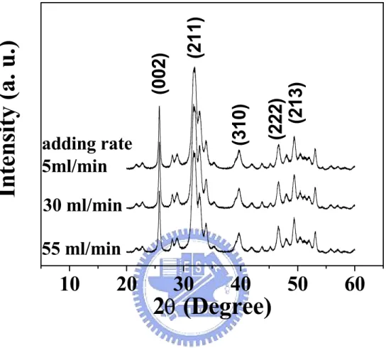
The calcium-deficient hydroxyapatite (CDHA) nano-particles with a Ca/P molar ratio of 1.5 were fabricated by co-precipitation method with Ca(CH3COO)2 and H3PO4 as Ca and P precursors, respectively. Prior to co-precipitation reaction, the pH value of H3PO4 solutions were kept above 12 by using NaOH solution. Subsequently, 0.001 wt% poly(acrylic acid) (PAA) (Aldrich Chemical, Mw 2000) was added into the alkalized H3PO4 solutions. In order to control the CDHA particle with different aspect ratios, three different adding rates of 5 ml/min, 30 ml/min, and 55 ml/min were used to dropCa(CH3COO)2 aqueous solution (0.5M) into H3PO4 solution (controlled at 0.33 M) at 60oC [86]. During the titration process, the temperature deviation of each mixture was monitored and kept less than 3oC. After filtration and washing by acetone for several times, the precipitate dough was then mixed in 80 ml acetone to form three kinds of CDHA suspensions. The solid content in each CDHA-acetone suspension was determined by measuring the weight difference between before and after the removal of the diluting medium at 105oC.
3.2.2. CDHA-PMMA composite mixtures
Various volume from 5% to 15% CDHA nano-particles with different aspect ratios (AR=length of particle/width of particle) were dispersed in the PMMA-Acetone (PMMA: Aldrich Chemical Corp. Mw 13,000) polymer solution to form PMMA-CDHA composite. After stirring for 24 h, the CDHA-PMMA suspensions were dried to remove the diluting solvent and then subjected to heat treatment in vacuum oven for the removal of the residual solvent. After that, the CDHA-PMMA nanocomposite powders can be obtained after pulverization.
3.2.3. Rheometrical measurement
The rheological behavior of the mixtures with different concentrations of CDHA nanoparticles was examined at a working temperature of 240oC using capillary rheometry
(Schimadzu, CFT-500D). The specimens were first prepared into cylindrical shape of 10 mm x 10 mm dimensions via a uniaxial molding at a compressive pressure of 30 MPaat 160oC. The cylindrical specimen was then placed into the cavity die of the capillary rheometer. The capillary die has a size of 1 mm in diameter, located at the bottom of the cavity. After the sample was stabilized at the testing temperature of 240oC for several minutes, the specimen was compressed with a pre-determined uni-axial pressure.
In order to eliminate the entrance and end effect of the rheological data, both the first and final 1-mm region of the 10 mm-height specimens were excluded. In addition, the Rabinowitsch equation was also adopted for shear rate correlation due to wall effect. The relationship between shear stress(τ) and pressure drop along the specimen dimension can be determined with the use of Bagley model as follows [87],
L PR 4 =
τ 3-(1)
where P, R and L represent the values of pressure drop, capillary radius and capillary length, respectively. The shear rate (γ) was determined by converting the ratio of the volume flow rate (F) to the capillary radius (R) through,
4 3 R
F
π
γ = 3-(2)
The shear viscosity (η) was then determined by;
γ τ
η = 3-(3)
Generally, three runs were conducted for each composition and aspect ratio to ensure the accuracy and the reproducibility of the rheological data. If needed, five runs were performed for more accurate results.
3.3. Results and discussion 3.3.1. Flow behavior
calcium-deficient hydroxyapatite (CDHA) phase irrespectively of the addition rate. According to TEM observation, the morphology of the precipitate crystals exhibits needle-like geometry with different aspect ratios (L/d, length/diameter), ranging from 7, 10, to 17 for the addition rate of 5, 30, and 55 ml/min, respectively. (TEM photographs were not shown here.)
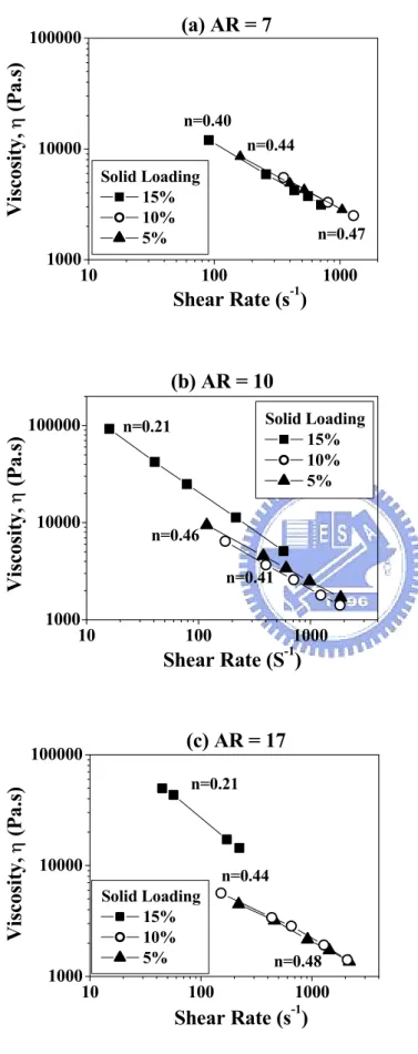
The flow behavior of the PMMA-CDHA mixtures can be characterized by a shear rate (γ)- viscosity (η) curve (derived simply from η= Kγn-1), as shown in a log-log plot of Figure 3-2(a), 3-2(b) and 3-2(c) for CDHA nano-needles with AR = 7, 10, and 17, respectively. It is evidenced that the mixtures are shear-thinning character and the flow behavior of the mixtures is as a function of both AR and concentration of the nano-needles. The flow index n is generally falling in the range between 0.4 and 0.5 for the mixtures with 5 and 10% concentration of nano-needles. Even increasing CDHA concentration up to 15%, the difference in the flow index for the composition with the smallest AR [Figure 3-2(a)] is essentially much less pronounced over the range of solid concentration employed. This may indicate that the flow behavior as well as the suspension structure of the mixtures prepared with the nano-needles of the smallest AR is similar. Accordingly, this could be true for the nanoparticles with a lower AR below a certain solid loading where the interparticle interaction, including surface attraction or mechanical interlocking, may remain similar in magnitude.
On the other hand, for the compositions with larger AR, a smaller n value was observed at a 15% solid loading as revealed from Figures 3-2(b) and 3-2(c). While comparing to Figure3-2(a), a considerable reduction in the flow index (n), appearing at 15% loading, may suggest that the nano-particles with a higher AR should exert strong influence onto suspension structure and flow behavior. From those measured data in Figures. 3-2(b) and 3-2(c) for high-AR particles, it seems to indicate that at lower solid loadings, i.e., 5 and 10%, the interactions between the nano-needles should be not as significantly strong as that at 15%.
However, a particle network structure with more extensive entanglement with random distribution of the nano-needles should arise because of increasing solid loadings, i.e., 15% in
the current system. If this assumption in structural development is correct, then, we can speculate that the mixtures with solid loading as high as 15% may have an open network structure with voids between the closely-connect framework filled with polymer melt. With such an interconnect structure, the flowability of the polymer melt was then effectively retarded and a higher shear viscosity can be expected under an identical shear rate (or shear stress), leading to a higher-shear-dependent character than that at lower solid concentrations.
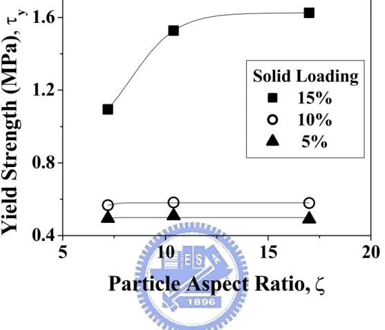
3.3.2. Yield strength
With the use of the Casson’s model [88], Eqn. 3-(4), the yield strength of the mixtures can be obtained by extrapolating the straight lines obtained from the
τ
1/2 -γ
1/2 plot to zero shear rate.τ
1/2 =τ
y1/2 + c
γ
1/2 3-(4) whereτ
y is the yield strength.Figure 3-3 shows Casson’s correlation for the mixtures with AR = 17, where straight lines for each solid loading can be obtained in Casson’s plot. After extrapolating to zero shear rate, the
τ
y was calculated and is also illustrated in the figure. It is observed thatτ
y first increases slightly with increasing solid loading from 5% to 10%, but considerably increased at 15% loadings. Similar behavior is also observed for other mixtures, as illustrated in Figure 3-4. However, as expected, theτ
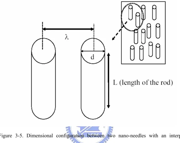
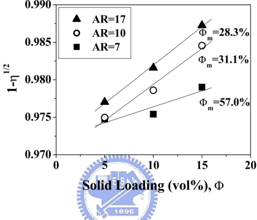
y at lower solid loadings, i.e., < 10%, does not show much difference for each category of AR, but the difference becomes more pronounced when the solid concentration is increased up to 15%.The yield strength is merely a direct indication of the strength of the particle network within the matrix, which has been well recognized due to net attractive interaction. For high-aspect-ratio particles, mechanical interlocking between neighboring particles should play some role. In a previous investigation [89], Liu experimentally verified that the yield strength is correlated linearly with the Van der Waals potential between two identical spheres; however,
![Table 2-5. Source and composition of the commercialized bone cement [20].](https://thumb-ap.123doks.com/thumbv2/9libinfo/8411817.179887/40.892.117.754.222.681/table-source-composition-commercialized-bone-cement.webp)
![Figure 2-1 Structural model of hydroxyapatite with the [0001] as projection axis [16]](https://thumb-ap.123doks.com/thumbv2/9libinfo/8411817.179887/41.892.171.594.190.501/figure-structural-model-hydroxyapatite-projection-axis.webp)