Bioavailability, Distribution and Anti-oxidative Effects of Triterpenes in Mice
MEI-CHIN YIN†,#,*, MEI-CHIN MONG†, CHIA-YU LIN†
†Department of Health and Nutrition Biotechnology, Asia University, Taichung City, Taiwan
#Department of Nutrition, China Medical University, Taichung City, Taiwan
Running title: tissue distribution of triterpenes
Corresponding Author: Dr. Mei-chin Yin, Professor, Department of Nutrition, China Medical University, 91, Hsueh-shih Rd., Taichung City, Taiwan
TEL: 886-4-22053366 ext. 7510 FAX: 886-4-22062891 Email: [email protected] 1 2 3 4 5 6 7 8 9 10 11 12 13 14 15 16 17
ABSTRACT
This study analyzed the content of eight triterpenes (oleanolic acid, ursolic acid, arjunolic acid, asiatic acid, boswellic acid, corosolic acid, madecassic acid and maslinic acid) in ten vegetables and eight fruits. These compounds at 0.5% were supplied to mice for 4 or 8 weeks. The bioavailability, tissue distribution and anti-oxidative protection of these triterpenes were examined. Results showed that triterpenes could be detected in eight vegetables and six fruits. Basil and brown mustard contained 7 test triterpenes, in the range of 14-102 mg/100 g dry weight. The level of each triterpene in plasma, brain, heart, liver, kidney, colon and bladder increased with increasing feeding period from 4 weeks to 8 weeks (P<0.05). Renal homogenates from mice with triterpenes intake had greater anti-oxidative effects against glucose induced glutathione loss, malondialdehyde and oxidized glutathione production when compared with those from control groups (P<0.05). These data support that these triterpenes could be absorbed and deposited in their intact forms, which in turn exerted in vivo anti-oxidative protection.
KEYWORDS: pentacyclic triterpenes; bioavailability; vegetable; anti-oxidative 18 19 20 21 22 23 24 25 26 27 28 29 30 31 32 33
INTRODUCTION
Oleanolic acid, ursolic acid, arjunolic acid, asiatic acid, boswellic acid, corosolic acid, madecassic acid and maslinic acid are pentacyclic triterpenes. It has been documented that oleanolic acid and ursolic acid possess many in vivo bioactivities including anti-oxidative, anti-inflammatory and anti-glycative activities (1, 2), which support their application in disease prevention and alleviation (3-5). Recently, more attention was paid to the bioactivities of other six triterpenes. Those studies suggest that these compounds are potent protective agents with multiple actions such as regulating immune function and nuclear factor-κB signaling pathway (6-8).
The recovery of oleanolic acid and ursolic acid in blood after i.v. injection in human subjects has been confirmed (9). However, tissue distribution of these compounds in their intact forms after oral intake remains unknown. Although the observed bioactivities of these triterpenes could be ascribed to their metabolites after absorbed and metabolized, the appearance of their intact forms in circulation and/or organs may further ensure their deposit and ability to exert local or systemic protective effects and actions. Thus, an animal study was designed to investigate the bioavailability and tissue distribution of these triterpenes after dietary intake.
These above triterpenes occurred naturally in many plants, especially herbs (10-12). Less information is available regarding their content in fresh vegetables and fruits, which are more easily for people to consume. In order to increase the source for obtaining these triterpenes, their content in several locally available vegetables and fruits was analyzed.
MATERIALS AND METHODS
Chemicals. Oleanolic acid (OA, 99%), ursolic acid (UA, 98%), arjunolic acid
(ARA, 98%), asiatic acid (ASA, 98.5%), boswellic acid (BOA, 98%), corosolic acid (CA, 34 35 36 37 38 39 40 41 42 43 44 45 46 47 48 49 50 51 52 53 54 55 56 57 58
99%), madecassic acid (MCA, 99%) and maslinic acid (MA, 98.5%) were purchased from Aldrich Chemical Co. (Milwaukee, WI, USA). Their structures are shown in Figure 1. All chemicals used in these measurements were of the highest purity commercially available.
Analysis of Triterpene Content in Vegetables and Fruits. Ten fresh vegetables
and eight fresh fruits were used to analyze the content of 8 triterpenes. Test vegetables included gynura (Gynura bicolor DC), basil (Ocimum basilicum), brown mustard (Brassica juncea), mahogany (Toona sinensis), madeira vine (Anredera cordifilia Moq.), daylily (Hemerocallis fulva L.), wild lettuce (Lactuca indica), balsam pear (Momordica
charantia), water convoevueus (Ipomoea aquatic) and spinach (Spinacia oleracea). Test
fruits were mulberry (Mours alba L.), carambola (Averrhoa carambola), waxapple (Syzygium samarangensem), mango (Mangifera indica L.), juiube (Zizyphus mauritiana), calamondin (Citrus microcarpa Bonge), guava (Psidium guajava) and loquat (Eriobotrya
japonica). These vegetables and fruits, harvested in Spring, 2012 were purchased from
four farms in Taichung City, Taiwan. The content of 8 triterpenes was analyzed by HPLC methods described in Li et al. (13). A 50 g edible portion was chopped, homogenized in a Waring blender, and freeze-dried to a fine powder. Five g dry sample was extracted with 10 mL of ethanol twice in an ultrasonic bath at room temperature. The combined extracts were filtered through an analytical filter paper, and dried under vacuum. After redissolved in 5 mL ethanol, sample was used for HPLC analysis. Agilent 1100 series HPLC system equipped with a Hypersil BDS C8 column (200 mm × 4.6 mm, 5 μm, ThermoQuest, Runcorn, UK), and a diode array and fluorescence detector was used. Mobile phase A and B were acetonitrile-water (30:70; v/v) and 100% acetonitrile, respectively. The detection limit was 0.5 mg/100 g dry weight.
Animal Experiments. Three-week-old male C57BL/6 mice were obtained from
59 60 61 62 63 64 65 66 67 68 69 70 71 72 73 74 75 76 77 78 79 80 81 82 83
National Laboratory Animal Center (National Science Council, Taipei City, Taiwan). Use of the mice was reviewed and approved by China Medical University animal care committee. Mice were housed on a 12-h light-12-h dark schedule, and fed with mouse standard diet for one week acclimation. Mice were then divided into nine groups, one consumed normal diet, and the other consumed normal diet plus 0.5% OA, UA, ARA, ASA, BOA, CA, MCA or MA prepared by mixing 0.5 g target compound with 99.5 g powder diet. All mice had free access to food and water at all times. Consumed water volume, feed and body weight were recorded. After 4- or 8-wk supplementation, mice were killed with carbon dioxide. Blood was collected, and plasma was separated from erythrocytes immediately. Brain, heart, lung, liver, kidney, spleen, colon and bladder were collected and weighted. Each organ at 0.1 g was homogenized in 2 mL ice cold phosphate buffer saline (PBS, pH 7.2). The homogenate was further passed through a Whatman No. 1 filter paper, and the filtrate was collected. The protein concentration of organ filtrate was determined by the method of Lowry et al. (14) using bovine serum albumin as a standard. In all experiments, the sample was diluted to a final concentration of 1 g protein/L using PBS, pH 7.2.
Determination of Triterpene Content in Plasma and Tissues. The content of
target compound in plasma or organ was analyzed by the method described in Gerbeth et al. (15). Briefly, tissue filtrate or plasma at 100 L was spiked with 25 μL methanol containing 1 μg fluoxymesterone, and followed by extraction with twice 2 mL tetrahydrofuran-n-hexane-ethyl acetate-2-propanol (160:160:160:15, v/v/v/v). Solvent was evaporated using a nitrogen stream, and the residue was reconstituted in 100 μL methanol:water (90:10, 400 mg/L ammonium formate). Glycyrrhetinic acid was used as the internal standard. Sample at 25 μL was injected into the HPLC/MS-system. Agilent 1100 series HPLC system equipped with a BDS RP C18 column (100 mm × 4 mm, 3 μm, 84 85 86 87 88 89 90 91 92 93 94 95 96 97 98 99 100 101 102 103 104 105 106 107 108
ThermoQuest, Runcorn, UK), and a diode array and fluorescence detector was used. Mobile phases contained methanol and water. This HPLC system was coupled on-line to an ion-trap mass spectrometer (Agilent Corp, Waldbronn, Germany) equipped with an electro-spray ionization source, and analysis was performed in a negative single ion mode. The limits of detections were 0.1 g/mL plasma or 0.1 g/g tissue.
Anti-oxidative Effects of Triterpenes in Renal Homogenate. Glucose at 50
mmol/L was used to initiate lipid oxidation in renal homogenate. After 3-day incubation at 37 °C, lipid oxidation was determined by measuring the level of malondialdehyde (MDA), glutathione (GSH) and oxidized glutathione (GSSG) via commercial colorimetric assay kits (OxisResearch, Portland, OR, USA).
Statistical Analysis. The effect of each treatment was analyzed from ten different
preparations (n=10). Data were reported as means standard deviation (SD), and subjected to analysis of variance (ANOVA). Differences among means were determined by the Least Significance Difference Test with significance defined at P<0.05.
RESULTS
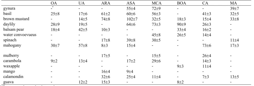
As shown in Table 1, basil and brown mustard contained 7 test triterpenes, in the range of 14-102 mg/100 g dry weight. Among test fruits, 5 triterpenes could be detected in calamondin and carambola, in the range of 7-32 mg/100 g dry weight. The content of test triterpenes in wild lettuce, madeira vine, juiube and loquat was too low to be detected.
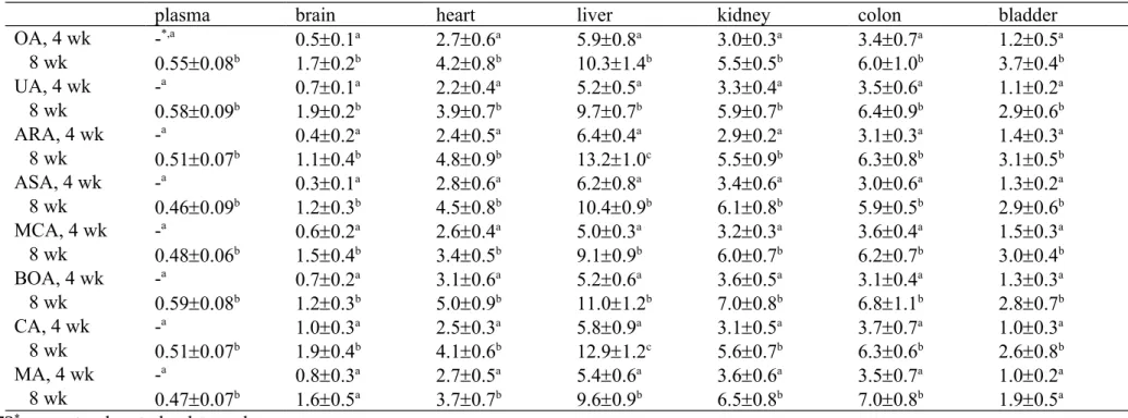
Besides lung and spleen, test triterpenes were detected in plasma, brain, heart, liver, kidney, colon and bladder (Table 2). The level of each triterpene in plasma and tissues could be increased with increasing feeding period from 4 weeks to 8 weeks (P<0.05). After 8-week supplementation, liver had the highest content of each triterpene.
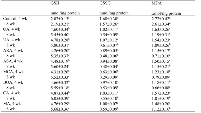
As shown in Table 3, glucose treatment led to GSH loss, GSSG formation and MDA 109 110 111 112 113 114 115 116 117 118 119 120 121 122 123 124 125 126 127 128 129 130 131 132 133
production in kidney homogenate of mice from control groups. Compared with control groups, renal homogenates from mice with target compound intake had higher GSH level, and lower MDA and GSSG production (P<0.05). There was a significant difference in GSH, GSSG and MDA levels between renal homogenates from 4 and 8 wk feeding period for each triterpene (P<0.05).
DISCUSSION
The presence of test triterpenes in herbs such as Ligustrum lucidum Ait. and
Cichorium intybus L. has been reported (10-12). Our present study further found that
several commonly available vegetables and fruits contained these triterpenes. Thus, these vegetables and fruits are also the sources of triterpenes. Furthermore, we notified that basil and brown mustard are rich in arjunolic acid, asiatic acid and madecassic acid. It has been indicated that these three triterpenes possess several protective functions including anti-oxidative and anti-inflammatory activities (7, 16, 17). It is possible that these two vegetables, based on their triterpenes levels, might be able to provide healthy benefits.
The recovery or organ distribution of ursolic acid and betulinic acid after i.v. injection in human and/or animals has been reported (9, 18, 19). The results of our present study revealed that the intact form of test triterpene could be recovered in blood and tissues after dietary intake. Furthermore, we found that the accumulated level of each triterpene in tissues could be increased with extending supplementary period. These findings indicate that dietary supplement of these compounds could be absorbed, metabolized and deposited in tissues, which in turn exerted their local and/or systemic actions. Many studies reported that dietary intake of these compounds exhibited cardiac, renal and hepatic protection
(20-22). Since intact forms of triterpenes could be available in tissues after oral intake, the
observed protective effects from triterpenes in those previous studies could be partially 134 135 136 137 138 139 140 141 142 143 144 145 146 147 148 149 150 151 152 153 154 155 156 157 158
ascribed to the recovery of these compounds in their intact forms. In addition, we notified that liver had the highest bioavailability for each triterpene compared with other tissues. It seems that liver is the major organ for triterpene storage and/or metabolism. This finding suggests that using these triterpenes for hepatic protection seems possible and feasible. Also, the appearance of test triterpenes in brain suggested that these compounds might be able to penetrate brain blood barrier after they were absorbed and metabolized. This finding supports the possibility of using these agents for brain protection.
In our present study, high glucose induced oxidative stress was markedly decreased in renal homogenate from mice with triterpene intake. The observed anti-oxidative effects could be partially ascribed to the existence of intact triterpenes in renal homogenate. It is reported that triterpenes possessed non-enzymatic activities such as reducing powder and scavenging free radicals. Our data revealed that these compounds could spare GSH. These results once again support that dietary intake of these compounds benefits tissue protection against oxidative injury. It is interesting to find that 8-wk ARA and BOA intake led to renal content of these compounds at 5.5 and 7.0 g/g tissue. However, their renal homogenates exhibited similar effects in retaining GSH, lowering GSSG and MDA levels. Apparently, the anti-oxidative effects observed in renal homogenate could not be solely ascribed to the intact forms of triterpenes. That is, the metabolite(s) of these triterpenes may also contribute to anti-oxidative protection.
Eight test compounds are pentacyclic triterpenes. Although they have similar structures, they exhibited different bioavailability, tissue distribution and anti-oxidative activity. It seems certain function group and/or the position of this group affect the absorption and/or metabolism of a triterpene. So far, more studies focused on the benefits of ursolic acid and oleanolic acid. Since other six triterpenes also provided substantial bioavailability, tissue distribution and anti-oxidative efficiency, the application of these six 159 160 161 162 163 164 165 166 167 168 169 170 171 172 173 174 175 176 177 178 179 180 181 182 183
triterpenes should be encouraged.
In conclusion, oleanolic acid, ursolic acid, arjunolic acid, asiatic acid, boswellic acid, corosolic acid, madecassic acid and maslinic acid could be detected in fresh vegetables and fruits locally available in Taiwan. The intake of these triterpenes increased their bioavailability and accumulation in circulation and tissues. The recovered triterpene in organ benefited organ’s anti-oxidative defense. These findings link dietary triterpenes and their application to prevent and/or alleviate diseases.
184 185 186 187 188 189 190
LITERATURE CITED
1. Tsai, S.J.; Yin, M.C. Antioxidative and anti-inflammatory protection of oleanolic acid and ursolic acid in PC12 cells. J. Food Sci. 2008, 73, 174-178.
2. Yin, M.C.; Chan, K.C. Nonenzymatic antioxidative and antiglycative effects of oleanolic acid and ursolic acid. J. Agric. Food Chem. 2007, 55, 7177-7181.
3. Wang, Z.H.; Hsu, C.C.; Huang, C.N.; Yin, M.C. Anti-glycative effects of oleanolic acid and ursolic acid in kidney of diabetic mice. Eur. J. Pharmacol. 2010, 628, 255-260.
4. Yamai, H.; Sawada, N.; Yoshida, T.; Seike, J.; Takizawa, H.; Kenzaki, K.; Miyoshi, T.; Kondo, K.; Bando, Y.; Ohnishi, Y.; Tangoku, A. Triterpenes augment the inhibitory effects of anticancer drugs on growth of human esophageal carcinoma cells in vitro and suppress experimental metastasis in vivo. Int. J. Cancer 2009, 125, 952-960.
5. Lu, J.; Zheng, Y.L.; Wu, D.M.; Luo, L.; Sun, D.X.; Shan, Q. Ursolic acid ameliorates cognition deficits and attenuates oxidative damage in the brain of senescent mice induced by D-galactose. Biochem. Pharmacol. 2007, 74, 1078-1090.
6. Manna, P.; Das, J.; Ghosh, J.; Sil, P.C. Contribution of type 1 diabetes to rat liver dysfunction and cellular damage via activation of NOS, PARP, IkappaBalpha/NF-kappaB, MAPKs, and mitochondria-dependent pathways: Prophylactic role of arjunolic acid. Free Radic. Biol. Med. 2010, 48, 1465-1484.
7. Won, J.H.; Shin, J.S.; Park, H.J.; Jung, H.J.; Koh, D.J.; Jo, B.G.; Lee, J.Y.; Yun, K.; Lee, K.T. Anti-inflammatory effects of madecassic acid via the suppression of NF-191 192 193 194 195 196 197 198 199 200 201 202 203 204 205 206 207 208 209 210 211 212
kappaB pathway in LPS-induced RAW 264.7 macrophage cells. Planta Med. 2010,
76, 251-257.
8. Chen, H.; Yang, J.; Zhang, Q.; Chen, L.; Wang, Q. Corosolic acid ameliorates atherosclerosis in apolipoprotein E-deficient mice by regulating the nuclear factor-κB signaling pathway and inhibiting monocyte chemoattractant protein-1 expression.
Circ. J. 2012, 76, 995-1003.
9. Xia, Y.; Wei, G.; Si, D.; Liu, C. Quantitation of ursolic acid in human plasma by ultra performance liquid chromatography tandem mass spectrometry and its pharmacokinetic study. J. Chromatogr. B Analyt. Technol. Biomed. Life Sci. 2011,
879, 219-224.
10. Yang, H.; Ding, C.; Duan, Y.; Liu, J. Variation of active constituents of an important Tibet folk medicine Swertia mussotii Franch. (Gentianaceae) between artificially cultivated and naturally distributed. J. Ethnopharmacol. 2005, 98, 31-35.
11. Kumari, R.; Ali, M.; Aeri, V. Two new triterpenoids from Cichorium intybus L. roots.
J. Asian Nat. Prod. Res. 2012, 14, 7-13.
12. Brendolise, C.; Yauk, Y.K.; Eberhard, E.D.; Wang, M.; Chagne, D.; Andre, C.; Greenwood, D.R.; Beuning, L.L. An unusual plant triterpene synthase with predominant α-amyrin-producing activity identified by characterizing oxidosqualene cyclases from Malus × domestica. FEBS J. 2011, 278, 2485-2499.
13. Li, G.L.; You, J.M.; Song, C.H.; Xia, L.; Zheng, J.; Suo, Y.R. Development of a new HPLC method with precolumn fluorescent derivatization for rapid, selective and 213 214 215 216 217 218 219 220 221 222 223 224 225 226 227 228 229 230 231 232 233
sensitive detection of triterpenic acids in fruits. J. Agric. Food Chem. 2011, 59, 2972-2979.
14. Lowry, O.H.; Rosebrough, N.J.; Farr, A.L. Protein determination with the Folin phenol reagent. J. Biol. Chem. 1951, 193, 265-275.
15. Gerbeth, K.; Meins, J.; Kirste, S.; Momm, F.; Schubert-Zsilavecz, M.; Abdel-Tawab, M. Determination of major boswellic acids in plasma by high-pressure liquid chromatography/mass spectrometry. J. Pharm. Biomed. Anal. 2011, 56, 998-1005.
16. Lee, M.K.; Kim, S.H.; Yang, H.; Lim, D.Y.; Ryu, J.H.; Lee, E.S.; Jew, S.S.; Park, H.G.; Sung, S.H.; Kim, Y.C. Asiatic acid derivatives protect primary cultures of rat hepatocytes against carbon tetrachloride-induced injury via the cellular antioxidant system. Nat. Prod. Commun. 2009, 4, 765-768.
17. Ghosh, J.; Das, J.; Manna, P.; Sil, P.C. Acetaminophen induced renal injury via oxidative stress and TNF-alpha production: therapeutic potential of arjunolic acid.
Toxicology 2010, 268, 8-18.
18. Udeani, G.O.; Zhao, G.M.; Geun, S.Y.; Cooke, B.P.; Graham, J.; Beecher, C.W.; Kinghorn, A.D.; Pezzuto, J.M. Pharmacokinetics and tissue distribution of betulinic acid in CD-1 mice. Biopharm. Drug. Dispos. 1999, 20, 379-383.
19. Chen, Q.; Luo, S.; Zhang, Y.; Chen, Z. Development of a liquid chromatography-mass spectrometry method for the determination of ursolic acid in rat plasma and tissue: application to the pharmacokinetic and tissue distribution study. Anal. Bioanal. Chem.
2011, 399, 2877-2884. 234 235 236 237 238 239 240 241 242 243 244 245 246 247 248 249 250 251 252 253 254
20. Du, Y.; Ko, K.M. Oleanolic acid protects against myocardial ischemia-reperfusion injury by enhancing mitochondrial antioxidant mechanism mediated by glutathione and alpha-tocopherol in rats. Planta Med. 2006, 72, 222-227.
21. Park, B.; Prasad, S.; Yadav, V.; Sung, B.; Aggarwal, B.B. Boswellic acid suppresses growth and metastasis of human pancreatic tumors in an orthotopic nude mouse model through modulation of multiple targets. PLoS One 2011, 6, e26943.
22. Guillen, N.; Acín, S.; Surra, J.C.; Arnal, C.; Godino, J.; García-Granados, A.; Muniesa, P.; Ruiz-Gutiérrez, V.; Osada, J. Apolipoprotein E determines the hepatic transcriptional profile of dietary maslinic acid in mice. J. Nutr. Biochem. 2009, 20, 882-893. 255 256 257 258 259 260 261 262 263 264
Figure1. Structure of eight triterpenes.
ursolic acid oleanolic acid
arjunolic acid asiatic acid
boswellic acid corosolic acid
madecassic acid maslinic acid
Table 1. Content (mg/100 g dry weight) of eight triterpenes in eight vegetables and six fruits harvested in Spring, 2012. Data are mean SD (n=10).
OA UA ARA ASA MCA BOA CA MA
gynura -* - - 554 729 - - 397 basil 258 176 612 606 563 - 413 325 brown mustard - 145 748 1027 325 183 154 338 daylily 289 195 - 646 733 909 263 -balsam pear 184 425 103 - - 334 162 -water convoevueus - - - - 458 265 144 -spinach - - 178 398 305 - - 114 mahogany 307 578 83 154 - - 736 173 mulberry - - 175 - 155 - 264 -carambola 92 134 - 172 296 - 143 -waxapple - - - 93 114 -mango - - 164 94 - - - -calamondin - - 326 254 114 - 73 135 guava - 122 153 - - 82 -
-*means too low to be detected. 266
267
268 269
Table 2. Eight triterpenes content in plasma (g/mL) or tissues (g/g) of mice consumed target compounds for 4 or 8 weeks. Data are mean SD (n=10).
plasma brain heart liver kidney colon bladder
OA, 4 wk -*,a 0.50.1a 2.70.6a 5.90.8a 3.00.3a 3.40.7a 1.20.5a 8 wk 0.550.08b 1.70.2b 4.20.8b 10.31.4b 5.50.5b 6.01.0b 3.70.4b UA, 4 wk -a 0.70.1a 2.20.4a 5.20.5a 3.30.4a 3.50.6a 1.10.2a 8 wk 0.580.09b 1.90.2b 3.90.7b 9.70.7b 5.90.7b 6.40.9b 2.90.6b ARA, 4 wk -a 0.40.2a 2.40.5a 6.40.4a 2.90.2a 3.10.3a 1.40.3a 8 wk 0.510.07b 1.10.4b 4.80.9b 13.21.0c 5.50.9b 6.30.8b 3.10.5b ASA, 4 wk -a 0.30.1a 2.80.6a 6.20.8a 3.40.6a 3.00.6a 1.30.2a 8 wk 0.460.09b 1.20.3b 4.50.8b 10.40.9b 6.10.8b 5.90.5b 2.90.6b MCA, 4 wk -a 0.60.2a 2.60.4a 5.00.3a 3.20.3a 3.60.4a 1.50.3a 8 wk 0.480.06b 1.50.4b 3.40.5b 9.10.9b 6.00.7b 6.20.7b 3.00.4b BOA, 4 wk -a 0.70.2a 3.10.6a 5.20.6a 3.60.5a 3.10.4a 1.30.3a 8 wk 0.590.08b 1.20.3b 5.00.9b 11.01.2b 7.00.8b 6.81.1b 2.80.7b CA, 4 wk -a 1.00.3a 2.50.3a 5.80.9a 3.10.5a 3.70.7a 1.00.3a 8 wk 0.510.07b 1.90.4b 4.10.6b 12.91.2c 5.60.7b 6.30.6b 2.60.8b MA, 4 wk -a 0.80.3a 2.70.5a 5.40.6a 3.60.6a 3.50.7a 1.00.2a 8 wk 0.470.07b 1.60.5a 3.70.7b 9.60.9b 6.50.8b 7.00.8b 1.90.5a
*means too low to be detected.
a,bMeans in a row without a common letter differ, P<0.05. 270
271
272 273 274
Table 3. Effect of test triterpenes in kidney homogenate from mice without (control) or with target compound intake for 4 or 8 weeks against
glucose induced oxidative stress. Data are mean SD (n=10). GSH nmol/mg protein GSSG nmol/mg protein MDA mol/mg protein Control, 4 wk 2.020.13a 1.680.30d 2.720.42d 8 wk 2.190.21a 1.570.26d 2.610.34d OA, 4 wk 4.600.34b 1.020.11c 1.630.26c 8 wk 5.450.40c 0.540.09b 1.190.31b UA, 4 wk 4.780.28b 1.070.12c 1.540.23c 8 wk 5.800.31c 0.610.07b 1.090.26b ARA, 4 wk 4.260.20b 0.890.05c 1.150.17b 8 wk 5.350.37c 0.480.06b 0.710.10a ASA, 4 wk 4.480.19b 0.940.08c 1.500.15c 8 wk 5.600.24c 0.480.04b 1.150.21b MCA, 4 wk 4.310.26b 0.630.06b 1.230.18b 8 wk 5.520.33c 0.290.09a 0.790.09a BOA, 4 wk 4.660.32b 0.970.10c 1.180.11b 8 wk 5.590.18c 0.530.09b 0.660.08a CA, 4 wk 4.870.44b 1.030.11c 1.570.23c 8 wk 6.050.39c 0.550.10b 1.010.19b MA, 4 wk 4.760.29b 1.080.07c 1.480.20c 8 wk 5.680.36c 0.590.09b 1.120.16b
a-dMeans in a row without a common letter differ, P<0.05. 275
276
TOC Graphic
MS Title: Bioavailability, Distribution and Anti-oxidative Effects of Triterpenes in Mice Structure of test triterpenes.
ursolic acid oleanolic acid
arjunolic acid asiatic acid
boswellic acid corosolic acid
madecassic acid maslinic acid
278 279 280