ELSEVIER
Journal of Chromatography A, 761 (1997) 307 313JOURNAL OF
CHROMATOGRAPHY A
Separation of retinoids by micellar electrokinetic capillary
chromatography
You-Zung Hsieh*, Kuang-Lung Kuo
Department of Applied Chemistry, National Chiao Tung University, Hsinchu, Taiwan Received 12 June 1996; revised 30 September 1996; accepted 30 September 1996
Abstract
In this study, we have developed a micellar electrokinetic capillary chromatography (MEKC) method with diode-array detection to separate five important retinoids, including retinol, retinal, retinoic acid, retinyl acetate and retinyl palmitate. These very hydrophobic retinoids are successfully separated with phosphate buffer containing sodium deoxycholate (SDC) and Brij 35 as the micellar pseudostationary phase. The effects of SDC and Brij 35 concentration on the migration behavior of the five retinoids are also discussed. The relative standard deviations of the migration times of the retinoids are less than 1.9% in the optimized separation condition. Moreover, a direct extraction method without saponification is employed to extract retinyl esters in pharmaceutical products. The ranges of recoveries are 95%-99% for retinyl esters with less than 2% relative standard deviations. Therefore, retinyl esters in pharmaceutical products can be determined by combining the direct extraction and the MEKC methods.
Keywords: Retinoids; Vitamin A
I. Introduction
Retinoids include those natural compounds having vitamin A activity and synthetic analogues of retinol, with or without biological activity [1]. Retinol is an essential element for normal growth, vision, re- production and epithelial differentiation. Retinal and retinoic acid are important metabolites of retinol. Retinal is the active form of retinoids in the vision process and retinoic acid performs important physio- logical functions [2]. The attention given to retinoids increases because of their antioxidant effects against damaging free radicals [3]. In addition, retinyl esters, including retinyl acetate and retinyl palmitate, are the esters of retinol and are used as pharmaceutical *Corresponding author.
products or food additives. Retinoids are labile compounds and subject to oxidation, isomerization and degradation when exposed to oxygen, heat or light [1,3]. Therefore, analytes must be stored at - 2 0 ° C and protected from light during the analysis process. Some retinoids have been separated and determined in foods and pharmaceutical products. For instance, retinyl acetate was extracted with a direct extraction method by stirring for more than 1 h and then analyzed by high-performance liquid chro- matography (HPLC) [3]. Most studies on retinoids also employed HPLC as the analysis method [4-7]. Capillary electrophoresis (CE), a highly effective analytical tool, has been extensively applied to separate various compounds, including ionic and neutral compounds [8-10]. High resolution, high separation efficiency and rapid analysis are the 0021-9673/97/$17.00 Copyright © 1997 Elsevier Science B.V. All rights reserved
308 E-Z. Hsieh, K.-L. Kuo / J. Chromatogr. A 761 (1997) 3 0 7 - 3 1 3
primary features of CE. Furthermore, compared with HPLC, only a small amount of sample and solvent is necessary and a small amount of waste solution is produced in CE. Terabe et al. first reported on micellar electrokinetic capillary chromatography (MEKC), which is a modified format of CE [11,12]. This method can provide highly efficient separation for neutral and hydrophobic compounds. The under- lying separation mechanism of MEKC is that ana- lytes have different partition coefficients between the aqueous phase and the micellar pseudostationary phase. Two surfactants can be used together in MEKC to increase selectivity, e.g., combining poly- oxyethylene (23) dodecanol (Brij 35) and SDS as a mixed micellar pseudostationary phase for MEKC separation [13]. The elution range in MEKC can be significantly altered [14]. In addition, selectivity and separation efficiency can also be changed when anionic and zwitterionic surfactants are used together to form a mixed micellar system for MEKC [15].
Chan et al. [16] employed the MEKC technique to separate retinoic acid isomers. Separation of vitamin A and other vitamins has been previously investi- gated by MEKC and MEEKC methods [17,18]. However, to our knowledge, retinoids, including retinol, its metabolites and its esters have not yet been analyzed by CE or MEKC. In the present study, a MEKC method is developed to analyze these important retinoids. The five retinoids are retinol, retinal, retinoic acid, retinyl acetate and retinyl palmitate, each having a long carbon chain. The MEKC method used herein has a micellar system containing bile salt and Brij 35. The effects of SDC concentration and Brij 35 concentration on the migration behavior of the five retinoids are dis- cussed. Furthermore, a direct extraction method for retinoids in pharmaceutical products is studied. The extracted retinoids are then determined by the MEKC method.
2. Experimental
2.1.1. Apparatus
A Beckman P/ACE 5500 capillary electrophoresis system (Beckman Instruments, Palo Alto, CA, USA) was used for capillary electrophoresis. A diode-array detector was employed for detection. Separation was
performed in a 47 cm (40 cm to detector)*50 Ixm I.D. fused-silica capillary tube (Polymicro Tech- nologies, Phoenix, AZ, USA). The capillary tube was assembled in the cartridge format of Beckman Instrument. The voltage of the electrophoresis sepa- ration was 20 kV and the temperature of the capillary tube was set at 25°C. A personal computer using System Gold software controlled the P/ACE instru- ment. Data analysis was also performed by System Gold software.
2.1.2. Chemicals
Retinyl acetate, retinyl palmitate, retinoic acid and retinal were obtained from Sigma (St. Louis, MO, USA). Retinol, sodium dihydrogenphosphate and disodium hydrogenphosphate were obtained from Fluka (Buchs, Switzerland). Polyoxyethylene (23) dodecanol (Brij 35) and ethanol were purchased from Merck (Darmstadt, Germany). Sodium deoxy- cholate (SDC), sodium cholate (SC), sodium taurocholate (STC) and sodium dodecyl sulfate (SDS) were purchased from Sigma. Centrum mul- tivitamin tablets and multivitamin syrup were made by Cyanamid Taiwan Corporation. Methanol was obtained from J. T. Baker (Phillipsburg, USA). Water was purified in a Milli-Q water system (Millipore, Bedford, MA, USA).
2.1.3. Sample preparation
An accurately weighted multivitamin tablet was formulated by the manufacturer to contain 5000 I.U. of retinyl acetate. One I.U. of retinyl acetate is equal to 0.344 ixg of retinyl acetate. The sample was transferred to a 50-ml beaker containing 15-ml of methanol. The sample solution was protected from light, extracted with methanol by ultrasonic agitation for 15 min and centrifuged at 2000 U/min for 5 min. The extract solution was directly injected into the capillary tube. One milliliter syrup was formulated by the manufacturer to contain 1000 I.U. of retinyl palmitate. One I.U. of retinyl palmitate is equal to 0.550 Ixg of retinyl palmitate. The sample was mixed with methanol by stirring. The mixed solution was transferred to a sample vial, then injected into the capillary tube.
2.1.4. Procedure
Y.-Z. Hsieh, K.-L. Kuo / J. Chromatogr. A 761 (1997) 3 0 7 - 3 1 3 309
were dissolved in ethanol. Sample solutions with various concentrations were prepared by diluting 2 mg/ml standard solutions with ethanol. The retinoid standards were stored at the - 2 0 ° C and were protected from light during the analysis process. Phosphate buffer for capillary electrophoresis was made ready by mixing 0.1 M sodium dihydrogen- phosphate and 0.1 M disodium hydrogenphosphate in deionized water. Running buffers for micellar electrokinetic capillary chromatography were pre- pared by dissolving Brij 35 and SDC in phosphate buffer. The capillary tube for separation was rinsed with 0.1 M sodium hydroxide, deionized water and running buffer before sample injection. The sample was injected into the capillary tube by pressure injection.
3. Results and discussion
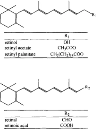
The five retinoids, including retinol, retinal, reti- noic acid. retinyl acetate and retinyl palmitate, are fat-soluble compounds with each having a long non- polar hydrocarbon chain. Fig. 1 illustrates their molecular structures. The non-polar parts in the molecular structures of the five analytes are identical and only the functional groups ( - O H , - C H O , - C O O H , - O C O C H 3 and - O C O ( C H 2) ~ 4 CH 3) at the end of the molecular structures are different. Based on their structures, dissolving these analytes in an
RI
retinol OH
retinyl acetate CH3COO retinyl palmitate CH3(CH2)I4COO
~
R2R2
retinal CHO
retinoic acid COOH
Fig. 1. M o l e c u l a r structures o f five retinoids.
aqueous solution is extremely difficult owing to their extremely hydrophobic properties.
The five analytes have different maximum ab- sorbance wavelengths, ranging from 325 nm to 365 nm. Therefore, a detector capable of performing a multi-wavelength detection must be used to detect the five analytes. Furthermore, retinoids are labile compounds and subject to oxidation. Thus, the analytes were protected from light during the analy- sis process to prevent a change in their absorbance spectra. The qualitative analysis could also be en- hanced by using the multi-wavelength detection system to discover any shift in the absorbance spectrum. Consequently, a diode-array detector in- stead of a single wavelength UV detector was employed in this application.
According to the experimental findings, only retinoic acid is detected by using aqueous buffers with various pH values ranging from pH 7.9 to pH 10.0 (containing only electrolytes). The migration velocity of retinoic acid is slower than that of electroosmotic flow. This result indicates that reti- noic acid can dissociate and become an anion in these buffers. Retinal, retinol, retinyl acetate and retinyl palmitate condense inside the capillary tube and do not elute through the detection window, The failure to detect the four analytes is due to the low solubility of the hydrophobic analytes in CE aqueous buffer solution. Therefore, surfactants would be added to CE buffer to form a pseudostationary phase to enhance solubility and separation.
In this study, SDS was first selected to separate the analytes. This compound can carry a negative charge in aqueous solution to form anionic micelles. The solubility of the retinoids in the running buffer are significantly enhanced because all of the analytes can be detected. However, these analytes coelute with the same migration velocity. They can not be separated by the aqueous buffer containing various SDS concentrations. According to these results, the partition coefficients of the analytes between the aqueous phase and the SDS micellar pseudo- stationary phase are similar. Hence, using SDS as a micellar pseudostationary phase is not a preferable option for separating these hydrophobic analytes. Thus, other surfactants are considered as a pseudo- stationary phase to provide different selectivity for separating the retinoids by MEKC.
310 Y.-Z. Hsieh, K.-L. Kuo / J. Chromatogr. A 761 (1997) 3 0 7 - 3 1 3 3.1. Migration behavior of the retinoids in bile
salt MEKC
(-CI5H31) at the ester group such that it is a more hydrophobic compound than the other four analytes.
Three types of bile salt were selected to form the pseudostationary phase: SDC, SC and STC. Bile salt has a hydrophilic face and a hydrophobic face in its molecular structure. Thus, bile salt monomer tends to combine together at the hydrophobic face in an aqueous solution. Sepaniak et al. indicated that bile salt monomer is more polar than SDS [19] and bile salt seems to have a relatively small solubilization effect compared with SDS micelles [20]. Therefore, bile salt can provide different interactions for the retinoids than SDS. The different interactions may enhance the separation of the retinoids. A detailed discussion follows regarding the effects of adding bile salt in MEKC for the analysis of retinoids.
SDC provides better separation results than SC and STC on the basis of our results in this study. Hence, SDC is employed herein to form a micellar pseudostationary phase for subsequent analyses. The analytes' migration times increase as the SDC con- centration increases. This phenomena can be ac- counted for by the fact that the analytes have more likelihood of interacting with micelles, thereby caus- ing lower migration velocities. Furthermore, the velocity of electroosmotic flow also decreases as the SDC concentration increases. Except for two re- tinoids, i.e., retinol and retinal, the other retinoids are completely separated when more than 50 mM SDC are added in the buffer. The resolution between retinol and retinal is nearly zero and is independent of the SDC concentration. Such results imply that these retinoids have identical interactions with SDC. Also, methanol, up to 20% (v/v), was added to the buffer to improve the resolution between the tyro retinoids. However, no significant improvement is observed except that the analysis time is extended.
The migration time of retinoic acid is longer than that of retinol, retinal and retinyl acetate because retinoic acid is an anion in the pH 7.9 running buffer. The migration velocity of retinyl palmitate is slower than those of other retinoids. This phenomena im- plies that the interaction between retinyl palmitate and SDC micelles is stronger than the others. The stronger interaction between retinyl pahuitate and SDC micelles is owing to the fact that retinyl palmitate has a non-polar long hydrocarbon chain
3.2. The effects of Brij 35 on the separation
The poor resolution for the separation of retinol and retinal is the main problem in analyzing the five retinoids. The composition of the buffer must be changed to successfully separate the five analytes. Because adding Brij 35 in the SDS buffer to form a mixed micellar pseudostationary phase can enhance MEKC separation, adding Brij 35 to the SDC buffer to enhance separation is under investigation.
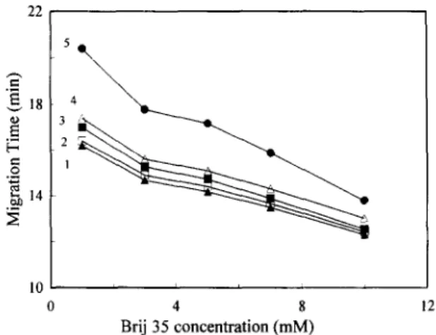
Fig. 2 illustrates the effect of the Brij 35 concentration on the retinoids' migration times when SDC is held at 75 mM. The migration times of the five retinoids decrease with an increasing Brij 35 concentration. This phenomenon indicates that all retinoids interact with Brij 35 micelles. As more Brij 35 is added, more neutral surfactants form the pseudostationary phase, thus leading to shorter mi- gration times of the retinoids. The satisfactory sepa- ration between retinol and retinal can be obtained by adding a suitable amount of Brij 35 to the running buffer containing 75 mM SDC. The migration times of retinal and retinyl acetate are closer to each other with an increase in Brij 35 concentration, whereas
22 54 N 2 ~ ~" 1 . _ 1 0 , ~ , 0 4 8 12 Brij 35 concentration (mM)
Fig. 2. Effect of Brij 35 concentration on the migration time of retinoids, l=retinol; 2=retinal; 3:retinyl acetate; 4=retinoic acid; 5=retinyl palmitate. Conditions: separation solution, Brij 35 in 0.02 M phosphate buffer containing 75 mM SDC, pH 7.9; capillary, 47 c m × 5 0 ~m I.D. (40 cm to the detector); applied voltage, 20 kV; detection wavelength, 330 nm.
Y.-z. Hsieh, K.-L. Kuo / J. Chromatogr. A 761 (1997) 307 313 311 the curves of retinyl acetate and retinoic acid are
further apart at higher Brij 35 concentration. How- ever, Fig. 2 does not clearly reveal the resolutions between these retinoids.
Fig. 3 presents the detailed resolutions o f the retinoids at various Brij 35 concentrations. As indi- cated in this figure, the resolutions o f the retinoids can be altered by changing the Brij 35 concentration. The resolution between retinol and retinal is op- timum at 3 m M Brij 35. The best resolution between retinyl acetate and retinoic acid reaches 2.5 at 10 mM Brij 35, but this condition leads to the worst resolution for retinal and retinyl acetate. Therefore, the optimum resolutions for all retinoids are achieved by mixing 3 m M Brij 35 and 75 m M SDC in the pH 7.9 phosphate buffer. Under such an optimized condition, the resolutions are all above 1. Fig. 4 shows that the five retinoids can be successfully separated in the optimized condition. The total analysis is completed within 18 min.
Table 1 lists the average migration times and reproducibility o f the retinoids. The relative standard deviations o f the migration times o f the retinoids are less than 1.9%, indicating a g o o d reproducibility of this method. Moreover, the qualitative analysis is also achieved by the diode-array detector system. The obtained spectrum o f the retinoids can be verified by their peak purity through performing the peak quality check. The dynamic range o f this
0 3 1 ! 0 3 6 9 12 Brij 35 concentration (mM)
Fig. 3. Effect of Brij 35 concentration on the resolution of retinoids. 1 =R~ of retinol and retinal; 2 - R of retinal and retinyl acetate; 3 - R of retinyl acetate and retinoic acid. Conditions as in Fig. 2. ,.Q < I 0.005 A I 0 2
1 1[ -3
i i 5 io 15 20 Time (rain)Fig. 4. Separation of five retinoids by MEKC. 1 retinol; 2 - retinal; 3=retinyl acetate; 4=retinoic acid; 5=retinyl palmitate. Separation solution, 0.02 M phosphate buffer containing 3 mM Brij 35 and 75 mM SDC. Other conditions as in Fig. 2.
method is larger than one order. The correlation coefficients of the linear calibration graphs for the retinoids are all above 0.995, in the range 2 0 - 5 0 0 p~g/ml for retinol, retinal, retinoic acid and retinyl acetate, except 100-500 i~g/ml for retinyl palmitate.
3.3. E x t r a c t i o n a n d d e t e r m i n a t i o n o f r e t i n o i d s
In this study, a direct extraction method was developed to extract different retinyl esters from multivitamin tablets and multivitamin syrup. The direct extraction allows relatively simple and rapid determinations of retinoids in various samples. Vita- mins in a tablet are protected by a matrix [3,4]. Extracting target analytes from the matrix is a prerequisite for a successful analysis. Therefore, selecting a proper solvent for extraction is a crucial step in the extraction process in order to obtain Table 1
Average migration times and reproducibility of the five retinoids Retinoids Migration time CN. (%)~
(min)" Intra-day Inter-day Retinol 14.7 0.76 0.93 Retinal 14.9 0.91 1.0 I Retinyl acetate t5.4 0.76 1.68 Retinoic acid 15.8 1.13 1.76 Retinyl palmitate 17.9 1.15 1.83 an=7.
312 Y.-Z. Hsieh, K.-L. Kuo / J. Chromatogr. A 761 (1997) 3 0 7 - 3 1 3
satisfactory results. Methanol and ethanol were used as test solvents for extraction because their solu- bilities in water are higher than other solvents, such as chloroform and ether, which could also dissolve retinoids. By using ultrasonic agitation, ethanol could not completely destroy the protective matrix even with a long extraction time. Increasing the tempera- ture of the extraction condition for a higher ex- traction efficiency would destroy retinal acetate. Consequently, methanol was chosen as the extraction solvent. The extraction process included a 15 rain ultrasonic agitation followed by centrifugation. The total extraction procedure was completed within 20 min. The range of recoveries are 9 5 % - 9 9 % for retinyl esters and the relative standard deviations are less than 2%.
Fig. 5a shows retinyl acetate separated from other components in a multivitamin tablet after a success-
<
o O
<
retinyl acetate
(a)
i i i 5 10 15 20 25 Time (min)
T o.oos Au
(b)
O O < i i i i 0 5 10 15 20 25 Time (rain)Fig. 5. Separation of retinoids' extracts from (a) a multivitamin tablet and (b) multivitamin syrup. Conditions as in Fig. 4.
ful extraction. Two other peaks appear in this electropherogram and they are well separated from the retinyl acetate peak. All other components in the tablet can not be detected at the current wavelength (330 nm). If the wavelength is changed from 330 nm to 210 nm, more peaks appear in this figure. How- ever, no other peaks have the same migration times as these retinoids. The labeled concentration of retinyl acetate in a multivitamin tablet is 5000 I.U. According to those results, there is 5100 I.U. (1754 p~g) per tablet and the relative standard deviation from triplicate analyses is 1.6%. This standard deviation is much better than that (8% R.S.D.) obtained from a previous HPLC analysis [3].
The extraction of retinyl ester in multivitamin syrup can also be achieved by using methanol as the extraction solvent. Fig. 5b displays the elec- tropherogram of the extract. The labeled concen- tration of retinyl palmitate in this multivitamin syrup is t000 I.U./ml. The concentration of retinyl palmi- tate on the basis of our experimental results from triplicate analyses is 965 I.U./ml (531 p.g/ml) with 3.8% relative standard deviation.
Our results demonstrate that this direct extraction method poses the advantages of high precision and a short extraction time. Therefore, this method is reliable and appropriate for extracting and determin- ing retinyl esters in various pharmaceutical products.
4. Conclusion
A combination of Brij 35 and an SDC MEKC system has been successfully developed for separat- ing five retinoids-retinol, retinal, retinoic acid, re- tinyl acetate and retinyl palmitate. The retinoids can
be completely resolved with the phosphate buffer containing 3 mM Brij 35 and 75 mM SDC. A relatively simple and fast direct extraction method is also provided to extract retinoids in pharmaceutical products. The retinoids are then determined by the high-precision and high-resolution MEKC technique. In the future, this MEKC method can also be applied to analyze retinoids in food and biological systems.
Acknowledgments
Y.-Z. Hsieh, K.-L. Kuo / J. Chromatogr. A 761 (1997) 3 0 7 - 3 1 3 313
2 1 1 3 - M - 0 0 9 - 0 0 7 f r o m t h e N a t i o n a l S c i e n c e C o u n c i l o f t h e R e p u b l i c o f C h i n a .
References
[1 ] A. Rizzolo and S. Polesello, J. Chromatogr., 624 (1992) 103. [2] R. Wyss, J. Chromatogr., 531 (1990) 481.
[3] C. Genestar and F. Grases, Chromatographia, 40 (1995) 143. [4] S.A. Barnett and L.W. Frick, Anal. Chem., 51 (1979) 641. [5] E. Brinkmann, L. Dehne, H.B. Oei, R. Tiebach and W.
Baltes, J. Chromatogr., 693 (1995) 271.
[6] M.M.D. Zamarrefio, A.S. Perez, M.C.G. Perez, M.A.F. Moro and J.H. Mendez, Analyst, 120 (1995) 2489.
[7] S. Scalia, G. Ruberto and F+ Bonina, J. Pharma. Sci., 84 (1995) 433.
[8] J.W. Jorgenson and K.D. Lukacs, Science, 222 (1983) 266. [9] T. Tsuda+ J.V. Sweedler and R.N. Zare, Anal. Chem., 62
(1990) 2149.
[10] F. Foret, L. Kriv~inkovti and E Bocek in B.J. Radola (Editor), Capillary Zone Electrophoresis, Cambridge University Press, New York, 1993.
[11] S. Terabe, K. Otsuka, K. lchikawa, A. Tsuchiya and T. Ando, Anal. Chem., 56 (1984) 111.
[12] S. Terabe, K. Otsuka and T. Ando, Anal. Chem., 57 (1985) 834.
[13] H.T. Rasmussen, L.K. Goebel and H.M. McNair. J. Chroma- togr., 517 (1990) 549.
[14] E.S. Ahuja, E.L. Little, K.R. Nielsen and J.E Foley, Anal. Chem., 67 (1995) 26.
[15] E.S. Ahuja, B.E Preston and J.E Foley, J. Chromatogr., B, 657 (1994) 271.
[16] K.C. Chen, K.C. Lewis, J.M. Phang and H.J. Issaq, J. High Resolut. Chromatogr., 16 (1993) 560.
[17] C.E Ong, NG.H.K. Lee and S.F.Y. Li, J. Chromatogr., 547 (1991) 419.
[18] R.L. Boso, M.S. Bellini, I. Mik,~fk and Z. Deyl, J. Chroma- togr. A, 709 (1995) 11.
[19] R.O. Cole, M.J. Sepaniak, W.L. Hinze, J. Gorse and K. OIdiges, J. Chromatogr., 557 (1991) 113.
[20] H. Nishi, T. Fukuyama, M. Matsuo and S. Terabe, J. Chromatogr., 513 (1990) 279.