國 立 交 通 大 學
生物醫學研究所
碩士論文
利用飽和定點突變方法針對酵母菌氧化鯊烯環化酵素內
Ile705 和豌豆氧化鯊烯-β-麥胚固醇環化酵素假設活性
區內的
Leu734 兩者的結構影響在環化及重組過程中的
功能性分析
Site-Saturated Mutagenesis on Isoleucine 705 from
Saccharomyces cerevisiae Oxidosqualene-Lanosterol Cyclase
and Leucine 734 from Pisum sativum β-amyrin Synthase
Generate Diverse Truncated Cyclization/Rearrangement
Products with Different Stereochemistry
研 究 生 : 張亦諄
指導教授 : 吳東昆 博士
中華民國 九十八年七月
Site-Saturated Mutagenesis on Isoleucine 705 from Saccharomyces
cerevisiae Oxidosqualene-Lanosterol Cyclase and Leucine 734 from
Pisum sativum β-amyrin Synthase Generate Diverse Truncated
Cyclization/Rearrangement Products with Different Stereochemistry
研究生:張亦諄 Student: Yi-Chun Chang
指導教授:吳東昆 博士 Advisor: Dr. Tung-Kung Wu
國 立 交 通 大 學
生物醫學研究所
碩士論文
A Manuscript of Thesis
Submitted to Department of Biological Science and Technology
College of Science
National Chiao Tung University
in partial Fulfillment of the Requirements
for the Degree of
Master
in
Biomedical Science
July, 2009
Hsinchu, Taiwan, Republic of China
利用飽和定點突變方法針對酵母菌氧化鯊烯環化酵素內
Ile705 和豌豆
氧化鯊烯-
β-麥胚固醇環化酵素假設活性區內的 Leu734 兩者的結構影
響在環化及重組過程中的功能性分析
學生: 張亦諄 指導教授: 吳東昆 博士 國立交通大學 生物醫學研究所碩士班 摘要在近半個世紀以來,讓許多有機生物化學家為之著迷的酵素-氧化鯊烯環化酵素, 以氧化鯊烯作為反應起始物,在不同生物體中經由各式各樣的氧化鯊烯環化酵素會形 成特定的環化產物。催化過程中包含氧化鯊烯上環氧基開環起始反應,經由複雜的環 化/重組反應以及最後高度專一性的去質子化步驟形成環化產物。為了比較不同類型 的氧化鯊烯環化酵素,我們選用了酵母菌氧化鯊烯-羊毛硬脂醇環化酵素和豌豆-β-麥 胚 固 醇 環 化 酵 素 兩 種 作 分 析 , 前 者 在 受 質 的 摺 疊 會 經 由 椅 形 - 船 形 - 椅 形 (chair-boat-chair) 形成原脂醇碳陽離子中間物 (Protosteryl cation intermediate) 而後者
則 會 經 由 椅 形 - 椅 形 - 椅 形 (chair-chair-chair) 形 成 達 瑪 烯 碳 陽 離 子 中 間 物
(Dammarenyl cation intermediate),兩種環化酵素分別會形成羊毛硬脂醇 (Lanosterol) 和 β-麥胚固醇 (β-Amyrin)。利用飽和定點突變的方式,分析存在於酵素假設活性區 中的相對應胺基酸,Ile705 及 Leu734。由於在細菌 SHC 酵素活性區相對位置 L607 的研究中,L607 的重要性不容被忽視,所以希望能從實驗分析找出它們的功能以及 重要性。 實驗結果方面,在 Ile705 位置突變過後的酵母菌氧化鯊烯-羊毛硬脂醇環化酵素, 產生了七種產物,除了原先就會產生的羊毛硬脂醇之外,產生五種已知的環化中間物 和一個先前未曾被發現過的未知物。四環的新產物較為被關注的焦點是它在 17 號碳 的型態擁有向上的氫,也就是長碳側鏈是向下的,此特殊構型我們將其定義為17α 構
型,同時在ERG7I705F 突變株可發現兩種擁有 17α 構型的產物,所以可以得知 ERG7I705 在決定 17 號碳向上或向下型態佔有一定的重要性。此外,除了新產物以外,其他環 化中間物皆和 ERG7F699X的分析相同,加上與離受質較遠的 I705 相比,F699 為活性 區第一層胺基酸且影響力大,可以推測I705 和第一層胺基酸的穩定性息息相關。 在豌豆-β-麥胚固醇環化酵素中 Leu734 的功能性分析結果方面,和 Ile705 的結果 大相逕庭,L734 的突變株沒有產生任何環化產物。而造成此結果的可能性是 L734 扮 演著穩定受質的腳色,因此在 L734 突變過後,由於立障、酸鹼度、極性等環境的改 變,從電腦模擬軟體的判斷推測鄰近胺基酸最有可能被影響是 F728,穩定受質環境 被破壞以致於沒有任何環化產物產生。另一方面,由於豌豆-β-麥胚固醇環化酵素活 性區胺基酸的分析非常稀少,對於 L734 的功能性判斷也無法深入,所以對於其鄰近 胺基酸的分析也是未來所必須面臨的。
cerevisiae Oxidosqualene-Lanosterol Cyclase and Leucine 734 from
Pisum sativum β-amyrin Synthase Generate Diverse Truncated
Cyclization/Rearrangement Products with Different Stereochemistry
Student: Yi-Chun Chang Advisor: Dr. Tung-Kung WuInstitute of Biomedical Science Naitonal Chiao Tung Unversity
Abstract
Oxidosqualene-lanosterol cyclase (S. cerevisiae ERG7) catalyzes the biotransformation of the linear form substrate, oxidosqualene, into tetracyclic lanosterol in yeast and mammals. Different species of organisms including S. cerevisiae OSC (ERG7) and P. sativum βAS (PSY) operate through different conformational intermediates within the oxidosqualene cyclization process. According to previous reports, by utilizing the diverse structural and stereochemical control in various catalytically important amino acid residue mutants, oxidosqualene cyclase produced diverse product profiles ranging from mono- to polycyclic triterpene alcohols. These data implied that the direction for the plastic enzyme was redesigned to obtain a novel reactivity from this complex enzyme, but with the characteristic of well-known high product specificity. Moreover, in order to further illustrate other critical amino acids involved in the catalytic significance and/or enzymatic plasticity of OSC and
PSY, we describe herein a series of site-saturated mutationsof the Ile705 residue of ERG7
and Leu734 of PSY.
In the mutations of I705, seven products including three known truncated cyclization tricyclic structures, three known tetracyclic structures, as well as one novel compound that
contains a tetracyclic scaffold with a 17α side chain and a △20/22 double bond, were
identified from various ERG7I705X mutants. From the product distribution of ERG7I705X
mutants, we deduce that the Ile705 residue may affect the first-tired residues and the
stereochemistry of exocyclic long side chain during the final step of cyclization, to produce either 17α or 17β side chain derivatives in different mutants. The relationship of the structure-function-mechanisms of Ile705 on the catalysis activity of OSC will be discussed. However, mutation of L734 causing disruption of catalytic cyclization, β-amyrin
synthase did not work in PSYL734X mutants. This result revealed that the L734 residue is
crucial within the putative active site of cyclase and significantly different with the
mutations of ERG7I705. PSYL734 may stabilize the substrate conformation with its neighbor
residues, but the detailed function should be investigated after the functional roles of the neighboring amino acids in the active site are confirmed.
誌謝
(Acknowledgement)
結束了冗長的論文完成,終於輪到了撰寫謝誌來為這本論文畫下句點。時間真的 過得很快,兩年的青春歲月就在這個實驗室渡過了,在這兩年經歷了很多,從一開始 很呆的小碩一,成長變成了普通呆的學姊,這段說長不長說短不短的日子,無論是對 我的未來走向,或是對人事物方面的學習,有很大的影響,這也都是我在大學時代所 沒有經歷過的。每個人的成長一定都伴隨著身邊的人無形的協助,這本論文的完成就 代表著有很多幫助、支持我的人,沒有你們,就沒有現在可以順利寫謝誌的我,雖然 只是簡單的文字,但全都是我發自內心的感謝。 最首先要感謝的是吳東昆老師,謝謝老師在兩年前大方的收留化學背景的我,進 入這個溫馨又活潑的大家庭,提供超好的實驗環境,讓我們能很自由的發展,且在實 驗上的思考、走向,論文撰寫的建議,都提供我很大的意見和幫助,平時也會告訴我 們一些研究生應該要有的精神、態度,讓我獲益良多,對我將來要延續下去的研究生 生涯有著很大的啟發。 接下來要感謝的是李耀坤老師、刁維光老師、林敬堯老師和鄭建中老師,感謝你 們百忙中抽空幫我們審核口試、論文,並且提供很多有利的建議,讓這本論文更加完 善。另外也感謝清華大學貴儀中心的彭菊蘭女士,在NMR光譜方面提供的協助。 再來感謝實驗室成員們,最要感謝媛婷學姐,聰明伶俐的妳總是很有耐心的指導、 幫助我的實驗,實驗低潮時有著妳樂觀的鼓勵真的倍感溫暖,小小隻的妳為OSC組成 員們提供了可靠的大肩膀,超謝謝妳;接下來感謝已畢業的程翔學長,學長對研究的 嚴謹態度是我的學習目標,有時也像大哥哥一樣關心著我的實驗和生活,是個亦師亦 友的好學長。感謝豪哥學長,在GC-MS操作上的協助,也提供我們平時消遣的娛樂來 源。感謝文鴻學長,總是麻煩你幫我搬重物,日常生活的關心、負責的實驗態度,讓 我學了很多。感謝搞笑又不失認真的晉源學長,謝謝你modeling操作和國外論文送改 的幫助。感謝小紅,和妳同時期進實驗室,想念一起修課、念書、逛街的日子,認真 的妳的實驗一定能很快的順利成功的。也感謝其他的博班學長姐,裕國、Mili和Allen, 因為有你們的廣博知識,讓我能增廣見聞。 再來是感謝一同打拼的同學們,禕庭、天昶、育勳,因為有你們讓我對實驗充滿 了動力。還有即將升上碩二的靜婷、小花、青山、奕齊,活潑、認真的你們為這個大 家庭帶來了活力和無限的希望。同樣的也感謝 2009 年夏天加入這個實驗室的新成員 們,有了你們在口試期間的幫忙,讓我們能專心的準備口試。另外,也要感謝已經畢 業的采婷學姐、文祥學長、文暄學姐、皓宇學長,在實驗室這期間的細心教導。 當然,也要感謝我的家人們,提供我衣食無缺的環境,讓我能無後顧之憂的專心 向學,你們永遠是我最大的精神支柱。感謝我的好室友姬瑩,總是耐心傾聽我的煩惱。 感謝小黃,總是在我無助和失落的時候陪著我,是我永遠的好朋友。感謝任逸,總是 一次又一次的包容我的任性,樂觀又聰明的你總會給我帶來不同的見解和想法,有你 真好。 謝謝所有曾幫助、關心過我的人,僅以此論文獻給你們。Table of Contents
Abstract (Chinese) ………. I
Abstract (English) ………... III
Acknowledgement……….. V
Table of Contents ……….…….... VI
List of Figures ………... IX
List of Tables ……...………. XI
Chapter1 Introduction ……….. 1
1.1 Triterpenoids and its biosynthetic pathway
……….... 11.2 Triterpene cyclases
………... 51.2.1 Product diversity ………..………...…. 5
1.2.2 Mechanism ………... 6
1.3 Oxidosqualene-lanosterol cyclase (OSC)
……….….. 81.3.1 The hypothesis of oxidosqualene cyclases ……..………... 8
1.3.2 The studies of mechanism and site-directed mutagenesis ………...10
1.3.3 Human oxidosqualene cyclase ……..………...18
1.4 Oxidosqualene cyclase in plants
………..…… 201.4.1 Cycloartenol synthase (CAS) .….….….….………..………... 20
1.4.2 β-amyrin synthase (βAS) .….….….……….………..………...24
1.5 Squalene-hopene cyclase (SHC)
………... 271.6 The amino acid sequence alignment of (oxido-)squalene cyclases
...30Chapter 2 Materials and Methods ………... 36
2.1 Materials
……….…. 362.1.1 Chemicals and reagents ....……….………...…. 36
2.1.2 Kits ....…...……….………...…. 37
2.1.3 Bacterial, yeast strains and vectors ....……….………....…. 38
2.1.4 Equipments ...……….………...…. 38
2.1.5 Solutions ...……….………...…. 39
2.2 Methods
………...… 432.2.1 The construction of recombinant plasmids ...………...…. 43
2.2.2 Preparation of competent cell (TKW14C2 and CBY57) ..…………... 46
2.2.3 Transformation of mutated plasmid into TKW14C2 …...……… 47
2.2.4 Ergosterol supplement ..…..………...……….. 47
2.2.5 Extracting lipids and silica gel column chromatography ……...……… 48
2.2.6 Acetylating modification and the alkaline hydrolysis reaction ... 48
2.2.7 AgNO3-impregnated silica gel chromatography ..………... 49
2.2.8 Deacetylation reaction of the modified compound …..……..……... 49
2.2.9 GC-MS column chromatography condition ………..………...…. 50
2.2.10 Molecular modeling .….….….….….….….….….….….….….….……... 50
Chapter 3 Results and Discussion ………...…….... 51
3.1 Functional analysis of ERG7
Ile705within S. cerevisiae
………. 513.1.1 Site-saturated mutagenesis of Ile705 …...………..…………. 51
3.1.2 The identification and characterization of novel product ………..……... 56
3.1.3 Proposed cyclization/rearrangement pathways of TKW14C2 expressing ERG7Ile705X ………..………... 60
3.1.4 Analysis of the ERG7Ile705X mutants with the ERG7 homology
modeling ...……… 62
3.1.5 Product analysis of the double mutant of ERG7I705F/F699X…………..…... 65
3.2 Functional analysis of PSY
Leu734within P. sativum
………... 683.2.1 Site-saturated mutagenesis of Leu734 ………..…… 68
3.2.2 Experimental result of PSYL734X mutants and its phenomenon ... 70
3.2.3 Analysis of the PSYL734X mutants with the PSY homology modeling ... 73
Chapter 4 Conclusions ... 76
4.1 Analysis of S. cerevisiae ERG7
I705Xmutations
………... 764.2 Analysis of P. sativum PSY
L734Xmutations
………....….77Chapter 5 Future prospects ... 78
Chapter 6 References ... 79
List of Figures
Fig. 1.1 Triterpenoid backbone...2
Fig. 1.2 Cholesterol...2
Fig. 1.3 Sterol biosynthetic pathway...3
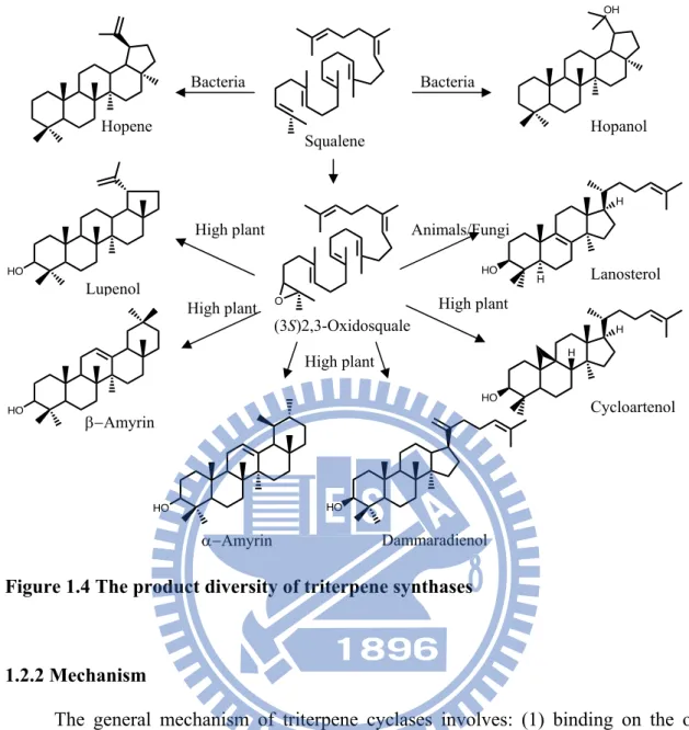
Fig. 1.4 The product diversity of triterpene synthases...6
Fig. 1.5 Cyclization of oxidosqualene to protosteryl and dammarenyl cation...7
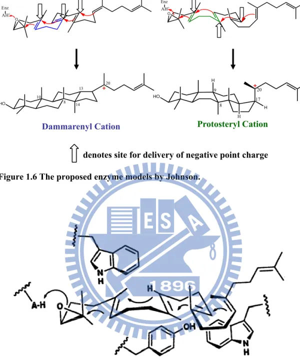
Fig. 1.6 The proposed enzyme models by Johnson...10
Fig. 1.7 Griffin’s hypothesis model...10
Fig. 1.8 The detailed mechanism of 2,3-oxidosqualene into lanosterol...11
Fig. 1.9 The proposed model for oxirane ring opening and cyclization initiation...13
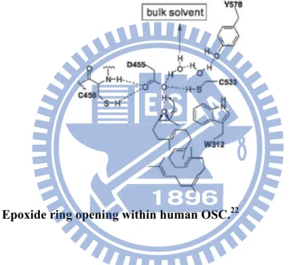
Fig. 1.10 Epoxide ring opening within human OSC...13
Fig. 1.11 Substrate analogue and product of oxidosqualene cyclase which are suggestive of five-membered C-ring intermediate...14
Fig. 1.12 Cyclization mechanisms in human OSC...15
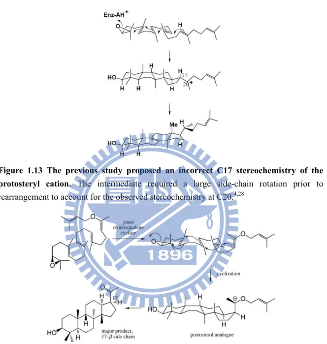
Fig. 1.13 The previous study of incorrect C17 stereochemistry of protosteryl cation intermediate...16
Fig. 1.14 The evidence for the stereochemistry of protosteryl cation intermediate...16
Fig. 1.15 Proposed mechanisms for C-ring expansion and D-ring formation were concerted by Hess...17
Fig. 1.16 Human OSC structure...18
Fig. 1.17 The difference between cyclization mechanisms of oxidosqualene-lanosterol cyclase and cycloartenol synthase...20
Fig. 1.18 Conservation pattern between CAS1 and ERG7...21
Fig. 1.19 The proposed mechanism of 2,3-oxidosqualene into β-amyrin...25
Fig. 1.20 Crystal structure of SHC...27
Fig. 1.21 The cyclization process of squalene-hopene cyclase (SHC)...28
Fig. 1.22 Amino acid sequence alignment of ERG7, PSY, CAS, and SHC genes...30
Fig. 2.1 QuikChange site-directed mutagenesis strategies...43
Fig. 2.2 The acetylation modification...49
Fig. 3.2 The GC data of ERG7I705F mutant...56
Fig. 3.3 The mass spectra of novel product from ERG7I705F mutant...57
Fig. 3.4 The structure and the NOE correlation of the novel compound...58
Fig. 3.5 Proposed cyclization/rearrangement pathway occurred in the ERG7I705X site-saturated mutants...61
Fig. 3.6 The homology models of wile-type ERG7 and ERG7I705F complexed with lanosterol and 17α-protosteryl cation...64
Fig. 3.7 The homology models of the double mutants ERG7F699M/I705F and ERG7F699T/I705F complexed with lanosterol...67
Fig. 3.8 The homology model of wild-type PSY complexed with β-amyrin...71
Fig. 3.9 The homology model of wild-type PSY complexed with β-amyrin...72
List of Tables
Table 1.1 Product profile of AthCAS Ile481, Tyr410 and His477 mutants...23
Table 2.1 QuikChange Site-Directed Mutagenesis Kit PCR composition...44
Table 2.2 QuikChange Site-Directed Mutagenesis PCR program...45
Table 2.3 QuikChange Site-Directed Mutagenesis PCR products diegestion...45
Table 3.1 The site-saturated mutants of S. cerevisiae ERG7I705X and their genetic analysis...53
Table 3.2 The product profiles of the S. cerevisiae ERG7I705X mutants...55
Table 3.3 NMR assignments for 17α-protosta-20(22),24-dien-3β-ol for dilute CD2Cl2 solution...59
Table 3.4 The distance of I705 to C-14 and C-17 complexed with different ligands in the homology models...65
Table 3.5 The products analysis of double mutants between I705F and F699X...67
Table 3.6 The site-saturated mutants of P. sativum PSYL734X and their genetic selection and products analysis...69
Chapter 1 Introduction
1.1 Triterpenoids and its biosynthetic pathway
Terpenoids, sometimes called isopenoids, are a large class of organic chemicals similar to terpenes, which are the combination of several isoprene units. Some chemists use the term “terpene” extensively more than terpenoids. The classification of terpenoids could accord the the number of isoprene units or the number of cyclic structures they contain. Triterpenoids were derived from six isoprene units, usually have tetracyclic or pentacyclic
structure and C30H50O formula. (Fig. 1.1) They could be found in many classes of living
things, and also are the largest group in the nature products. Moreover, the interest in various aspects of the biological activities on triterpenoids and triterpenoids saponins was
increasing.1 Belongs to triterpenoids, sterols have the same general ring structure and
known as steroid alcohols with a hydroxyl group at the C-3 position of the A-ring. Thus, the hydroxyl group on the A-ring is polar, whereas the rest of aliphatic chain is non-polar. Sterols were vital constituents of cell membranes. In animals, cholesterol (Fig. 1.2) was found in the cell membranes and transported in the blood plasma. It was required to build and maintain cell membranes and regulate the membrane fluidity on the range of physiological temperatures. Also, cholesterol was the precursor molecule in many biochemical pathways. The phytosterols include campesterol, stigmasterol and β-sitosterol, and act as a sterol component in the membrane. Furthermore, food additive, medicines and cosmetics were all applications of phytosterols. Although sterols were usually absent in bacteria, some bacteria and protozoan could produce triterpenes such as hopene which was
regarded as the sterol substitute.2 Because these compounds play the important role in
many ways, the complete understanding of the specific enzyme during their biosynthetic pathway was necessary.
HO R
A B
C D
H HOFigure 1.1 Triterpenoid backbone Figure 1.2 Cholesterol
The sterol biosynthesis was generally through the mevalonic acid (MVA) pathway. Acetyl coenzyme A is to be the starting material, it is converted into acetoacetyl-CoA by acetoacetyl-CoA thiolase. After the catalysis of 3-hydroxy-3-methylglutaryl-CoA synthase (HMG-CoA synthase) and 3-hydroxy-3-methygulutary-CoA reductase (HMG-CoA reductase), acetoacetyl-CoA is converted into mevalonate. Mevalonate is phosphorylated by 2 sequential Pi transfers from ATP, yielding the pyrophosphate derivative. ATP-dependent decarboxylation, with dehydration, yields isopentenyl pyrophosphate. Isopentenyl pyrophosphate in the pathway is referred to as isoprenoid, by a series of chemical reactions including isomerization and condensation, a linear molecule with 30 carbons, squalene, was produced. Oxidation of squalene by squalene synthase yields an acyclic polyolefin substrate, (3S)-2,3-oxidosqualene (OS). The common substrate oxidosqualene solely proceeded the biotransformation for the production of tetracyclic lanosterol in animals and fungi, whereas a variety of polycyclic triterpene alcohols including cycloartenol, lupeol, α-amyrin and β-amyrin are simultaneously generated in higher plants. (Fig. 1.3)
3 Acetyl CoA
C2Acetoacetyl CoA
3-Hydroxy-3-methylglutaryl-CoA
Figure 1.3 Sterol biosynthetic pathway
Isopentenyl tRNA
Cholesterol
Mevalonate
Isoprenoid intermediate
Ubiquinone Dolichol Heme ASqualene
(3S)2,3-Oxidosqualene
O
O
S
S
C
C
C
C
A
A
S
S
Lanosterol
Cycloartenol
Ergosterol
HMG-CoA reductase C5 C30 HMG-CoA synthase geranyl pyrophosphate farnesyl pyrophosphateSHC
Hopene
Bacteriaβ
β
A
A
S
S
High plant High plant
β-Amyrin
Fungi AnimalOleanane-type
triterpene saponins
Phytosterol
One of the triterpenoids that is crucial in our life is cholesterol. In the study of the treatment on hypercholesterolemia was targeting at the step catalyzed by HMG-CoA reductase, which is the rate-limiting step in the early part of the pathway. But the inhibition of HMG-CoA reductase not only affects the production of cholesterol, but also have the side effect that influence the isoprenoid intermediates and its derivatives. It may cause the adverse clinical results. On the other hand, in the downstream of the biosynthetic pathway, inhibitors of oxidosqualene-lanosterol cyclase (OSC) as anticholesteraemic drugs might provide insight of the development of a safer, more effective treatment. Furthermore, the highly-complex and specific reaction of oxidosqualene-lanosterol cyclase include protonation, cyclization, rearrangement of carbon skeleton, and deprotonation, have bring to the interest of chemists. Therefore, current research for OSC is increasing, and the structure-function relationship of this enzyme is to be worth to discuss in depth.
1.2 Triterpene cyclases
1.2.1 Product diversityThe triterpenoids are a large diverse group of natural products derived from squalene, oxidosqualene or related acyclic 30-carbon precursors. Nearly 200 tertripene compounds are known from nature or enzymatic reactions which are cyclization products of olefins
such as squalene, oxidosqualene, or bis-oxidosqualene.3 Several triterpene cyclase enzymes
transform these olefins into complex and biologically important polycyclic products such as hopene (6-6-6-6-5 pentacycles), hopanol (6-6-6-6-5 pentacycles), lanosterol (6-6-6-5 tetracycles), cycloartenol (6-6-6-5 tetracycles), lupenol (6-6-6-6-5 pentacycles), and β-amyrin (6-6-6-6-6 pentacycles). (Fig. 1.4) The reason for triterpene diversity is that triterpene cyclases are groups of plastic enzymes and have novel catalytic properties from mutational alteration. The triterpenes serve as the precursors of all hopanoids and steroids, including cholesterol, glucocorticoid, estrogens, and hormones. Moreover, the mechanism of action of triterpene cyclases stimulated the interest of many biologists and chemists nearly a half century ago. Early studies on triterpene cyclases had used substrate analogs to provide important mechanistic information, the range of structures which cyclases can
transform, as well as the nature of any resulting products.4 However, in-depth and detailed
understanding the structure-function-reaction mechanism relationships of the plastic triterpene cyclases, such as the role of cyclases in substrate prefolding and cation stabilization is required to develop new methods for use in protein expression and crystallization research.
Figure 1.4 The product diversity of triterpene synthases
1.2.2 Mechanism
The general mechanism of triterpene cyclases involves: (1) binding on the olefin substrate in a folded conformation; (2) initiated by protonation of double bond (squalene) or one oxirane ring (oxidosqualene); (3) four or five-membered stereochemically controlled ring formations; (4) nucleophilic hydride and methyl group rearrangement; and, (5) termination either by deprotonation or water addition. Thus, the cyclized process involved sixteen bonds broken, and sixteen new bonds formed, and through the cation-olefin cyclization, the unstable cationic intermediates are formed. The transient cationic intermediates are stabilized by the putative active site of cyclases, hence, it could not be isolated by scientists. Due to different mechanisms, the diversity of triterpenes could be explained by different hydride / methyl shifts, and the early deprotonated truncations.
H HO H H HO H (3S)2,3-Oxidosquale Squalene Bacteria Bacteria Hopene Hopanol Lanosterol Cycloartenol O OH Animals/Fungi High plant High plant High plant High plant HO Lupenol HO β−Amyrin HO HO Dammaradienol α−Amyrin
In contrast to oxidosqualene cyclase, squalene-hopene-cyclase (SHC) allows all-chair conformation of squalene, and there are two pre-folded conformational types in the oxidosqualene cyclization. One is so-called “chair-boat-chair” conformation of oxidosqualene, as a substrate. The “chair-boat-chair” conformation would form lanosterol in animal/ fungi or cycloartenol in higher plants. On the other way, oxidosqualene would pre-fold into “chair-chair-chair” conformation. This conformation is usually found in the plant species such as Olea europaea (lupenol synthase) and Pisum sativum (β-amyrin
synthase).5 These two different conformations via two different cation intermediates,
cyclization of “chair-boat-chair” conformation would give the protosterol cation which had a positive charge at C-20, whereas the “chair-chair-chair” conformation would give the dammarenyl C-20 cation. In the protosteryl cationic pathway, B-ring boat products were produced, while B-ring chair products were formed in the dammarenyl cationic pathway. (Fig. 1.5)
(3S)-2,3-Oxidosqualene O
Figure 1.5 Cyclization of oxidosqualene to protosteryl and dammarenyl cation. Chair-Boat-Chair Chair-Chair-Chair Protosteryl Cation Dammarenyl Cation HO HO Lupenol H HO H Lanosterol H HO H Cycloartenol High plant Animals&Fungi High plant High plant O Enz AH+ O Enz AH+ HO HO β-Amyrin
1.3
Oxidosqualene-lanosterol cyclase (OSC)
1.3.1 The hypothesis of oxidosqualene cyclasesThe family of oxidosqualene cyclases extensively exist in the organism, and the common cyclases such as oxidosqualene-lanosterol cyclases (OSC) in animals and fungi, plant oxidosqualene cyclases including cycloartenol synthase (CAS), lupenol synthase (LUS), and β-amyrin synthase (βAS). These different enzyme systems convert the oxidosqualene, as a substrate, into various polycyclic triterpenes. And the relationship between the enzyme structure and cyclization mechanism is extremely interesting.
Research on oxidosqualene cyclases was remarkable for nearly half a century. Woodward and Bloch first proposed that in the cyclization of squalene in cholesterol
synthesis followed by rearrangement to lanosterol in 1953.6 In 1958 and 1965, Maudgal
and Cornforth groups evidenced for 1.2-methyl and hydride shifts during lanosterol
formation by incorporation experiments.7,8 Corey and van Tamelen showed that
2,3-oxidosqualene is more efficiently incorporated in sterol synthesis than squalene,
demonstrated intermediacy of 2,3-oxidosqualene in lanosterol biosynthesis.9,10 Also, their
laboratories had many substrate analogue compounds for synthesis and test. In addition, van Tamelen also showed the nonenzymatic cyclization of 2,3-oxidosqualene resulted in truncated cyclization to produce a tricyclic product, suggesting that direct enzymatic control is necessary for the prevention of the chemical tendency in the formation of five-membered C-ring, and for emergence of the biologically required six-membered
C-ring.11 In 1975, Barton confirmed that eukaryotic oxidosqualene cyclases accepted only
(3S)- but not (3R)-enantiomer of 2,3-oxidosqualene as a substrate in the formation of lanosterol, demonstrating the highly substrate-specific property of oxidosqualene cyclases
rather than squalene cyclases.12 Guy Ourisson and his co-workers proposed the possible
molecular evolution from the primitive squalene cyclases to oxidosqualene cyclases in
cyclization of the A-ring are concerted and it is essential for electrophilic activation of the
oxirane function.14,15
In previous research, oxidosqualene would be stable in the neutral condition at room temperature for the whole day. And the more powerful acid, such as trichloroacetic acid, is
required for strong epoxide activation to promote the cyclization step.16 Then, it would
form many unstable cation intermediates in the cyclization process, where there are two hypotheses that illustrated how the enzyme functions to stabilize the high-energy cation intermediate. In 1987, Johnson proposed the “cation-stabilizing auxiliary” model, which proposed that the Lewis acid residues on the active site provide a proton for initiation of the epoxide group, and a number of anionic sites in the cyclase enzyme would lead the cation generation and the formation of the proper ring system. The axial negative charge residues would face toward the transition states or the intermediates for cation stabilization, and it would demonstrate the ring formation of B-boat/chair ring. Therefore the B-boat ring could be promoted by delivery of a point charge to the α-face at pro-C-8, and lowering the
activation energy of the boat form rather than the chair skeleton.17,18 (Fig. 1.6) In addition,
Griffin and co-workers proposed the “aromatic hypothesis” model which the electron-rich aromatic side chain such as Trp and Tyr, might stabilize the positively charged transition states or high-energy intermediates during the process of cyclization and rearrangement
steps.19 (Fig. 1.7) The hypothesis is that aromatic residues play the role of the anions group,
just like that proposed in the Johnson model mechanism. Cation-π interactions are common features in enzyme-substrate complexes.
O Enz AH+ O Enz AH+ HO H H H 20 17 9 8 HO 20 13 14 8 10 Protosteryl Cation Dammarenyl Cation
denotes site for delivery of negative point charge Figure 1.6 The proposed enzyme models by Johnson.
Figure 1.7 Griffin’s hypothesis model for involvement of electron-rich aromatic side chains from Trp and Tyr residues in the cyclization of oxidosqualene to the protosteryl cation.19
1.3.2 The studies of mechanism and site-directed mutagenesis
Oxidosqualene-lanosterol cyclases (OSC), also called lanosterol synthases, are widely found in animals and fungi, and usually convert oxidosqualene into the tetracyclic
product, lanosterol. In Saccharomyces cerevisiae, the cyclases were encoded from ERG7 gene, as a membrane protein. The protein contains 731 amino acids and the molecular mass is approximately 83 kDa. Because of the difficulty in purifying membrane protein, the crystal structure of S. cerevisiae oxidosqualene-lanosterol cyclases (SceERG7) enzyme has not been attained until now. In order to understand the structure-function relationships of oxidosqualene cyclase-catalyzed reactions in depth, site-directed saturated mutagenesis coupled with genetic selection and products analysis for illustrating the functional importance of these mutated residues, and describing the carboncationic intermediates in the reaction cascade.
Briefly, the mechanism of oxidosqualene-lanosterol cyclases include one oxirane ring opening, four stereochemically controlled ring formations, four nucleophilic hydride and methyl group migrations, and the final deprotonation reaction at C-8 or C-9. (Fig. 1.8)
Figure 1.8 The detailed mechanism of conversion of 2,3-oxidosqualene into lanosterol.
(3S)-2,3-Oxidosqualene O Enz AH Chair-Boat-Chair conformation HO 6 HO H 10 HO H 14 13 HO 14 H HO H H H 20 17 9 8 Lanosterol O A ring B ring C ring H C D B HO A H D ring Protosteryl Cation
In the detailed aspect, the enzyme catalyzed conversion of (3S)-2,3-oxidosqualene into a pre-folded chair-boat-chair conformation, a single aspartic acid residue (D456) in
SceERG7 provided a proton to induce the epoxide opening. Before the structure of human
OSC was crystallized, scientists used an affinity-labeling strategy of the substrate analogs,
trying to identify the important residues within oxidosqualene cyclases.20 In 1997, Corey
and Matsuda used an “alanine scanning site-directed mutagenesis” method to identify the highly-conserved residues in the putative active site within SceERG7, revealing that
His146, His234 and Asp456 are essential for catalytic activity.11,14 Furthermore, these
researchers also showed that His146 assists in forming hydrogen bonds with Asp456, and it
would enhance the acidity of Asp456, to induce the oxirane ring opening.21 (Fig. 1.9)
Similarly, in human OSC research, Asp455 (which corresponds to Asp456 within SceERG7) was confirmed as the general acid that protonates the epoxide group of oxidosqualene. By hydrogen bonding association with Cys456 and Cys533, the acidity of Asp455 was increased and then activated the initial catalyzed reaction. After completing the entire reaction cycle, Asp455 would be reprotonated either from the bulk solvent through the carboxylate group of Glu459 and the water molecules, or by shifting the proton from the
final deprotonation step back to the Asp455 residue.22 (Fig. 1.10) In addition, some
research has shown evidence to support concerted epoxide opening and A-ring closure by oxidosqualene-lanosterol cyclases, and these studies usually used oxidosqualene analogs to
demonstrate the concerted C-O cleavage and A-ring formation.14 In addition, some
computational results agreed with these experimental findings.23 Therefore, these two steps
Figure 1.9 The proposed model for oxirane ring opening and cyclization initiation.21
Figure 1.10 Epoxide ring opening within human OSC.22
The formation of the B-boat ring probably occurs very rapidly when the positive charge appears on the C-6 of the A-ring. The closure of the C-ring is puzzling because of the direct formation of the six-membered C-ring would represent the anti-Markovnikov form. In 1995, E. J. Corey and his co-workers first proposed the five-membered C-ring Markovnikov cyclization against the direct formation of the six-membered C-ring. By oxidosqualene analogs, 20-oxaoxidosqualene had been reacted with oxidosqualene cyclase, and the resulting products suggested that there was a five-membered C-ring intermediate
appearing through an intramolecular reaction.24 (Fig. 1.11) Moreover, incubations of
products, providing more evidence for a five-membered C-ring closure followed by a ring
expansion.4 In the molecular biological aspects, the highly-conserved residue Val454 within
SceERG7 had been proven to have a role in the B-ring formation by its steric side chain in
Matsuda’s lab.25 They used a series of SceERG7 mutants such as glycine, alanine,
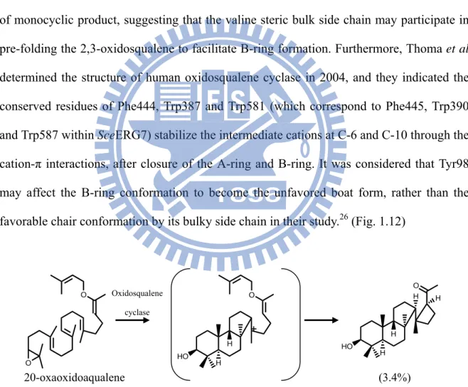
phenylalanine, leucine, and isoleucine to substitute for the Val454 residue. The Val454Phe mutant was inactive, while Val454Lue and Val454Ile mutations produced only lanosterol as the sole product. However, the Val454Ala and Val454Gly produced not only lanosterol by also achilleol A as a minor product. Decreasing the amino acid side chain allows formation of monocyclic product, suggesting that the valine steric bulk side chain may participate in pre-folding the 2,3-oxidosqualene to facilitate B-ring formation. Furthermore, Thoma et al determined the structure of human oxidosqualene cyclase in 2004, and they indicated the conserved residues of Phe444, Trp387 and Trp581 (which correspond to Phe445, Trp390 and Trp587 within SceERG7) stabilize the intermediate cations at C-6 and C-10 through the cation-π interactions, after closure of the A-ring and B-ring. It was considered that Tyr98 may affect the B-ring conformation to become the unfavored boat form, rather than the
favorable chair conformation by its bulky side chain in their study.26 (Fig. 1.12)
Figure 1.11 Substrate analog and product of oxidosqualene cyclase which are suggestive of a five-membered C-ring intermediate.24
O O 20-oxaoxidoaqualene HO H H O HO H H O Oxidosqualene H H cyclase (3.4%)
Figure 1.12 Cyclization mechanisms in human OSC. Phe444, Trp387 and Trp581
stabilize the intermediate cations at C-6 and C-10. In addition, the Tyr98 side chain
contributes the energetically unfavorable boat conformation of the B-ring.26
The final step of the mechanism in lanosterol synthesis is protosterol cation formation and its rearrangement. Early studies of sterol biosynthesis assumed that the conversion of oxidosqualene into lanosterol proceeded via a protosterol cation where its
cationic side chain at C-17 is α-oriented.27 After hydride migration of the 17α- protosterol
cation from C-17 to C-20, it does not form the natural sterol 20R skeleton, but the unnatural 20S configuration. Another explanation for the mechanism was the occurrence of a 120° rotation of C17-C20 bond via the hydride migration, in order to produce the 20R skeleton
of lanosterol, whereas only a 60° rotation produces the unnatural 20S configuration.28 (Fig.
1.13) In addition, it had been proposed that the 17α- protosterol cation may not be the intermediate, but a covalent adduct formed by the reaction of the 17α-protosterol cation with the nucleophilic group within the cyclase. In 1992, Corey proposed the evidence that the stereochemistry at C-17 of protosterol cation prefers the β rather than α orientation by the enzyme-catalyzed cyclization of 20-oxaoxidosqualene. (Fig. 1.14) This result overthrows the large rotation around the C17-C20 bond prior to rearrangement, and the
17β-protosterol cation was also demonstrated to occur by the catalysis of
20,21-dehydrooxidosqualene.29
Figure 1.13 The previous study proposed an incorrect C17 stereochemistry of the protosteryl cation. The intermediate required a large side-chain rotation prior to
rearrangement to account for the observed stereochemistry at C20.4,28
Figure 1.14 The evidence for the stereochemistry of the protosteryl cation intermediate at C17 prefers the β rather than α orientation by incubation of substrate
analog.4,28
In theoretical calculation research, Hess performed the 6-6-5-tricyclic cation, which was the first intermediate during the formation of the protosterol cation, and suggested that
the SHC study, Gao showed both none, and fewer intermediates of tricyclic and tetracyclic
cations, suggesting that the formation of the C, D and E-ring were concerted.31
Figure 1.15 Proposed mechanisms for C-ring expansion and D-ring formation were concerted by Hess.30
The human OSC structure showed that the Phe696 and His232 (which correspond to Phe699 and His234 in SceERG7) could stabilize the positive charge at C-20 of the protosteryl cation through cation-π interactions, and then the highly conserved aromatic residues such as Trp192, Trp230, His232, Tyr237, Tyr503, Phe521 and Phe696 could stabilize the methyl/ hydride rearrangement process. Ultimately, His232 is the nearly basic residue that could accept a proton in the deprotonation of the C-8 or C-9 lanosterol cation, and complete the final step of catalysis. It is also possible that the Tyr503 residue which is hydrogen-bonded with His232 could be in the appropriate position for the final cyclization step. On the other hand, in the mutation of Phe699 within SceERG7, we suggested that it could stabilize the anti-Markovnikov 6-6-5-tricyclic C-14 cation and lanosteryl C-17 cation based on its product profiles. Unlike SHC, oxidosqualene cyclase lacks particular specific electron-rich residues that could promote the D-ring expansion and E-ring formation.
1.3.3 Human oxidosqualene cyclase
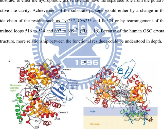
In 2004, Thoma and his co-workers solved the X-ray structure of human OSC which is in complex with lanosterol, published in Nature. Human OSC is a monomer which consists of two barrel domains that are connected by loops and three smaller β-structures, and the large active-site cavity is located in the center of the molecule between domains 1 and 2. It is a monotopic membrane protein that attached to the membrane from one side, and the membrane-inserted surface consists of a plateau 25 Å in diameter and a channel that leads to the active-site cavity. The channel is considered to admit oxidosqualene, as a substrate, to enter the hydrophobic active site and have the separated role from the putative active-site cavity. Achievement of the substrate passage would either by a change in the side chain of the residue such as Tyr237, Cys233 and Ile524 or by rearrangement of the
strained loops 516 to 524 and 697 to 699.26 (Fig. 1.16) Because of the human OSC crystal
structure, more relationship between the functional residues could be understood in depth.
Figure 1.16 Human OSC structure. (a) The ribbon diagram of human OSC. The C and N
termini and several sequence positions are labeled. The inner barrel helices are colored yellow. The bound inhibitor, Ro48-8071 (black), indicates the location of the active site. (b) The orientation of OSC relative to one leaflet of the membrane, Ro 48-8071 bind in the
Before the human OSC was solved, the homologue structure of SHC was the model for understanding the OSC catalysis. But SHC and OSC have different substrate, different B-ring conformation, further expandsion of D-ring, and cyclization of E-ring. To gain more information for the highly stereo selective cyclization, the highly sequence identity of human OSC with SceERG7 would be more useful than the SHC structure.
1.4 Oxidosqualene cyclase in plants
1.4.1 Cycloartenol synthase (CAS)Cycloartenol is the sterol precursor in higher plants and its skeleton is similar to lanosterol found in animals and fungi. Cycloartenol synthase converts oxidosqualene into cycloartenol via protosteryl cation intermediate, following a series of methyl and hydride shifts to form the C-9 cation, and then terminating the reaction by cyclopropyl ring formation and deprotonation at C-19 to form cycloartenol. Whereas lanosterol synthesis via the extremely similar reaction intermediate that deprotonated from either C-8 or C-9 carbocationic intermediate after methyl and hydride shifts of protosteryl cation, and then yields lanosterol. (Fig. 1.17) Both cycloartenol synthase and lanosterol synthase have more than 700 amino acids, but about 400 amino acids are different in the sequence alignment, and therefore suggested the catalytic distinction for specific deprotonation or cyclopropyl ring formation of these two enzymes. Cycloartenol synthase was first cloned from
Arabidopsis thaliana (AthCAS1) and expressed and characterized in the yeast lanosterol
synthase system by the Matsuda group.32 The system used the mutated lanosterol synthase
as an expression strain, incubated it by exogenous sterol for growth, and then analyzed the details of cycloartenol biosynthesis.
The study of cycloartenol synthase (AthCAS1) by site-directed mutagenesis showed (3S)-2,3-Oxidosqualene O Enz AH H HO H H H Protosteryl Cation H H H HO H H C-8 Cation H HO H H C-9 Cation ERG7 CAS1
Figure 1.17 The difference between
cyclization mechanisms of oxidosqualene-lanosterol cyclase and cycloartenol synthase.
H H HO H Lanosterol HO H Cycloartenol
some important residues such as Tyr410, His477 and Ile481. These residues are highly-conserved within cycloartenol synthase in many plant species, whereas these
residues are totally different within ERG7 just like Thr, Cys, Gln and Val amino acids.33
(Fig. 1.18) Speculation on this phenomenon, these residues may promote the formation of cyclopropyl ring within AthCAS1. Corresponding to Ile481 that residue in the SceERG7 active site is Val454. Mutation on Ile481 into Val would have a subtle steric change by subsyituting the isopropyl side chain instead the sec-butyl side chain. The little steric change cause 25% lanosterol, 21% parleol and 54% cycloartenol formation, indicate that Ile481 is not catalytically necessary but may influence the substrate and active site base by its steric effect. Moreover, in order to understand the steric effect at Ile481, the Matsuda group constructed AthCAS1 Ile481Phe, Ile481Leu, Ile481Ala and Ile481Gly mutations. The Ile481Phe was inactive, and suggested that Phe residue may occludes the active site to cause this result, whereas the similar size but different shape of Ile481Leu produces cycloartenol, parkeol and a little lanosterol. In addition, Ile481Ala and Ile481Gly mutations yielded lanosterol and parkeol, also produce monocyclic compounds achilleol A and camelliol C. These experiments indicated the steric bulky residue at this position is important in controlling cyclization and deprotonation. Also, the native cyclases may have
less sterically bulky group at this position in order to form the monocyclic products.21
(Table 1.1)
Figure 1.18 Conservation pattern between CAS1 and ERG7.33
performed a search for similar conservation patterns between CASs and ERG7s. Tyr410, Gly488, Phe717, and Met731 were strictly conserved within CASs, but only Tyr410Thr mutation altered catalysis, and produced lanosterol, parkeol, and 9β-lanosta-7,24-dien-3β-ol at the ratio of 65:2:33 instead of cycloartenol. Tyr410Thr abolishes cycloartenol formation that implicates Tyr410 is essential for cyclopropyl ring formation. Because Tyr410 mutant produced not only lanosterol but also the other by-products, thereby additional mutations are necessary to abolish parkeol and 9β-lanosta-7,24-dien-3β-ol formation for converting cycloartenol synthase into accurate oxidosqualene-lanosterol synthase. The previous study showed Ile481Val also induce the formation of lanosterol, therefore Tyr410Thr Ile481Val was constructed and the products profile was lanosterol, parkeol, and 9β-lanosta-7,24-dien-3β-ol at the ratio of 75: 0.6: 24. The double mutants yielded lanosterol more efficiently than single mutant. However, the
Tyr410Thr and Ile481Val mutation have synergistic effects.21 (Table 1.1)
The other research indicated that the second-tired residue His477 in the putative active site of AthCAS1 which hydrogen bonding with Tyr410. His477 is considered to pull Tyr410 out of the active site and allow reorientation of the intermediate cation to form
other products.21,33 Corresponds to His477 within AthCAS1, the residues within ERG7s is
Cys or Gln. The His477Gln mutant is the most accurate parkeol synthase because the ratio of parkeol is 73%, whereas His477Asn is currently the most accurate lanosterol synthase at the ratio of 88% that had been yielded by mutating cycloartenol synthase. Due to the result of double mutant Tyr410Thr Ile481Val generated by-products 9β-lanosta-7,24-dien-3β-ol, converting a cycloartenol synthase to an accurate lanosterol synthase would require at least one additional mutant to preclude deprotonation from C-7. Therefore combining the Tyr410Thr Ile481Val double mutants with His477Asn and His477Gln mutation to construct the triple mutants. Both the result of Tyr410Thr Ile481Val His477Asn and Tyr410Thr Ile481Val His477Gln triple mutants have lanosterol, and 9β-lanosta-7,24-dien-3β-ol at the
ratio of 78: 22, and the percentages of parkeol is less than 1%. Compare to the single mutant of His477Gln, the Tyr410Thr Ile481Val mutations may block the hydride shift from C-9 to C-8 and then preclude the opportunity for His477 mutant to promote the formation of parkeol which is derived from the C-9 cation. Furthermore, Table 1.1 showed that the
double mutants Ile481Val His477Asn had the maximum yield of lanosterol.21 (Table 1.1)
AthCAS1 mutant Cycloartenol Lanosterol Parkeol 9β-△7-
Lanosterol Achilleol A Camelliol C
CAS1I481 99 — 1 — — — CAS1I481L 83 1 16 — — — CAS1I481V 54 25 21 — — — CAS1I481A 12 54 15 — 13 6 CAS1I481G 17 23 4 — 44 12 CAS1Y410T — 65 2 33 — — CAS1H477N — 88 12 — — — CAS1H477Q — 22 73 5 — — CAS1I481V/Y410T — 75 <1 24 — — CAS1I481V/H477N/Y410T — 78 — 22 — — CAS1I481V/H477Q/Y410T — 78 — 22 — — CAS1I481V/H477N — 99 1 — — —
Table 1.1 Product profile of AthCAS Ile481, Tyr410 and His477 mutants.21
Moreover, our group also shows that the combination of random mutation coupled with in vivo selection and provided an effective method of indentifying single residue alteration could lead to the change of product specificity. We found the Y532H mutant could alter the product specificity, a new position that be indentified in the A. thaliana CAS active site. Furthermore, A469 and H477 residues were considered that would not in direct contact with the substrate because of their positions are not in the putative active site of CAS. H477Y mutant generated lanosterol as the dominant product and the A469 mutant yielded lanosterol and achilleol A. This is the first example of nonactive site mutations that
alter the product specificity in a triterpene synthase. Thus the A469 and H477 residues may be the nearest neighbor to the putative active site residues and provide the indirect effect on
the active site structure.34
1.4.2 β-amyrin synthase (βAS)
The remarkable cyclization of 2,3-oxidosqualene into β-amyrin has interested organic chemists for over a half century since the biogenetic isoprene rule was proposed by Eschenmoser and co-workers. β-amyrin synthase cyclizes oxidosqualene into β-amyrin, which initially pre-folds the substrate into a chair-chair-chair conformation, and then forms the 6-6-6-5-fused tetracyclic dammarenyl C-20 cation. In contrast to the biosynthesis of lanosterol or cycloartenol, the expansion of the D-ring occurs, then the electrophilic addition of the baccharenyl cation on to the terminal double bond generates the lupenyl cation with a five-membered E-ring, which then yields the six-membered E-ring oleanyl cation by ring expansion. Finally, the oleanyl cation is produced via a hydride shift and deprotonation at C-12 to yield the 6-6-6-6-6-fused pentacyclic β-amyrin. (Fig. 1.19)
Figure 1.19 The proposed mechanism of 2,3-oxidosqualene into β-amyrin.
β-amyrin synthase (βAS) originating from several plant species, including Pisum
sativum and Panax ginseng, and Pisum sativum β-amyrin synthase (PSY), have been
cloned and functionally expressed in Saccharomyces cerevisiae.35 To investigate the
catalytic motifs within cyclases that form the dammarenyl cation, the Ebizuba group first generated chimeras of the A. thaliana lupenol synthase (LUP1) and Panax ginseng β-amyrin synthase (PNY). They determined the functions of the regions by using a domain swapping strategy, and only relatively small portions of the protein were found to control the production of either lupenol or β-amyrin. One chimera in which only one fourth of the protein was β-amyrin sequence but made four times as much β-amyrin as it did lupenol, and a mixed PCR method further confirmed the important region of chimeras and narrowed it down to an 80 amino acid span. Later, Ebizuba and co-workers looked within the 80 amino acid region to define specific residues responsible for the product specificity of
(3S)-2,3-Oxidosqualene Chair-Chair-Chair conformation O O Enz AH+ HO Dammarenyl Cation HO Baccharenyl Cation HO Lupenyl Cation HO HO Oleanyl Cation β−Amyrin
β-amyrin and lupenol synthases. Results from alignment analysis showed that Trp259 within Panax ginseng β-amyrin synthase (PNY), and Leu256 within Olea europa lupenol synthase (OEW) might control the product specificity, therefore PNY Trp259Leu and OEW Leu256Trp mutants were contructed. Lupenol occurred twice as much as β-amyrin in the product profile of the PNY Trp259Leu mutant, whereas β-amyrin was the major product in the product profile of the OEW Leu256Trp mutant. These two results demonstrate that this position plays a critical role in directing either β-amyrin or lupenol formation. Furthermore, the PNY Tyr261His mutant was also created, and the product analysis result showed that Tyr261 stabilized one of the cationic intermediates formed after the dammarenyl
cation.21,36,37 In addition, one research study for Pisum sativum β-amyrin synthase (PSY)
showed the expansion of the D-ring could take place in the absence of the terminal double bond. Thus, the formation of the anti-Markovnikov six-membered D-ring does not depend on the terminal π-electrons. In summary, the aromatic residues within putative active site
1.5 Squalene-hopene cyclase (SHC)
Squalene-hopene cyclase is a homodimeric enzyme and it is organized in two α-helical domains, which together form a dumbbell-shaped molecule. (Fig. 1.20) It contains 631 amino acids per subunit and the molecular mass are about 71.5kDa. In 1997, the X-ray crystal structure of Alicyclobacillus acidocaldarius SHC was first reported at 2.9
Å resolution, later refined to 2.0 Å resolution in 1999.38,39 Moreover, Reinert et al reported
another X-ray crystal structure which the squalene-hopene cyclase was cocrystallized with
2-azasqualene and its resolution is 2.13 Å in 2004.40 These structures, which combine with
the biological studies, provide more mechanistic insight into squalene-hopene cyclases and oxidosqualene cyclases.
Figure 1.20 Crystal structure of A. acidocaldarius squalene-hopene cyclase (SHC).38
Different from the cyclization of oxidosqualene, squalene-hopene cyclase pre-fold squalene into an all-chair conformation, and the lower basicity of the double bond relative to the oxirane ring suggests that SHC provide a catalytic acid at least as strong as that used
to initiate the reaction of oxidosqualene. Thus, this property induce the bacterial squalene cyclase to accept oxidosqualene and its analogues as substrates for cyclization. The cyclization process of SHC is simpler than oxidosqualene cyclase, and then convert squalene into hopanyl C-22 cation with 6-6-6-6-5-fused pentacyclic ring. Without the rearrangement step, deprotonation of hopanyl C-22 cation yield hopene or addition of water to produce hopanol. (Fig. 1.21)
Figure 1.21 The cyclization process of squalene-hopene cyclase (SHC).
In the site-directed mutagenesis studies, the catalytic acid Asp376 residue (which corresponds to Asp456 within SceERG7) hydrogen-bonded with His451 to initiate the reaction by protonation, and then Asp376 is likely to be stabilized by His451 residue that in
turn is electrostatically stabilized by the solvent accessible Glu454.35,40 Following
protonation, the process of cyclization undergo small conformational change during A to D-ring formation. Several cation-stabilizing residues have been subjected to mutational studies to evaluate the role in different stage of squalene cyclization. Substitution of Asp377 with Cys or Asn result in the formation of monocyclic product, and it suggested that Asp377 may stabilize C-10 cation, and then the other mutation of Tyr420Ala yielded
Squalene
Enz AH
22
Chair-Chair-Chair conformation Hopanyl C-22 cation
OH
Anti-Markovnikov addition
two bicyclic products and one tricyclic compound, it was the evidence of truncating the C-8 B-ring cation reaction to form the bicyclic products, indicating Tyr420 may stabilized C-8 cation intermediate within the putative active site of SHC. In addition, the Phe365Ala and Tyr609Phe mutants also induced the formation of bicyclic products, indicated that Phe365 and Tyr609 residues could stabilized C-8 cation intermediate as well as Tyr420. Furthermore, Phe601, Phe605 and Trp169 are considered to stabilize the concerted C/D ring formation and to generate the 6-6-6-5-membered tetracyclic cation intermediate by cation-π interactions. Also, the Phe605 residue is considered as a key amino acid to form the E-ring. Finally, a polarized water molecule abstracts the proton or attacks the E-ring cation, in order to terminate the reaction, and Glu45 residue has been proposed that it
1.6 The amino acid sequence alignment of (oxido-)squalene cyclases
The amino acid sequence alignments of the enzyme cyclases could provide much information and led us understand these cyclases in depth. In the evolutionary history, some highly-conserved residues indicated that they may play some specific function and it is necessary for the organisms, and therefore it would be retained in different species, whereas some residues are not conserved or even be deleted through the variation of many species. These differences could help us figure out the cyclized mechanism of enzyme cyclases. Such as the SceERG7 showed about 40 and 37% sequence indentity to human OSC (H. sapiens ERG7) and Arabidopsis thaliana CAS (AthCAS1) respectively; their similar structures, stereoselectivity and the catalytic mechanism could be investigated and
deeply realize the relationship between their function and structure.41
In order to understand the functional residues within the putative active site of oxidosqualene cyclase, we used the program of Clustal W to produce multiple sequence alignment of the following enzymes: H. sapiens ERG7: P48449, S. cerevisiae ERG7: P38604, A. thaliana CAS: NP_178722, A. acidocaldarius SHC: BAA25185, P. sativum βAS: BAA97558. All of them were obtained from the Protein Data Bank (PDB) in NCBI, and the result of sequence alignment was shown below. (Fig. 1.22)
H.sapiens ERG7 MTEGTCLRRRGGPYKTEPATDLG--RWRLN-CERGRQTWTYLQDER---AGREQT 49 S.cerevisiae ERG7 MTEFYSDTIG---LPKTDPR--LWRLRTDELGRESWEYLTPQQ---AANDPP 44 A.thaliana CAS MWKLKIAEGGS-PWLRTTNNHVGRQFWEFDPNLGTPEDLAAVEEARKSFSDNRFVQKHSA 59 P.sativum bAS MWRLKIAEGGNDPYLFSTNNFVGRQTWEYDPEAGSEEERAQVEEARRNFYNNRFEVKPCG 60 A.acidocaldarius SHC ---
H.sapiens ERG7 GLEAYALGLDTKNYFKDLPKAH---TAFEGALN----GMTFYVGLQAED-GHWTGDY 98 S.cerevisiae ERG7 STFTQWLLQDPK-FPQPHPERNKHSPDFSAFDACHN----GASFFKLLQEPDSGIFPCQY 99 A.thaliana CAS DLLMRLQFSRENLISPVLPQVKIEDTDDVTEEMVETTLKRGLDFYSTIQAHD-GHWPGDY 118 P.sativum bAS DLLWRFQVLRENNFKQTIGGVKIEDEEEITYEKTTTTLRRGTHHLATLQTSD-GHWPAQI 119 A.acidocaldarius SHC ---MAEQLVEAPAYARTLDRAV---EYLLSCQKDE-GYWWGPL 36
H.sapiens ERG7 GGPLFLLPGLLITCHVAR---IPLPAGYREEIVRYLRSVQLP-DGGWGLHIEDKSTVFGT 154 S.cerevisiae ERG7 KGPMFMTIGYVAVNYIAG---IEIPEHERIELIRYIVNTAHPVDGGWGLHSVDKSTVFGT 156 A.thaliana CAS GGPMFLLPGLIITLSITGALNTVLSEQHKQEMRRYLYNHQNE-DGGWGLHIEGPSTMFGS 177 P.sativum bAS AGPLFFMPPLVFCVYITGHLDSVFPPEHRKEILRYIYCHQNE-DGGWGLHIEGHSTMFCT 178 A.acidocaldarius SHC LSNVTMEAEYVLLCHILDR----VDRDRMEKIRRYLLHEQRE-DGTWALYPGGPPDLDTT 91
H.sapiens ERG7 ALNYVSLRILGVGPDDP---DLVRARNILHKKGGAVAIPSWGKFWLAVLNVYSWEGLNTL 211 S.cerevisiae ERG7 VLNYVILRLLGLPKDHP---VCAKARSTLLRLGGAIGSPHWGKIWLSALNLYKWEGVNPA 213 A.thaliana CAS VLNYVTLRLLGEGPNDG-DGDMEKGRDWILNHGGATNITSWGKMWLSVLGAFEWSGNNPL 236 P.sativum bAS ALNYICMRILGEGPDGGEDNACVRARNWIRQHGGVTHIPSWGKTWLSILGVFDWLGSNPM 238 A.acidocaldarius SHC IEAYVALKYIGMSRDEE---PMQKALRFIQSQGGIESSRVFTRMWLALVGEYPWEKVPMV 148
H.sapiens ERG7 FPEMWLFPDWAPAHPSTLWCHCRQVYLPMSYCYAVRLSAAEDPLVQSLRQELYVEDFASI 271 S.cerevisiae ERG7 PPETWLLPYSLPMHPGRWWVHTRGVYIPVSYLSLVKFSCPMTPLLEELRNEIYTKPFDKI 273 A.thaliana CAS PPEIWLLPYFLPIHPGRMWCHCRMVYLPMSYLYGKRFVGPITSTVLSLRKELFTVPYHEV 296 P.sativum bAS PPEFWILPSFLPMHPAKMWCYCRLVYMPMSYLYGKRFVGPITPLILQLREELHTEPYEKI 298 A.acidocaldarius SHC PPEIMFLGKRMPLNIYEFGSWARATVVALSIVMSRQPVFPLPERARVP--ELYETDVPPR 206
H.sapiens ERG7 DWLAQRNNVAPDELYTPHSWLLRVVYALLNLYEHHHS---AHLRQRAVQKLYEHIVA 325 S.cerevisiae ERG7 NFSKNRNTVCGVDLYYPHSTTLNIANSLVVFYEKYLRN---RFIYSLSKKKVYDLIKT 328 A.thaliana CAS NWNEARNLCAKEDLYYPHPLVQDILWASLHKIVEPVLMRWPG-ANLREKAIRTAIEHIHY 355 P.sativum bAS NWTKTRHLCAKEDIYYPHPLIQDLIWDSLYIFTEPLLTRWPFNKLVRKRALEVTMKHIHY 358 A.acidocaldarius SHC RRGAKGG---GGWIFDALDRALHGYQKLSVHP---FRRAAEIRALDWLLE 250
H.sapiens ERG7 DDRFTKSISIGPISKTINMLVRWYVDGPASTAFQEHVSRIPDYLWMGLDGMKMQGTNGSQ 385 S.cerevisiae ERG7 ELQNTDSLCIAPVNQAFCALVTLIEEGVDSEAFQRLQYRFKDALFHGPQGMTIMGTNGVQ 388 A.thaliana CAS EDENTRYICIGPVNKVLNMLCCWVED-PNSEAFKLHLPRIHDFLWLAEDGMKMQGYNGSQ 414 P.sativum bAS EDENSRYLTIGCVEKVLCMLACWVED-PNGDAFKKHIARVPDYLWISEDGMTMQSF-GSQ 416 A.acidocaldarius SHC RQAGDGSWGGIQPPWFYALIALKILDMTQHPAFIKGWEGLELYGVELDYGGWMFQASISP 310
H.sapiens ERG7 IWDTAFAIQALLEAGGHHRPEFSSCLQKAHEFLRLSQVP-DNPPDYQKYYRQMRKGGFSF 444 S.cerevisiae ERG7 TWDCAFAIQYFFVAGLAERPEFYNTIVSAYKFLCHAQF---DTECVPGSYRDKRKGAWGF 445 A.thaliana CAS LWDTGFAIQAILATNLVE--EYGPVLEKAHSFVKNSQVLEDCPGDLNYWYRHISKGAWPF 472 P.sativum bAS EWDAGFAVQALLATNLIE--EIKPALAKGHDFIKKSQVTENPSGDFKSMHRHISKGSWTF 474 A.acidocaldarius SHC VWDTGLAVLALRAAGLPAD---HDRLVKAGEWLLDRQIT--VPGDWAVKRPNLKPGGFAF 365
H.sapiens ERG7 STLDCGWIVSDCTAEALKAVLLLQEK--CPHVTEHIPRERLCDAVAVLLNMRNPD----G 498 S.cerevisiae ERG7 STKTQGYTVADCTAEAIKAIIMVKNSPVFSEVHHMISSERLFEGIDVLLNLQNIGSFEYG 505 A.thaliana CAS STADHGWPISDCTAEGLKAALLLSKVP-KAIVGEPIDAKRLYEAVNVIISLQNAD----G 527 P.sativum bAS SDQDHGWQVSDCTAEGLKCCLLLSLLP-PEIVGEKMEPERLFDSVNLLLSLQSKK----G 529 A.acidocaldarius SHC QFDNVYYPDVDDTAVVVWALNTLRLPD---ERRRRDAMTKGFRWIVGMQSSN----G 415
H.sapiens ERG7 GFATYETKRGGHLLELLNPSEVFGDIMIDYTYVECTSAVMQALKYFHKRFPEHRAAEIRE 558 S.cerevisiae ERG7 SFATYEKIKAPLAMETLNPAEVFGNIMVEYPYVECTDSSVLGLTYFHKYF-DYRKEEIRT 564 A.thaliana CAS GLATYELTRSYPWLELINPAETFGDIVIDYPYVECTSAAIQALISFRKLYPGHRKKEVDE 587 P.sativum bAS GLAAWEPAGAQEWLELLNPTEFFADIVVEHEYVECTGSAIQALVLFKKLYPGHRKKEIEN 589 A.acidocaldarius SHC GWGAYDVDNTSDLPNHIPFCDFG--EVTDPPSEDVTAHVLECFG---SFGYDDAWK 466
H.sapiens ERG7 TLTQGLEFCRRQQRADGSWEGSWGVCFTYGTWFGLEAFACMGQTYRDGTACAEVSRACDF 618 S.cerevisiae ERG7 RIRIAIEFIKKSQLPDGSWYGSWGICFTYAGMFALEALHTVGETYEN---SSTVRKGCDF 621 A.thaliana CAS CIEKAVKFIESIQAADGSWYGSWAVCFTYGTWFGVKGLVAVGKTLKN---SPHVAKACEF 644 P.sativum bAS FIFNAVRFLEDTQTEDGSWYGNWGVCFTYGSWFALGGLAAAGKTYTN---CAAIRKGVKF 646 A.acidocaldarius SHC VIRRAVEYLKREQKPDGSWFGRWGVNYLYGTGAVVSALKAVGIDTREP----YIQKALDW 522
H.sapiens ERG7 LLSRQMADGGWGEDFESCEERRYLQSA--QSQIHNTCWAMMGLMAVRHPDIE--AQERGV 674 S.cerevisiae ERG7 LVSKQMKDGGWGESMKSSELHSYVDSE--KSLVVQTAWALIALLFAEYPNKE--VIDRGI 677 A.thaliana CAS LLSKQQPSGGWGESYLSCQDKVYSNLDGNRSHVVNTAWAMLALIGAGQAEVDRKPLHRAA 704 P.sativum bAS LLTTQREDGGWGESYLSSPKKIYVPLEGNRSNVVHTAWALMGLIHAGQSERDPTPLHRAA 706 A.acidocaldarius SHC VEQHQNPDGGWGEDCRSYEDPAYAGKG--ASTPSQTAWALMALIAGGRAESE--AARRGV 578
H.sapiens ERG7 RCLLEKQLPNGDWPQENIAG-VFNKSCAISYTSYRNIFPIWALGRFSQLYPERALAGHP 732
S.cerevisiae ERG7 DLLKNRQEESGEWKFESVEG-VFNHSCAIEYPSYRFLFPIKALGMYSRAYETHTL---- 731
A.thaliana CAS RYLINAQMENGDFPQQEIMG-VFNRNCMITYAAYRNIFPIWALGEYRCQVLLQQGE--- 759
P.sativum bAS KLLINSQLEQGDWPQQEITG-VFMKNCMLHYPMYRDIYPLWALAEYRRRVPLP--- 758
A.acidocaldarius SHC QYLVETQRPDGGWDEPYYTGTGFPGDFYLGYTMYRHVFPTLALGRYKQAIERR--- 631
Figure 1.22 Sequence alignment of H. sapiens ERG7, S. cerevisiae ERG7, A. thaliana CAS,
P. sativum βAS and A. acidocaldarius SHC. The black markers mean the Ile705 and
1.7 Research motive
The diversity of triterpene products, which were generated by oxidosqualene-lanosterol cyclase have fascinated scientists. Oxidosqualene-lanosterol cyclases convert (3S)-2,3-oxidosqualene into lanosterol in animals and fungi. The high stereoselectivity, species specificity, and reaction complexity led us to investigate the multiple enzyme functions or to treat as a tool for chemical synthesis. In addition, the crystal structures of A. acidocaldarius SHC and human OSC, and the homology model of S.
cerevisiae ERG7 provide more insight into the structural basis for observed altered product
specificity utilizing the mutants.41
In the sequence alignment of SceERG7, there is a highly-conserved residue, Ile705, which has hydrophobic properties. According to previous published research, the role of aromatic residues is implicated in stabilizing the high-energy cation intermediate. Different from the π-electron-rich amino acids including Tyr99, Trp232, His234, Trp390, Trp443, Phe445, Tyr510, Phe699, and Tyr707 with occur in the vicinity of the putative active site, the I705 which is hydrophobic, is a special amino acid different from the other aromatic acids. The minimum distance from Ile705 to the lanosterol molecule within the SceERG7 modeling structure is 5.61 Å. It effectively means that Ile705 is the second-tired residue of
SceERG7. Although Ile705 is not in close proximity to the substrate, there are some
examples where second-tired residues are also important in the active site. The His477 (a second-sphere residue) within AthCAS1 is the best example, where hydrogen is bonded to
Tyr410, thus it is essential for cycloartenol biosynthesis.33 In a previous study, abnormal
bicyclic products were isolated from the Leu607Lys (which corresponds to Ile705 in S.
cerevisiae) mutation of a squalene-hopene cyclase of Alicyclobacillus acidocaldarius.42
Furthermore, in 2002, Tsutomu Sato showed the steric bulk size at position 607 was a key factor during the polycyclization process. Folding squalene into a boat conformation has
probably has some interaction with the substrate and may be involved in influencing the first-tired residues.
It is interesting that the B-chair/boat ring and the 17α/17β mechanistic transitions
have occurred rarely in evolutionary history.44 In 2003, Matsuda’s group published a
review paper, collected nearly 200 triterpene compounds and classified them into many groups. The chair-boat-chair 17α-derivatives were not discovered, while the chair-chair-chair conformation has both α and β configurations at C-17, and then some products were proposed to form via either 17α or 17β dammarenyl cation, but these were
not classified specifically.3 Subsequently, they proved that formation of the 17β-
dammarenyl cation occurred in LUP1-catalyzed biosynthesis, not the 17α configuration. Although the C-17 configuration may be lost during D-ring expansion, the configurational information could be gained in C-20. They found a rule that the 17β-dammarenyl cation would always migrate from C-16 to C-20 via a five-membered D-ring expansion step, whereas the C-13 migration would only occur for the 17α-dammarenyl cation. Nearly all of the common higher plant species would form 17β tetracyclic intermediates in their catalytic processes, while 17α dammarenyl cation intermediates appeared only in bacteria or rare plant species. The multiple products of plant species include the B-chair ring or the 17α/17β skeleton, whereas the ERG7s usually yield the B-boat ring and the 17β structural compounds. For understanding the evolutionary divergence of cyclases from different species, especially the stereochemistry of enzymatic control, we choose to study Leu734
(which corresponds to Ile705 in SceERG7) in Pisum sativum β-amyrin synthase (PSY)
through different conformational intermediates within oxidosqualene cyclization. By a series of site-saturated mutations on Ile705 and Leu734 residues, the structure-function relationships between these conserved amino acids in different species could be addressed in depth.
Chapter 2 Materials and Methods
2.1 Materials
2.1.1 Chemicals and reagents
Acetic acid (Merck) Anisaldehyde (Mecrk) Acetone (Merck) 95% Alcohol (Merck) Acetic anhydride (Sigma) Ampicillin sulfate (Sigma) Adenine (Sigma)
Agarose-LE (USB)
BactoTMAgar (DIFCO)
Dichloromethane (Merck) D-Sorbitol (Sigma)
Dimethyl sulfoxide (MP Biomedicals) DNA 10Kb Ladder (Bio Basic Inc., Tanwan) Ethyl acetate (Merck)
Ether (Merck) Ergosterol (Sigma) Glycerol (Merck) Glucose (Sigma) G418 (Gibco)
Hemin Chloride (Merck) Hexane (Merck)