行政院國家科學委員會專題研究計畫 成果報告
磁振造影流速分析於腦脊髓液生產率與顱內壓量測之研究
(2/2)
計畫類別: 個別型計畫 計畫編號: NSC92-2320-B-002-059- 執行期間: 92 年 08 月 01 日至 93 年 07 月 31 日 執行單位: 國立臺灣大學電機工程學系暨研究所 計畫主持人: 鍾孝文 計畫參與人員: 鍾孝文、陳震宇、吳明龍 報告類型: 完整報告 處理方式: 本計畫可公開查詢中 華 民 國 93 年 11 月 8 日
行政院國家科學委員會專題研究計畫成果報告
磁振造影流速分析於腦脊髓液生產率與顱內壓量測之研究(2/2)
MR flow analysis in the measurements of CSF production rate
and intracranial pressure
計畫編號:NSC92-2320-B-002-059
執行期限:92 年 8 月 1 日至 93 年 7 月 31 日
主持人:鍾孝文副教授 台大電機系
[email protected]
一、中文摘要 本 計 畫 之 目的在於發展影像分析技 術,用於高空間時間解析度之腦脊髓液動 態流速分析,以估計腦脊髓液產生率。十 九位受試者參與實驗,其中九位分別在下 午與深夜重複兩次實驗。掃瞄影像後以相 關係數分佈圖及區域成長法自動選取大腦 導水管區域進行腦脊髓液產生率自動計 算。所求得健康受試者腦脊髓液產生率約 為 332±137 ul/min,與侵入式量測法結果相 符。日間與夜間的腦脊髓液產生率之變化 則不具統計顯著性(p = 0.16)。根據相位 對比磁振造影經過解析度最佳化以及分析 自動化之技術評估,吾人推論以往所報告 之人體腦脊髓液產生率在一日當中的變異 應不明顯。 關鍵詞:腦脊髓液產生率、每日變化、磁 振造影、相位對比。 AbstractThe purpose of this study was to determine the CSF production rate and its possible circadian variations in healthy young adults using cine phase-contrast MR imaging with high temporal and spatial resolutions. 19 healthy adults (15 males, 4 females, 21-39 years of age) were imaged axially perpendicular to the distal third of the aqueduct of Sylvius on a 1.5T system, with
30 cardiac phases at 0.39 mm in-plane resolution. Nine underwent two scans at 1500~1900h and 2100~2400h. Correlation coefficient maps were generated based on pixel-by-pixel analysis of temporal flow patterns, from which the cross-sectional aqueduct area was defined using a region-growing algorithm. CSF production rates were calculated using numerical integration. The automatic segmentation approach yielded average CSF production rate of 332±137 ul/min, which is in good agreement with literature values obtained with invasive ventriculolumbar perfusion measurements. Diurnal change was not statistically significant (292±79 ul/min at ~1700h and 344±179 ul/min at ~2230h, respectively, p = 0.16). Based on MR imaging measurements, a systematic pattern in circadian variations of CSF production rate is not evident in humans.
Keywords: CSF production rate, circadian variation, magnetic resonance imaging, phase contrast.
二、計畫緣由與目的
The production of cerebrospinal fluid (CSF) is known to be associated with intracranial homeostatic response to changes in the physiological environment such as the blood pressure or heart rate. Hence, clinical assessment of the CSF production rate may
help in understanding its auto-regulatory alterations in response to certain pathophysiological states. A typical example is shown in a recent study, which demonstrated that patients with Alzheimer dementia had reduction in the CSF production rate (1). Differences in CSF production, on the other hand, may influence treatment design for pediatric patients with hydrocephalus (2).
Velocity-sensitive magnetic resonance (MR) imaging based on phase-contrast technique has recently evolved as an attractive alternative in quantitative assessment of the CSF production rate, due primarily to its noninvasive nature. A previous investigation employing phase- contrast MR imaging has reported circadian variations of the CSF production rate in healthy adults, with minimum production rate (12±7 ml/h around 1800h) at about 30% of the maximum values (42±2 ml/h at 0200h) (3). Along with experimental evidence provided by follow-up studies including a report of inhibition of CSF production by administration of a beta 1-receptor antagonist, atenolol (4), the phase-contrast MR method seems to have won increasing promise in the quantification of CSF production.
In this study, we attempted a re-evaluation of the quantitative assessment of the CSF production rate, using two-dimensional cine phase-contrast MR imaging of the aqueduct of Sylvius at high temporal and spatial resolutions, combined with dedicated automatic image analysis. With this high-resolution approach, we report the values of CSF production rate measured from healthy young adults. The possible presence of circadian variations for these healthy subjects was also examined.
Nineteen healthy young adults (15 males, 4 females, 21-39 years of age) underwent MR velocity mapping examinations using a two-dimensional through-plane phase- contrast technique on a 1.5 Tesla MR system
(Siemens Vision+, Erlangen, Germany). Of these subjects, nine (5 males, 4 females) were scanned twice at two prearranged time periods, i.e., 1500~1900h and 2100~2400h, respectively, to investigate diurnal changes in the CSF production rate. Care was taken such that the nine subjects were in similar physiological conditions (e.g., similar heart rate and blood pressure) for the two imaging periods. A 6-mm slice was selected perpendicular to the distal third of the aqueduct of Sylvius by referencing a mid-sagittal image. A matrix size of 256x256 was used with 10-cm field-of-view to yield 0.39 mm in-plane spatial resolution. A cine phase-contrast gradient-echo sequence with TR of 45 ms was used to obtain phase images with bipolar velocity encoding gradients set at a maximum encoding velocity (VENC) of 20 cm/s. Sixty-four
cardiac phases were acquired and re-arranged via retrospective ECG gating to form thirty images representing sequential phases in a cardiac cycle. The total acquisition time with one signal average was about 10 minutes, well within the tolerance limits for all our subjects.
Derivation of CSF production rate was performed on a personal computer following digital transfer of the phase images from the MR console. The velocity values were first corrected with respect to a nearby static tissue. A rectangular region-of-interest was then selected on the velocity map to encompass the aqueduct (Fig.2), after which all the analysis steps were executed automatically. Automatic segmentation of the aqueduct was performed using a method modified from a pulsatility-based segmentation approach (5). Using this principle, the temporal velocity profile of the pixel showing maximum CSF flow velocity was first derived, following which the Pearson’s correlation coefficients with the above reference velocity profile were calculated for the temporal velocity profiles of all the other pixels. In this manner a correlation coefficient map, which exhibited
better immunity to noise fluctuations than the original phase-contrast velocity map, was obtained. The area of aqueduct was subsequently defined from the correlation coefficient map using a region growing algorithm. The CSF production rate was obtained as the net CSF volume flowing through the aqueduct in the craniocaudal direction in one cardiac cycle, multiplied by the heart rate. One also notices that since all the measurements were performed in the aqueduct, the CSF production rate reported in this study reflects CSF secretion from the lateral and third ventricles only. Having determined the CSF production rates, possible presence of diurnal changes for the nine subjects undergoing two scans were examined using paired student’s t-test.
三、結果與討論
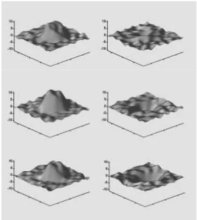
The spatial CSF flow profile in a 20x23-pixel region for one male subject (25 years of age) was shown excerpted from the phase-contrast velocity maps (Fig.1). Automatic outlining of the aqueduct of Sylvius using the pulsatility-based segmentation algorithm was found to be successful with little difficulty. Operator dependency was thus obviously absent.
The CSF production rate measured using the automatic analysis algorithm from the nineteen subjects was 332±137 ul/min. The nine subjects underwent two imaging examinations showed averaged CSF production rates of 292±79 ul/min at ~1700h and 344±179 ul/min at ~2230h, respectively. These estimation values were in good agreement with the CSF production rate of 347~370 ul/min reported from ventriculolumbar perfusion measurements (6). Figure 2 plotted the time-dependent variations in the meausred CSF production rates for all nine subjects. Paired student t-test showed that there was no significant diurnal difference in the CSF prduction rate (p = 0.16).
The CSF production rates measured from healthy young adults in our study are slightly less than, but in good agreement with the value of 347~370 ul/min reported from ventriculolumbar perfusion measurements (6). The slight discrepancy is within reasonable expectation in that measurement of the net CSF outflow through the aqueduct of Sylvius only registered CSF secretion in the lateral and third ventricles, while CSF produced from choroid plexus in the fourth ventricle may not be taken into account. Previous studies employing phase-contrast MR technique have reported values much larger than ours (3,4). We anticipate that these previous investigations may be prone to resolution imprecision plus operator-related inaccuracy, compared with the high temporal and spatial resolution in addition to the sophisticated image analysis software used in our study. Further studies performed by other independent investigators are needed to resolve the disagreement between MR-based measurements of the CSF production rate.
Using our high resolution approach, we re-examine the circadian variations of the CSF production for nine healthy adults. Contrary to previous reports (3,4), however, we find no fixed pattern of diurnal changes between 1700h and 2230h. Paired student t-test for the data in Fig.5 shows the diurnal difference to be statistically insignificant (p = 0.16). In addition to the issue of lower resolution employed in these previous reports as stated above (3,4), it has also been pointed out that the reported huge diurnal change (difference by a factor of 3) in human CSF production (3) was inconsistent with the very small variations observed experimentally in a very large number of animals. On the other hand, the intra-individual change shown in our study (5 increasing and 4 decreasing from 1700h to 2230h, Fig.2) implies that a measurement of the CSF production rate using one single imaging examination may be subject to extrapolation error, because production of CSF is likely to be affected by many other physiological factors such as the
blood pressure. Such phenomena point out yet another intrinsic difficulty in estimating CSF production, that is, the rate of production may itself be highly variable in a day, even if an optimized acquisition and analysis protocol can be employed. Another study aiming for a larger population with rigorous control of the physiological status is currently underway.
In conclusion, for quantitative studies on the physiological and pathological mechanism of human CSF secretion, it is important to establish a reliable procedure to measure the CSF production rate. Our results show that two dimensional cine phase-contrast MR imaging can be used for estimation of the CSF production rate, if objective analysis can be performed on images with sufficiently high temporal and spatial resolution. Using our approach we report CSF production rates in good agreement with literature values obtained using invasive ventriculolumbar perfusion measurements, and that no statistically significant circadian variations have been found. Phase-contrast MR imaging is an effective technique that can potentially help resolving the unanswered issues in human CSF secretion as well as in its relation to intracranial pathophysiology.
四、 計畫成果自評
Our efforts spent in this project have created results substantially greater than that mentioned in this brief report. Overall, the two-year project has generated two journal papers published in the prestigious Radiology (7) and Magnetic Resonance in Medicine (8), one journal article submitted to American
Journal of Radiology, two journal articles
under preparation, plus more than twenty international and domestic conference papers. Achievements from this project have raised the attention of other domestic medical centers, including Taipei Veteran General Hospital who approached us aggressively for mutual cooperation. In short, we have
confidence that our results from this project should benefit both the medical centers as well as the patients suffering from CSF-related diseases such as hydrocephalus. 五、參考文獻
1. Yasuda T et al. Measurement of cerebrospinal fluid output through external ventricular drainage in one hundred infants and children: correlation with cerebrospinal fluid production.
Pediatr Neurosurg 2002;36:22-28.
2. Silverberg GD et al. The cerebrospinal fluid production rate is reduced in dementia of the Alzheimer's type.
Neurology 2001;57:1763-1766.
3. Nilsson C et al. Circadian variation in human cerebrospinal fluid production measured by magnetic resonance imaging.
Am J Physiol 1992;262:R20-R24.
4. Nilsson C et al. The nocturnal increase in human cerebrospinal fluid production is inhibited by a beta 1-receptor antagonist.
Am J Physiol 1994;267:R1445-R1448.
5. Alperin N et al. PUBS: Pulsatility-based segmentation of lumens conducting non-steady flow. Magn Reson Med 2003; 49:934-944.
6. Rubin RC et al. Cerebrospinal fluid perfusion for central nervous system neoplasms. Neurology 1966;16:680-692. 7. Huang TY et al. Supratentorial
cerebrospinal fluid production rate in healthy adults: quantification with two-dimensional cine phase-contrast MR imaging with high temporal and spatial resolution. Radiology 2004;233:603-608. 8. Lin YR et al. Comparison of arterial spin
labeling and first-pass dynamic contrast-enhanced MR imaging in the assessment of pulmonary perfusion in human: the inflow spin-tracer saturation effect. Magn Reson Med, 2004, accepted.
六、圖表
Figure 1. Graphs demonstrate CSF hydrodynamic analysis in a 25-year-old healthy male subject. Spatial profiles of the CSF velocity show velocity distribution at six specific cardiac phases. Positive velocity stands for flow in the craniocaudal direction.
15~19H 20~24H 0 100 200 300 400 500 600 700 Measurement period C S F pr oduc ti on r a te (u l/m in)
Figure 2. Plot shows the CSF production rates measured from 9 healthy subjects at two prearranged time periods (left column: 1500~1900h; and right column: 2100~2400h). The plot shows no statistical evidence of systematic trends (p = 0.16) in the diurnal variation of CSF production as reported in the literature.