A Pilot Learning/Training/Teaching System of Mammography Screening
for Radiologists
Mann-Li Chang, Chien-Peng Chuang, Tai-Long Kuo
Department of Industrial Education, National Normal University, Taipei, Taiwan, R.O.C.
Ching-Chi Wu
Bureau of Education, Taipei City Government, Taiwan, R.O.C.
Giu-Cheng Hsu
Department of Radiology, Tri-Service General Hospital and National Defense Medical Center
Taipei, Taiwan, R.O.C.
Chia-Pei Huang, Yang Lang Chang
Department of Electrical Engineering National Taipei University of Technology, Taipei, Taiwan
San-Kan Lee
Yuan-Shan and Suao Veterans Hospitals, Yilan, Taiwan, R.O.C.
Abstract
Mammography is by far is the only effective means to be able to detect breast cancer in early stage. It has been widely used as a screening procedure to provide radiologists with a second opinion and further help radiologists improve their diagnosis. An experienced and well-trained radiologist in mammography can significantly reduce fatality rate in breast cancer by 20-30%. As a result, how to effectively use mammography to detect early breast cancer becomes increasingly important in Twomen’s health screening
procedureT. In order to meet this need, this paper
presents a learning system that can be used to train and teach radiologists to improve their diagnosis. It adopts Microsoft Visual Studio Net 2003 to develop a new software viewing tool, called ImageViewer that allows radiologists to self learn and teach themselves through biopsy-proven mammograms in a well-designed data base via easily used commands such as zoom-in, zoom-out, contrast enhancement, filtering in a friendly
environment so that radiologists can manipulate functions provided by the ImageViewer at their discretion to increase their experience and further improve their diagnosis.
1.
Introduction
Breast cancer is a second leading cause of cancer in American women. Although breast cancer can be fatal, it has been reported by many study groups that its survival rate can be very high if radiologists can detect the cancer at very early stage. Therefore, early detection and treatment plays a key and critical role in increasing the survival rate. The mammography screening has been recommended as the most effective method for breast cancer early detection. A screening program generally involves a huge number of mammograms, which require a limited number of radiologists for examinations. As a result, misdiagnosis often occurs due to human errors and visual fatigue. To address these issues, two
approaches are usually taken. One is to train more radiologists ready for the job. The other is to develop a Computer Aided Diagnostic (CAD) system to assist radiologists in providing a second opinion.
According to the randomized controlled trail: RCT mammography in breast cancer detection has been widely considered to be very effective via. Due to high-rise on incident rates the American Cancer Society has recommended that all women between the ages of 40 and 49 should get their mammograms every two years and every year thereafter. In the Northern America and Europe mammography has become a standard screening procedure for women with high risks and women whose ages are over 50. In particular, since 1995, the Department of Health in Japan has been pouring government funding to support research and conduct studies in breast cancer and has also implemented the same policy which also recommends that women with ages older than 50 should have annual mammography screening and physical examination. [1]
Like western world, the incident rate on breast cancer has grown rapidly in recent years in Taiwan. In the past 20 years, this number has doubled and tripled, and the tendency has not been slowed down, rather the number is continuously growing. [6] In order to cope with this increase many hospitals have included mammography screening as a routine check-up in women’s physical examination. When the mammography is used as a screening method to detect breast cancer, radiologists are often looking for small, low-contrast objects in breast images which are likely microcalcifications or masses related to cancer, particularly small tumors in dense breasts. Statistics have demonstrated that 70% of cases found by the mammography screening turned out to be suspicious lesions and required for further follow-up. Despite that not all small objects detected by the mammography are breast carcinomas, clinical studies
have shown that the mammography remains the only effective means in early detection of breast cancer. [8]
Bureau of Health Promotion Department of Health in Taiwan began its project of mammography screening in July 2002 for women with ages between 50-69 and received very good results. As a consequence, it decided in July 2004 to make free offer of mammography screening to women whose ages are from 50 to 69 every other year. Based on the finding from the survey conducted by Bureau of Health Promotion Department of Health for more than 80 hospitals that participated this project, the image quality of mammograms is not up to expectation. If the standard set by American College of Radiology is applied, many hospitals would be disqualified to perform mammography screening. [2]
Most importantly, due to poor image quality of mammograms, the ability of radiologists’ diagnosis can be significantly impaired. Therefore, misreading and poor diagnostic accuracy occur in many cases of suspicious abnormality. In order to resolve this issue, the work presented in this paper is to develop a system with two goals in mind. One is to provide radiologists with learning and training environments from which they can self improve their diagnosis experience. As indicated, due to shortage of experienced radiologists in mammography many radiologists must learn their skills and knowledge from various sources such as taking classes. However, due to nature in their job, one of most effective and easy ways to accomplish this goal is to provide radiologists with easy access and learning tools, which allow them to do anytime and any place whenever they would like to do it. Our system is developed to meet such need. The other is to provide a vehicle of conducting survey for the purpose of teaching and education and a forum of exchanging opinions among radiologists for the purpose of research.
2. Materials and Methods
According to reports by the American College of Radiology and its 2003 published Breast Imaging Reporting And Data Systems (BI-RADS), the classification of mammography screening can be categorized into 6 categories, Category 0: No findings, Category 1: Negative, Category 2: Benign Finding, Category 3: Probably Benign Finding, Category 4: Suspicious Abnormality, Category 5: Highly Suggestive of Malignancy. This 6-category categorization described below will be used as a guide to develop our system.
[3-5]
The system to be developed comprises of three component modules, a software module, called ImageViewer which includes many functions to allow users for manipulation of mammograms, a computer-aided diagnostic (CAD) module which enables users to learn and educate themselves via a provided CAD system through custom-designed data bases and tele-mammography module which enables users to communicate and consult at remote sites via internet. Each of these three modules can be developed separately and jointly used for various applications. This paper only presents the preliminary report of the first module, ImageViewer in our currently developed system. It is designed based on the Microsoft Visual Studio.NET 2003 coupled with the BI-RADS guideline to provide an environment with which radiologists can learn various cases from a data base which can be either obtained from the public domain or selected from well-documented cases.
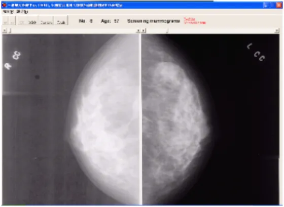
The current system, ImageViewer is basically a software-driven tool. It allows users to perform various functions such as zoom-in, zoom-out, contrast adjustment, Region of Interest (ROI) finding, position adjustment of mammograms, etc. via maneuvering a mouse. Fig. 1 provides an illustrative example to
demonstrate some of its functionalities. Fig. 1(a) shows two views of left and right breasts brought up from the database. Fig. 1(b-d) shows the zoom-in view, ROI view and contrast enhancement view of the breast image at the left of Fig. 1(a) respectively.
(a) Two views of left and right breasts
(b) Zoom-in view
(d) View of contrast enhancement
Figure 1. Illustration of ImageViewer
3. Results
In order to justify its utility of our developed ImageViewer, it has recently been used for certification of radiologists in mammography where several requirements must be satisfied. First of all, a radiologist to be certified must read mammograms at least for 240 cases within most recent 6 months. Second of all, he/she must read mammograms at least 1000 cases within most recent two years. However, due to various reasons, radiologists may go aboard for trips or studies or small hospitals that radiologists work for may not have sufficient mammograms for radiologists to read. Under circumstances, the requirements for 6-month and two-year mammogram reading cannot be met. On way to mitigate this dilemma is to use the ImageViewer to train themselves by reading mammograms custom-designed and collected for this purpose so as to increase their experience in clinical diagnosis. In the mean time, the ImageViewer can facilitate radiologists to be familiar with the guideline provided by the BI-RADS. [9]
In the initial phase, 77 radiologists who request to be certified were provided with 12 custom-designed cases for examination. Two scenarios were conducted. The first scenario was conducted twice and required the participants to only use a given set of magnified views to answer questions according to the BI-RADS’s guideline.
The second scenario was provided with the ImageViewer to examine cases of mammograms. Since the ImageViewer offers many functionalities that allow the participants to make various maneuvers at their discretion to answer the questions, it was expected that the diagnosis accuracy of the second scenario was 79% on average which was significantly better than 61% and 63% using the first scenario, while the two results using the same first scenario, 61% and 63% did not change much. This experiment demonstrated two key improvements of using the ImageViewer over the traditional method. It was clear evidence that the ImageViewer provides great benefits to radiologists who can take advantage of the software to improve their diagnosis. This was witnessed by the fact that the participants could not improve their diagnosis accuracy very much using the same given set of magnified views very much even if they were another chance to do it again and the benefit gained from repetitions was very limited. The second improvement is the versatility of the ImageViewer, which can be very flexible to be used by radiologists without time and location constraints. Additionally, the database will be also updated and custom-designed to meet different criteria and needs.
Finally, two concluding remarks are noteworthy. 1. In the current developed certification system, the
criteria to measure accuracy of diagnosis are based on the six categories recommended by the BI-RADS. The ground truth will be used to verify participants’ reading skills on the mammograms provided by the program authority.
2. Due to unavailability of digital mammograms this time, all the mammograms to be used for the test were digitized to 0.05mm (or 50μm) per pixel. The ImageViewer allows participants to adapt their viewing machines up to the image resolution of 0.05mm/pixel. It is believed that this year may be the last year to use digitized mammograms. In the
future, digital mammograms will be used by this program.
4. Conclusions and Future work
Despite the fact that mammography provides an effective screening method to detect breast cancer. It has been shown that it can be effective in detection of microcalcifications, but may not effective in detection of masses. In this case, the ultrasound must be used in conjunction with mammography. [7]
Due to differences in the breast structure such as size, density, shape and a variety of reasons such as, giving a birth, breast feeding, eating habits, breasts appear in many different forms and no gold standard for reading mammograms. Furthermore, there are no clear mammographic differences for radiologists to discriminate malignant and benign lesions, which make extremely difficult for them in their diagnosis. However, the accuracy of diagnosis in mammography can be improved if radiologists are well-trained by their experience or learning and studying more cases. The ImageViewer is developed to help less-experienced radiologists learn and teach themselves by working on many cases from data bases to accumulate their knowledge and increase their experience.
The mammography in Taiwan is still in its infancy. Many resources have not been allocated to implement the mammography screening as a routine procedure. One of most important issues is the shortage of experienced radiologists. In order to correct this problem, one most effective way is to train new radiologists through a learning process that is well-designed by experienced radiologists in a form of teaching classes or offering short courses. Our proposed ImageViewer can serve this purpose and provide access to users who can self learn and self teach themselves using their own time at free of locations.
The current ImageViewer is a preliminary pilot system that is developed to work on off-line to evaluate its efficacy and utility for clinical value. Several improvements are currently being investigated. Due to enormous image files, the image transmission is a major issue if the ImageViewer is implemented for the Internet use. The data storage and transmission speed are key issues that must be resolved. Second, due to filmless requirement for radiology department, the Picture Archiving and Communication Systems (PACS) has been used for radiologists to make diagnosis and writing reports. The ImageViewer has great potential integrated with the PACS. Third, it can use in conjunction with a CAD system that can be used as a teacher while the ImageViewer is used as a platform that allows radiologists to interact with the CAD-based teacher. Finally, the ImageViewer can be implemented as a vehicle for tele-mammography to make tele-consultation possible.
References
T [1]HUhttp://www1.cgh.org.tw/content/healthy/magazin e/medi/01-04_04.htmUHT,T T“乳房X光檢查”, 輻射防護 委員會.[2] HTUhttp://www.imaging.com.tw/center15.htmUTH, “乳房
X光可疑異常疑似癌症誤判率七成八”, June 7 2004聯合報.
[3] C. Zhou, H.P. Chan, N. Petrick, M.A. Helvie, M.M. Goodsitt, B. Sahiner and L.M. Hadjiiski, “Computerized image analysis: Estimation of breast density on mammograms,” Med. Phys, vol. 2, no. 6, pp. 1056–1069, 2001.
[4] K. Bovisand S. Singh, “Classification of mammographic breast density using a combined classifier paradigm,” in Int. Work. On Dig. Mammography, 2002, pp. 177–180.
Kegelmeyer, “The digital database for screening mammography,” in Int. Work. On Dig. Mammography, June 2000. [6] 李三剛, “乳房攝影檢查於乳癌篩檢的重要性與現 況”, 95年度防癌防治「蘭陽護波月」活動 [7] 李三剛、李覃、劉自嘉, “乳癌病例女性親屬的早 期乳癌偵測:臨床乳房檢查、乳房攝影與乳房超音波 的比較”, 世界醫用超音波大會2000. [8] 許居誠, “乳房X光攝影 偵測細微鈣化利器”, 中 華民國癌症希望協會, June 2004. [9] 許居誠, “乳房X光攝影醫療機構認證制度”, 中華 民國放射線醫學會舉辦「95年度乳房X光攝影品質提 升教育訓練課程」, May 2005.