利用蛋白質分析法比較氣喘病人的CD4+T淋巴球在穩定控制期與不穩定期間的差異; Proteomic Analysis of CD4+ T-lymphocytes in Patients with Asthma between Controlled and Uncontrolled Level
27
0
0
全文
(2) 摘要 背景:氣喘屬慢性發炎疾病,多數研究焦點放在引發發炎的機制。在氣喘病因學中,T-淋巴球衍生 的細胞激素被廣為討論。在後基因領域中,蛋白質分析技術快速發展且成為基因領域裡重要的輔助 工具。我們利用蛋白質分析法中兩個重要的技術:二維式膠片電泳分析法與蛋白質身份鑑定質譜分 析,來比較氣喘病人從不穩定期到穩定控制期時 T-淋巴球蛋白質間的差異。 材料與方法:對連續 6 位到院表現為不穩定氣喘患者抽血並萃取到 CD4+ T-淋巴球,患者在三個月 內經藥物控制為穩定性氣喘,再次抽血取得 CD4+ T-淋巴球。接著利用二維式膠片電泳分析法比較 不穩定期氣喘與穩定控制期氣喘時 T-淋巴球蛋白質間的差異。對於在膠片上有明顯差異的小點, 我們再取出並利用蛋白質身份鑑定質譜儀分析與比對資料庫來確定為何種蛋白質。 結果:氣喘患者體內的 CD4+ T-淋巴球有超過一百個以上的小點顯現在二維式膠片上。這些小點經 由考馬斯亮藍染色液染色後,兩組患者的小點分佈呈現類似情況,但是其中有 13 個小點卻出現有 意義的差異表現。若以從不穩定控制組到穩定控制組來看,其中有 6 種蛋白質減少而 7 種蛋白質增 加。 結論:經由蛋白質分析法的應用,確實讓我們發現在氣喘患者體內 CD4+ T-淋巴球在穩定控制期與 不穩定控制期間的差異,我們需要更多數量與不同範疇的研究來確認何者為氣喘發作的標記或作為 治療不穩定控制的方法。 關鍵字:氣喘、T-淋巴球、蛋白質分析法. i.
(3) Abstract Background. As a chronic inflammatory disease, much of the research related to asthma has focused. on proinflammatory mechanisms. T-lymphocyte (T-LC)-derived cytokines have been implicated in asthmatic pathogenesis. Proteomic technology has rapidly developed in the postgenomic era, and it is now widely accepted as a complementary technology to genetic profiling. We investigated the changes of proteins in T-LC of asthmatic patients from the uncontrolled to controlled level by using standard proteome technology: two-dimensional polyacrylamide gel electrophoresis (2D-PAGE), liquid chromatography/mass spectrometry (LC/MS), and a database search.. Methods. The proteins of CD4+ T-LC were isolated from the whole blood of six asthmatic patients. with uncontrolled to controlled levels over three months. 2D-PAGE was performed and coomassie blue stained protein spots were comparatively analyzed between the uncontrolled and controlled groups using an image analyzer. Some differentially expressed spots were identified by LC-MS/MS and database search.. Results. More than 100 spots were identified in the 2D-PAGE gels from the CD4+ T-LC of the. asthmatic patients. The general distribution pattern of the spots in the Coomassie blue-stained gel was similar in both groups, and 13 proteins showed different intensity, suggesting differential expression. Six protein spots in the CD4+ T-LC of the uncontrolled asthmatic patients were increased and 7 spots were decreased compared to those of the controlled subjects.. Conclusions. The proteomic examination of the CD4+ T-LC revealed some differentially expressed. proteins in the uncontrolled and controlled asthmatic patients. The possibility of using the differentially expressed proteins as important biomarkers and therapeutic targets in uncontrolled asthmatic patients warrants further study.. ii.
(4) Keywords: asthma, T-lymphocyte, proteomic analysis. iii.
(5) Acknowledgements This study was supported by grants from the Institutional Review Board (IRB) of St. Martin De Porres Hospital.. We thank the medical staffs and primary care doctors at Division of Pulmonary and Critical Care Medicine, Department of Internal Medicine, St. Martin De Porres Hospital. We thank the study staff (Chia-Guan Chen, and Mau-Lin Song) from Institute of Biochemical Science at National Chia-Yi university for data handling.. iv.
(6) 目錄 第一章. 前言 Introduction. 頁1. 第一節 研究背景 Background. 頁1. 第二節 研究目的 Purpose. 頁3. 第二章 研究方法 Methods. 頁4. 第一節 研究材料 Materials. 頁4. 第二節 研究設計 Design. 頁6. 第三節 統計方法 Statistic Methods. 頁8. 第三章 研究結果. Results. 頁9. 第四章 結論 Conclusion 第一節 討論. 頁 10. Discussion. 頁 10. 第二節 研究限制 Limitations. 頁 14. 第三節 建議 Suggestions. 頁 15. 表一. 頁 16. 表二. 頁 17. 圖一. 頁 18. 參考文獻 References. 頁 19. v.
(7) Chapter 1 Introduction 1.1. Background The recognition of asthma as a chronic inflammatory disease in the 1980s led to new paradigms for asthma pathogenesis and treatment. Asthma is characterized by recurrent dyspnea, coughing, wheezing, and reversible airway obstruction mediated by airway inflammation. 1-3. in which a variety of cell types,. including eosinophils 4, mast cells 5, and T lymphocytes 6 contribute to a complex pathologic process that causes airway remodelling and ultimately leads to compromised lung function. It has been estimated that 5% to 10% of patients with asthma have severe disease that is not effectively controlled by typical therapies, and these patients are at high risk of asthma-related death 7. However, there is still little information on the pathophysiological mechanisms responsible for uncontrolled asthma. It is well known that T-lymphocytes (T-LC) play a critical role in the initiation, progression and persistence of allergic disease, including asthma 8. The critical role of TH2 cells in asthma is also now widely accepted 9. Natural killer T (NKT) cells are also believed to play a role in allergen sensitization and pathologic states in asthma. 10. . Recently, advances have been made in defining mechanisms that control inflammation and. induce immune tolerance to specific antigens. Subsets of CD4+ cells known as T regulatory cells still play an important role in directing these processes. 11. , and recent experiments have begun to define crucial. molecular and signalling pathways. However, the immunological basis of this disease is still controversial. To understand the pathophysiologic mechanisms of disease, it is important to know which genes, gene transcripts, proteins, and metabolites are specifically expressed in the disease. Nair et al. 12. stressed the. importance of the protein synthesis from the expression because the proteins produced by genes are ultimately responsible for most biological functions. Especially, because proteins undergo many post-translational modifications that affect their structures and functions, their ultimate phenotypes often differ from genetic information. Use of proteomic techniques to identify disease-specific protein biomarkers is a powerful tool for defining prognosis of disease and gaining deep insights into disease 1.
(8) mechanisms in which proteins play a major role. Protein profiling has often been performed by the classical two-dimensional sodium dodecyl sulfate polyacryamide gel electrophoresis (2D-PAGE) based on the densitometric quantification of proteins visualized using dyes on gel. After in-gel enzymatic digestion of the subject protein spots, the resulting peptides are subjected to liquid chromatography/ mass spectrometry (LC/MS). Several proteome profiles of bronchial asthmatic lungs have been reported; however, most of them were from animal studies 13,14.. 2.
(9) 1.2. Purpose The purpose of this study is to explore the differentially expressed proteins of the T-lymphocytes in patients with asthma between uncontrolled and controlled level with using 2D-PAGE and LC/MS.. 3.
(10) Chapter 2 Methods 2.1. Materials Six consecutive patients from 21 to 54 years of age (3 females, 3 males; mean age, 34 years; Table 1) with asthma were enrolled on presentation to the pulmonary medicine clinic at ST. Martin De Porres hospital, a regional hospital in central Taiwan from June through December 2008, which satisfied the American Thoracic Society criteria. 15. , and had asthmatic symptoms within 1 week before the first visit.. Uncontrolled asthma was diagnosed according to the Global Initiative for Asthma guidelines. 16. . Upon. enrolment, all asthmatic patients underwent pulmonary function test, blood test for eosinophil count, chest radiography, and some necessary examinations for ruling out other pulmonary diseases. All subjects were non-smokers, and those who suffered from heart disease, autoimmune disease, endocrine disease, chronic liver and renal disease, and cancer were excluded. We also excluded both viral and bacterial infection-induced acute asthma according to the following signs: (i) no significant elevation of serum antibodies for influenza A and B virus; parainfluenza 1, 2, and 3 virus; and respiratory syncytial virus; (ii) no fever elevation; (iii) no purulent sputum; and (iv) no increased peripheral neutrophils and serum C-reactive protein. Concomitant with the initial evaluation was the initiation of management with nebulized [beta]-agonists with or without inhaled or systemic corticosteroids, oral theophylline, nedocromil, or cromolyn for at least 1 month. A blood specimen was obtained from each patient, and a standardized questionnaire of an action plan to maintain asthma control was administered by a trained investigator. The subjects who suffered from airway infection within 4 weeks immediately after the examinations were also excluded. The characteristics of the subjects are detailed in Table 1. Written informed consent was obtained from each subject before the study, and the study protocol was approved by the Institutional Review Board (IRB) of St. Martin De Porres Hospital. For all participants, initial and subsequent therapies were ordered by the examining pulmonary medicine physicians. Three months later, these subjects had been free of symptoms of a respiratory infection and of any asthma exacerbation for at least the previous 4 weeks and had not used either inhaled 4.
(11) or systemic corticosteroids, nedocromil, or cromolyn for at least 1 month before the second evaluation. Data from these six patients that satisfied these criteria for controlled asthma were included in the analysis and blood specimens were obtained again for proteomics.. 5.
(12) 2.2. Design Heparinized peripheral venous from each subject was collected in a sterile vacuum tube as enrolled in uncontrolled asthma and subsequently controlled subjects three months later. Peripheral blood mononucleated cells were separated from 40 ml heparinized blood after washed and centrifuged by 40 ml of Histopaqu1-1077 solution (Sigma-Aldrich, Inc.) and 80 ml of normal saline. CD4+ TLC were independently purified from each blood sample by the methods of a T-cell-negative isolation procedure with using monoclonal antibody (MACS; Miltenyi Biotec; J&H Technology Co. Ltd.). Twelve samples of TLC from six persons were prepared using the processes described below. For protein extraction, the CD4+ TLC pellet of each person was washed and centrifuged with 12000 rpm, 10 min for three times to remove the serum. The cells were then treated with lysis buffer containing 7 M urea; 2 M thiourea; 4% CHAPS; and protease inhibitor. The mixture was then stored at -80℃ until analysis after protein concentration was determined using a commercial reagent (2-D Quant Kit, Amersham Biosciences, Cat. No. 80-6486-22). Samples containing approximately 280 μg of total protein were precipitated with 2-D Clean-Up Kit (Amersham Biosciences, Cat. No. 80-6484-51) and resuspended in 250μl rehydration buffer (7 M urea, 2 M thiourea, 4% CHAPS, 20 mM DTT, 1% IPG buffer, 0.002% Bromophenol Blue). The first-dimensional gel separation was carried out with 13 cm pH 3-10 IPG strips and iso-electric focusing (IEF) was performed using Ettan IPGPhor II (Amersham Biosciences) for 70 kVhr at 20℃. After IEF, strips were equilibrated for 15 min in 6 mmol/L urea, 2% sodium dodecyl sulfate, 50 mmol/L Tris-Cl (pH 8.8), and 20% glycerol containing 1% dithiothreitol, and then equilibrated again for 15 min in the same buffer containing 2.5% iodoacetamide. Equilibrated IPG strips were placed on top of vertical slabs of 12% polyacrylamide gels and run in a PROTEAN II xi Cell tank (Bio-Rad) at 35 mA per gel. The gels were visualized using Coomassie Brilliant Blue R250 staining. After staining, 2-D gels were scanned using Powerlook 1120 (UMAX, Fremont, CA, U.S.A). NanoLC-MS/MS analysis was performed on an integrated nanoLC-MS/MS system (QSTAR XL) 6.
(13) comprising a LC Packings NanoLC system with an autosampler, and a QSTAR XL Q-Tof mass spectrometer (Applied Biosystems) fitted with nano-LC sprayer. Online nanoESI-MS survey scan and data dependent acquisition of CID MS/MS were fully automated and synchronized with the nanoLC runs under the full software control of AnalystQS. After data acquisition, the individual MS/MS spectra acquired for each of the precursors within a single LC run were combined and output as a single Mascot-searchable peak list file. The peak list files were used to query the NCBI database using the Mascot program with the following parameters: peptide mass tolerance, 150 ppm; MS/MS ion mass tolerance, 0.15 Da; allowing up to one missed cleavage. Only significant hits as defined by Mascot probability analysis will be considered initially. In addition, a minimum total score of 20 comprising of at least a peptide match of ion score more than 20 was arbitrarily set as the threshold for acceptance.. 7.
(14) 2.3. Statistic methods The images of 2-D gels were analyzed and compared using ImageMasterTM2D Platinum version 5.0 (Amersham Biosciences). Analyses of spot-intensity calibration, spot detection, background abstraction, and matching of approximately 12 2D-PAGEs were performed using software (ProteomWeaver; Definiens; Munich, Germany). All selected spots were present in all gels. The differentially expressed spots were analyzed with using Mann-Whitney U test (SPSS, version 10.0.05; SPSS; Chicago, IL). Correction for multiple comparisons was done according to the false discovery rate (FDR) method and p values larger than the FDR α value of 0.05 that was calculated using the software were considered statistically nonsignificant.. 8.
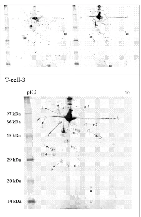
(15) Chapter 3 Results More than 100 spots were identified in the 2D-PAGE gels from the CD4+ TLC of the asthmatic patients from uncontrolled to controlled levels. The general distribution pattern of the spots in the coomassie blue-stained gels was similar in both groups (Fig. 1, top left and top right, percent matches: 95.91%). The spots in the area of pI 3 to 10, and molecular weight of 10 to 100 kd were analyzed by an image analysis program (ProteomWeaver). Protein spots of the uncontrolled and controlled asthmatic groups were compared, and 13 proteins showed different intensity, suggesting the differential expression. Among them, the intensities of 6 spots were significantly increased and the intensities of 7 spots were decreased in the uncontrolled group compared to the controlled group. The 13 selected spots of the 2D-PAGE of an asthmatic, non-smoking, 32-year-old woman are shown in Fig. 1, bottom, C. After destaining, extraction, and lysis with trypsin, the individual spots were identified by LC/MS and this was followed by the MASCOT database search algorithm. On the list of 13 candidate proteins for each spot, the final protein was determined by comprehensively considering the corresponding experimental isoelectric point, the molecular masses, the number of matched peptides, and the sequence coverage. Six up-regulated and 7 down-regulated proteins in the uncontrolled asthmatic group were identified (Table 1).. 9.
(16) Chapter 4 Conclusion 4.1. Discussion In 2007, Jeong et al.. 17. reported that proteomic examination of the peripheral T-lymphocytes revealed. some differentially expressed proteins in the asthmatic patients. However, this is the first proteomic approach using the CD4+ TLC of blood in asthmatic patients and defining the difference in severity. It is known that the asthmatic process that triggers the immune system can lead to excessive release of various cytokines and inflammatory mediators, which are produced by T-cells, infiltrated mononuclear cells, eosinophils, and local mast cells into the lung. Subsets of CD4+ cells known as T regulatory cells still play an important role in directing these processes. 11. , and recent experiments have begun to define. crucial molecular and signalling pathways. But the aetiology of airway hyperresponsiveness, a cardinal feature of asthma, has not been fully elucidated. Therefore, we performed proteomic analysis of the CD4+ TLC of controlled and uncontrolled asthmatic patients. In the study, we identified several proteins that were previously unknown or known to be associated with the pathogenesis of asthmatic exacerbation. Our study showed the increase of the heat shock protein (HSP)-70 and HSP-90 in CD4+ TLC from the uncontrolled asthmatic group to controlled level. HSP is a ubiquitous, abundant and conserved protein whose rate of synthesis is increased in response to cellular stress. It is often the target of humoral and T-cell-mediated immune responses to infection. 18. and could. protect tissue from the deleterious effects of numerous mediators, reactive oxygen species, or tumor necrosis factor-α. Several studies 19,20 have suggested that HSP is correlated with the severity of asthma exacerbation. It has also been proposed that the HSP-70 and HSP-90 chaperone associate with a range of cellular steroid receptor and may modulate the effectiveness of steroid in asthmatics 21. Tyrosine 3-monooxygenase/tryptophan 5-monooxygenase activation protein (YWHA) is highly conserved molecule that functions as intracellular adaptors in a variety of biological processes, such as signal transduction, cell cycle control, and apoptosis. It is also a HSP that protects cells against physiological stress as its new cellular function 22. Our study also showed the up-regulated expression of 10.
(17) YWHA from the uncontrolled asthma group to controlled level. Several cytoskeletal proteins were associated with the T-lymphocytes of asthma patients. Vimentin is a type III intermediate filament protein normally expressed in cells of mesenchymal origin and it attaches to the nucleus, endoplasmic reticulum, and mitochondria for the control of shape, motility and migration of mesenchymal cells. 23. . The increased expression of vimentin was observed in the uncontrolled asthma. patients in our study. The cytoskeletal change may illustrate the functional change in the T-lymphocytes of asthma exacerbation. β-actin, also a cytoskeletal protein, functions in cellular shape and anchorage where transmembrane glycoproteins link fibronectin in the extracellular matrix with actin microfilaments on the cytoplasmic side of the membrane. 24. . It is not surprising therefore that cellular proliferation,. activation, and/or differentiation induces up-regulation ofβ-actin expression in CD4+ TLC of the uncontrolled asthmatic group and subsequent cytoskeletal remodelling. Lung alveolar epithelial cells could produce fibrinogen when induced with proinflammatory mediators. 25. Fibrin is formed when thrombin, generated by the coagulation cascade factors and cofactors, cleaves. fibrinopeptides A and B from fibrinogen. The most powerful protein inactivator of surfactant, to the best of our knowledge, is fibrin. 26. and immunohistochemically detected fibrin was seen along the luminal. surface of the distal airways in a patient who died of status asthmatics.. 27. Fibrinogen β-chain was also. significantly increased in CD4+ TLC of the uncontrolled asthmatic group in this study. The enolase 1 is a key glycolytic enzyme with highly conserved amino acid sequences from microbial organism to mammal. 28. . This enzyme is ubiquitously expressed in nearly all cell types, including. epithelial cells, endothelial cells, and hematopoietic cells. 28. . Although enolase 1 is mainly a cytosolic. protein, it is also expressed on cell surfaces and functions as a plasminogen receptors was ever reported as an autoantigen associated with severe asthma. 29. 28. . The enolase 1. and upregulation of Enolase 1 has. been detected in many types of human cancer. Its activity also increased in the uncontrolled asthmatic group in our study. Peroxiredoxins are antioxidant enzymes involved in protein and lipid protection against oxidative 11.
(18) injury and in cellular signalling pathways regulating apoptosis. Peroxiredoxin 2 is a member of the mammalian peroxiredoxin family of thiol proteins that is important in antioxidant defences and redox signalling. The decreased expression of peroxiredoxin 2 was reported to be associated with enhanced sensitivity of Down syndrome neurons to reactive oxygen species. 30. . In our study, the up-regulation of. peroxiredoxin 2 was also observed in CD4+ TLC from the uncontrolled asthmatic patients to controlled level. Rho GTPases are molecular switches that regulate many essential cellular processes, including actin dynamics, gene transcription, cell-cycle progression and cell adhesion. To date most progress has been made in the cytoskeleton field, and several biochemical links have now been established between GTPases and the assembly of filamentous actin. Rho GDP dissociation inhibitor beta (Rho-GDI beta), an inhibitor of Rho GTPase, is primarily expressed by hematopoietic cells. Rho-GDI beta was also observed to be down-regulated in interleukin-4(IL-4) treated T cells and IL-4 regulates the subsequent stages of T helper 2 cell mediated disease, such as allergies 31. Our study also demonstrated that its activity increased in CD4+ TLC from the uncontrolled asthmatic patients to controlled level. Nuclear DNA helicase II (NDH II) belongs to the DEXH helicase superfamily and is able to unwind both double-stranded DNA and RNA in the presence of one of the four ribo- or deoxyribonucleoside triphospates. In addition to transcription, NDH II has multiple physiological functions, such as in RNA processing and transport, in DNA repair, and also in tumorigenesis 32. The helicase containing protein complexes may also facilitate the entry of transcriptional apparatus to the IL-4 responsive promoters 33. However, change of NDH II might modify the exacerbation of asthma in a still unknown manner. Tropomyosins (Tm) are a group of proteins with multiple isoforms found in both muscle and nonmuscle cells and in association with the troponin complex play a central role in the actin-linked calcium regulatory system of muscle contraction 34. The predominant colonic epithelial Tm isoform, hTm5, can induce both humoral (B-cells) and cellular (T-cells) response in patients with UC. Tm has also been described as relevant allergens 35. Change of Tm 3 in CD4+ TLC was also observed in this study, but the relation of this protein with the asthmatic exacerbation is not yet clear. Calreticulin, a major Ca2+ binding 12.
(19) (storage) chaperone in the endoplasmic reticulum, is a key component of the calreticulin/calnexin cycle which is responsible for the folding of newly synthesized proteins and glycoproteins by binding monoglucosylated carbohydrate and for quality control pathways in the endoplasmic reticulum. Immunogenic death show that apoptotic cells also displaying calreticulin on their surface are processed by dendritic cells that induce a specific T cell–mediated immune response against these apoptotic cells. The expression of calreticulin, which is also formed in vascular smooth muscle cells and vascular endothelial cells exposed to hyperglycaemic conditions, underlies the process of down-regulation of glucose transport in vascular cells under high-glucose conditions 36. The adaptive mechanism protects vascular cells against damaging effects of an uncontrolled influx of glucose in face of hyperglycemia. The expression of calreticulin precursor increased in CD4+ TLC from the uncontrolled asthma patients to controlled level in our study, the change could possibly interfered glucose metabolism to response to asthmatic exacerbation.. 13.
(20) 4.2. Limitations From the above, it is apparent that proteomic analysis reveals differences in protein expression that come from the change in level of the asthma patients. Both strategies have unique strengths and limitations. 2D-PAGE resolves thousands of proteins in a single run, and provides crucial molecular weight and isoelectric point information on intact protein; however, identification of some proteins may be hindered by their expression in various forms and the data may be skewed toward the more abundant proteins in the sample. Otherwise, although we selected non-smokers and relatively young participants, it is known that depending on the different physiologic conditions, such as age, gender, physical activity and the disease status, 2D-PAGE can show different panels. 12. . Finally, whether the changes of such. proteins are pathognomonic markers of asthma or of a reactive phenomenon is questionable.. 14.
(21) 4.3. Suggestions In summary, we have used 2D-PAGE/LC/MS to examine the protein profiles and expression in CD4+ TLC from uncontrolled and controlled asthma patients. Identification of these proteins revealed significant increase in expression for eight proteins, whereas six showed a significant reduction. These differences provide novel information highlighting proteins that may be linked to the mechanism(s) that defines why the asthma patients develop acute deterioration. The possibility of using the differentially expressed proteins as important biomarkers and therapeutic targets in uncontrolled asthma patients warrants further study.. 15.
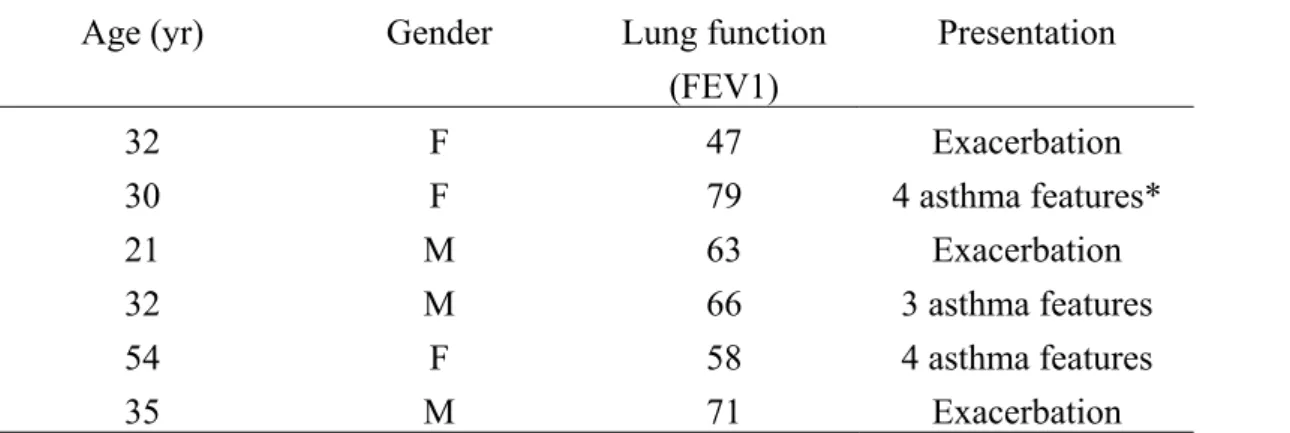
(22) Table 1. Demographics of Patients from Uncontrolled Asthmatics to Controlled Level Age (yr). Gender. Lung function (FEV1). Presentation. 32 30 21 32 54 35. F F M M F M. 47 79 63 66 58 71. Exacerbation 4 asthma features* Exacerbation 3 asthma features 4 asthma features Exacerbation. Definition of abbreviations: F = female; M = male; FEV1 = forced expiratory volume in the first second, expressed as the percentage of normal. *Asthma features: daytime symptoms, limitations of activities, nocturnal symptoms/awakening, need for reliever/rescue treatment or abnormal lung function.. 16.
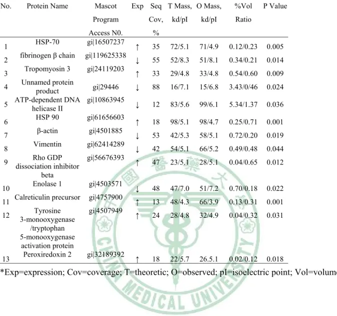
(23) Table 2. Identification of the Proteins with Altered Expression Levels in the Human CD4+ T-Lymphocytes from the Uncontrolled Asthma Patients to Controlled Level* No.. 1 2 3. Protein Name. Mascot. Exp Seq T Mass, O Mass,. Program. Cov,. Access N0. gi|16507237. %. HSP-70 fibrinogen β chain. gi|119625338. Tropomyosin 3. gi|24119203. Unnamed protein 4 gi|29446 product ATP-dependent DNA gi|10863945 5 helicase II HSP 90 gi|61656603 6 β-actin gi|4501885 7 Vimentin gi|62414289 8 Rho GDP gi|56676393 9 dissociation inhibitor beta Enolase 1 gi|4503571 10 Calreticulin precursor gi|4757900 11 Tyrosine gi|4507949 12 3-monooxygenase /tryptophan 5-monooxygenase activation protein Peroxiredoxin 2 gi|32189392 13. %Vol. kd/pI. kd/pI. Ratio. P Value. ↑. 35. 72/5.1. 71/4.9. 0.12/0.23. 0.005. ↓. 55. 52/8.3. 51/8.1. 0.34/0.21. 0.014. ↑. 33. 29/4.8. 33/4.8. 0.54/0.60. 0.009. ↓. 88. 16/7.1. 15/6.8. 3.43/0/46. 0.024. ↓. 12. 83/5.6. 99/6.1. 5.34/1.37. 0.036. ↑. 18. 98/5.1. 98/4.7. 0.25/0.71. 0.001. ↓. 53. 42/5.3. 58/5.1. 0.72/0.20. 0.019. ↓. 42. 54/5.1. 66/5.2. 0.49/0.48. 0.044. ↑. 47. 23/5.1. 28/5.1. 0.04/0.65. 0.012. ↓. 48. 47/7.0. 51/7.2. 0.70/0.18. 0.022. ↑. 13. 48/4.3. 66/3.9. 0.13/0.31. 0.001. ↑. 24. 28/4.8. 32/4.9. 0.04/0.32. 0.031. ↑. 18. 22/5.7. 26.5.1. 0.02/0.12. 0.018. *Exp=expression; Cov=coverage; T=theoretic; O=observed; pI=isoelectric point; Vol=volume.. 17.
(24) Figure 1. Representative 2D-PAGE gels of the CD4+ T-lymphocytes from uncontrolled asthma patients (top left) to controlled level (top right). Each image shows a similar expression pattern of spots between top left and top right. Thirteen selected and identified spots are shown by the numbered arrows in a 2D-PAGE gel (bottom) of an uncontrolled, asthmatic, 32-year-old woman.. 18.
(25) 1 2 3 4 5. 6 7. 8. 9 10 11 12 13. 14 15. 16 17. References Dunnill MS. The pathology of asthma, with special reference to changes in the bronchial mucosa. J Clin Pathol 1960; 13:27-33 Bousquet J, Chanez P, Lacoste JY, et al. Eosinophilic inflammation in asthma. N Engl J Med 1990; 323:1033-1039 Laitinen A, Laitinen LA. Cellular infiltrates in asthma and in chronic obstructive pulmonary disease. Am Rev Respir Dis 1991; 143:1159-1160; discussion 1161 Vignola AM, Chanez P, Campbell AM, et al. Airway inflammation in mild intermittent and in persistent asthma. Am J Respir Crit Care Med 1998; 157:403-409 Wardlaw AJ, Dunnette S, Gleich GJ, et al. Eosinophils and mast cells in bronchoalveolar lavage in subjects with mild asthma. Relationship to bronchial hyperreactivity. Am Rev Respir Dis 1988; 137:62-69 Robinson DS, Hamid Q, Ying S, et al. Predominant TH2-like bronchoalveolar T-lymphocyte population in atopic asthma. N Engl J Med 1992; 326:298-304 The ENFUMOSA cross-sectional European multicentre study of the clinical phenotype of chronic severe asthma. European Network for Understanding Mechanisms of Severe Asthma. Eur Respir J 2003; 22:470-477 Bian T, Yin KS, Jin SX, et al. Treatment of allergic airway inflammation and hyperresponsiveness by imiquimod modulating transcription factors T-bet and GATA-3. Chin Med J (Engl) 2006; 119:640-648 Busse WW, Lemanske RF, Jr. Asthma. N Engl J Med 2001; 344:350-362 Kim JO, Kim DH, Chang WS, et al. Asthma is induced by intranasal coadministration of allergen and natural killer T-cell ligand in a mouse model. J Allergy Clin Immunol 2004; 114:1332-1338 Seroogy CM, Gern JE. The role of T regulatory cells in asthma. J Allergy Clin Immunol 2005; 116:996-999 Nair KS, Jaleel A, Asmann YW, et al. Proteomic research: potential opportunities for clinical and physiological investigators. Am J Physiol Endocrinol Metab 2004; 286:E863-874 Roh GS, Shin Y, Seo SW, et al. Proteome analysis of differential protein expression in allergen-induced asthmatic mice lung after dexamethasone treatment. Proteomics 2004; 4:3318-3327 Fajardo I, Svensson L, Bucht A, et al. Increased levels of hypoxia-sensitive proteins in allergic airway inflammation. Am J Respir Crit Care Med 2004; 170:477-484 Standards for the diagnosis and care of patients with chronic obstructive pulmonary disease (COPD) and asthma. This official statement of the American Thoracic Society was adopted by the ATS Board of Directors, November 1986. Am Rev Respir Dis 1987; 136:225-244 Bateman ED, Hurd SS, Barnes PJ, et al. Global strategy for asthma management and prevention: GINA executive summary. Eur Respir J 2008; 31:143-178 Jeong HC, Lee SY, Lee EJ, et al. Proteomic analysis of peripheral T-lymphocytes in patients with asthma. Chest 2007; 132:489-496 19.
(26) 18 19 20 21 22. 23. 24. 25 26 27. 28 29 30 31. 32. 33 34 35. Shingai R, Maeda T, Onishi S, et al. Autoantibody against 70 kD heat shock protein in patients with autoimmune liver diseases. J Hepatol 1995; 23:382-390 Aron Y, Busson M, Polla BS, et al. Analysis of hsp70 gene polymorphism in allergic asthma. Allergy 1999; 54:165-170 Tong W, Luo W. Heat shock proteins' mRNA expression in asthma. Respirology 2000; 5:227-230 Qian X, Zhu Y, Xu W, et al. Glucocorticoid receptor and heat shock protein 90 in peripheral blood mononuclear cells from asthmatics. Chin Med J (Engl) 2001; 114:1051-1054 Yano M, Nakamuta S, Wu X, et al. A novel function of 14-3-3 protein: 14-3-3zeta is a heat-shock-related molecular chaperone that dissolves thermal-aggregated proteins. Mol Biol Cell 2006; 17:4769-4779 Katsumoto T, Mitsushima A, Kurimura T. The role of the vimentin intermediate filaments in rat 3Y1 cells elucidated by immunoelectron microscopy and computer-graphic reconstruction. Biol Cell 1990; 68:139-146 Blatti SP, Foster DN, Ranganathan G, et al. Induction of fibronectin gene transcription and mRNA is a primary response to growth-factor stimulation of AKR-2B cells. Proc Natl Acad Sci U S A 1988; 85:1119-1123 Haidaris PJ. Induction of fibrinogen biosynthesis and secretion from cultured pulmonary epithelial cells. Blood 1997; 89:873-882 Seeger W, Stohr G, Wolf HR, et al. Alteration of surfactant function due to protein leakage: special interaction with fibrin monomer. J Appl Physiol 1985; 58:326-338 Wagers SS, Norton RJ, Rinaldi LM, et al. Extravascular fibrin, plasminogen activator, plasminogen activator inhibitors, and airway hyperresponsiveness. J Clin Invest 2004; 114:104-111 Pancholi V. Multifunctional alpha-enolase: its role in diseases. Cell Mol Life Sci 2001; 58:902-920 Nahm DH, Lee KH, Shin JY, et al. Identification of alpha-enolase as an autoantigen associated with severe asthma. J Allergy Clin Immunol 2006; 118:376-381 Sanchez-Font MF, Sebastia J, Sanfeliu C, et al. Peroxiredoxin 2 (PRDX2), an antioxidant enzyme, is under-expressed in Down syndrome fetal brains. Cell Mol Life Sci 2003; 60:1513-1523 Rautajoki KJ, Marttila EM, Nyman TA, et al. Interleukin-4 inhibits caspase-3 by regulating several proteins in the Fas pathway during initial stages of human T helper 2 cell differentiation. Mol Cell Proteomics 2007; 6:238-251 Friedemann J, Grosse F, Zhang S. Nuclear DNA helicase II (RNA helicase A) interacts with Werner syndrome helicase and stimulates its exonuclease activity. J Biol Chem 2005; 280:31303-31313 Valineva T, Yang J, Silvennoinen O. Characterization of RNA helicase A as component of STAT6-dependent enhanceosome. Nucleic Acids Res 2006; 34:3938-3946 Smillie LB. Preparation and identification of alpha- and beta-tropomyosins. Methods Enzymol 1982; 85 Pt B:234-241 Aki T, Kodama T, Fujikawa A, et al. Immunochemical characterization of recombinant and native 20.
(27) 36. tropomyosins as a new allergen from the house dust mite, Dermatophagoides farinae. J Allergy Clin Immunol 1995; 96:74-83 Totary-Jain H, Naveh-Many T, Riahi Y, et al. Calreticulin destabilizes glucose transporter-1 mRNA in vascular endothelial and smooth muscle cells under high-glucose conditions. Circ Res 2005; 97:1001-1008. 21.
(28)
數據
相關文件
The purpose of this research is to explore the important and satisfaction analysis of experiential marketing in traditional bakery industry by using Importance-Performance and
By using Balanced Scorecard (BSC), the purpose of this study is to construct indicators of school management with Analytic Hierarchy Process (AHP) for L junior high school in
The purpose of this study was to explore the effects of learning organization culture on teachers’ study and teaching potency in Public Elementary Schools.. The research tool of
The main purpose of this study is to explore the status quo of the food quality and service quality for the quantity foodservice of the high-tech industry in Taiwan;
The main purpose of this study is to explore the work enthusiasm of the Primary School Teachers, the attitude of the enthusiasm and the effect of the enthusiasm.. In this
Evaluation of the association between t he characteristics of physicians and th eir practices with the availability of electronic health records.. Association with the availability
Reading Task 6: Genre Structure and Language Features. • Now let’s look at how language features (e.g. sentence patterns) are connected to the structure
Teachers may consider the school’s aims and conditions or even the language environment to select the most appropriate approach according to students’ need and ability; or develop
