行政院國家科學委員會補助專題研究計畫
基因體醫學國家型科技計畫
高惡性度胃 MALToma 之分子分類法:尋求可預測幽門桿菌依賴性腫瘤之基因 表現圖譜
Molecular Classification of High-Grade Gastric MALToma: identification of gene expression profile predicting H. pylori-dependence of the tumor
計畫類別:■ 個別型計畫 □ 整合型計畫 計畫編號:NSC-91-3112-B-002-009 執行期間:民國 91 年 5 月 1 日起至民國 94 年 4 月 30 日,共 3 年 計畫主持人:鄭安理教授 共同主持人:董馨蓮, 陳健尉, 張啟仁, 郭頌鑫 計畫參與人員:許惠貞, 高明, 曾怡欣 成果報告類型(依經費核定清單規定繳交):□精簡報告 ■完整報告 本成果報告包括以下應繳交之附件: □赴國外出差或研習心得報告一份 □赴大陸地區出差或研習心得報告一份 ■出席國際學術會議心得報告及發表之論文各一份 □國際合作研究計畫國外研究報告書一份 處理方式:除產學合作研究計畫、提升產業技術及人才培育研究計畫、列管計畫 及下列情形者外,得立即公開查詢 □涉及專利或其他智慧財產權,□一年□二年後可公開查詢 執行單位:
台大醫學院內科
中 華 民 國 92 年 7 月 23 日■ 成 果 報 告
□期中進度報告
目錄部分
內容 頁數
英文摘要 3-4
中文摘要 5-6
前言 7
研究目的 8
文獻探討 9-11
研究方法 12-14
結果與討論 15-22
計畫成果自評部份 23
參考文獻 24-27
附錄 (出席國際學術會議心得報告及發表之論文) 28-33
英文摘要 (ENGLISH ABSTRACT)
Keywords: High-grade gastric MALToma, HP-independence, BCL10, NF-B, CD86, and CD56 (+)NK cells
Mucosa-assocaited-lymphoid tissue lymphoma (MALToma) of stomach is the most common extranodal lymphomas of humans. Gastric MALToma is classified into high-grade (HG) and low-grade (LG) subtypes by histological criteria. LG gastric MALToma is characterized by its closely association with Helicobacter
pylori (HP) infection; and eradication of HP by antibiotics cures 50-70% of this
tumor. However, HG gastric MALToma, in contrast to its LG counterpart, was once believed to consist of highly-transformed cells, of which growth is independent of HP. We were the first group of investigators who demonstrated that nearly 60% of early-stage HG gastric MALToma remains HP-dependent and can be cured by HP eradication (J Clin Oncol 2001; 19:4245-51). Clinically, it is most
important to predict HP-independence state of freshly diagnosed HG gastric
MALToma, since the latter may progress rapidly if unresponsive to anti-HP therapy. However, histologic or molecular features, which predict HP-dependence of this tumor, remain elusive. We hypothesize that molecular mechanisms of
HP-dependence may be different between LG and HG gastric MALToma. For
example, genetic aberration such as t(11;18)(q21;q21), which results in chimeric protein API2-MALT1 and is closely associated with HP-independent state of LG gastric MALToma, dose not seem to exist in HG gastric MALToma.
This study aims to identify molecular and cellular markers, as well as gene expression profiles, which may help predict HP-dependence state of HG gastric MALToma. Further clarification of the biologic significance and function of novel
HP-dependence-relevant genes will be done.
In the first year of the project, we have already identified two molecular markers
and two immunologic cellular markers that are closely associated with
HP-independence state of HG gastric MALToma. We have demonstrated that
nuclear expression of BCL10 has a sensitivity of 85.7%, and a specificity of 100% to predict HP-independence of HG gastric MALToma. In addition, confocal immunofluorescence microscopy revealed that nuclear expression of NF-B
correlated well with the aberrant BCL-10 nuclear expression; and nuclear expression of NF-B alone predicts HP-independence with a sensitivity of 85.7% and a
specificity of 86.4%. We confirmed that t(11;18)(q21;q21) rarely occur in HG gastric MALToma. Therefore, BCL10 nuclear translocation appears to be a major independent event in predicting HP-independence of HG gastric MALToma. We
are currently working on clarification of the mechanism and biologic significance of BCL10 nuclear translocation in HP-independent HG gastric MALToma.
Two immunologic cellular markers were found to be relevant to
HP-independence state of HG gastric MALToma. We showed that non-expression
of CD86 of MALToma cells is a useful marker for predicting HP-independence of HG gastric MALToma. The expression of CD86 was detected in 9 (64%) of 14
HP-dependent high-grade gastric MALTomas but in none of 6 HP -independent
cases (p=0.010). We reason that loss of co-stimulatory markers may preclude tumor cell/reactive T-cell interaction and contribute to the transition to
HP-independent state of HG gastric MALToma. Finally, we discovered that
decreased infiltration of NK cells in tumor tissues is associated with
HP-independence of HG gastric MALToma. We found that HP -dependent HG
gastric MALToma contained significantly higher numbers of CD56 (+) NK cells than
HP -independent cases (2.7 ±1.2% versus 0.80±0.70%; p=0.005). CD56 (+) nature
killer (NK) cells are thought to suppress the growth of HP- related autoreactive and neoplastic B lymphoid cells of the stomach. These molecular and immunologic cellular markers will be immediately useful for the physicians to select the right modality of treatment for patients with HG gastric MALToma.
We have thus far collected 20 freshly frozen tissue samples of HG gastric MALToma, of which the HP-dependence state has already been verified by prospective clinical study. Microarray approaches to identify gene expression profiles of HP-dependent and HP-independent tumors are ongoing. Among 20 freshly frozen tissue samples of HG gastric MALToma, 16 samples containing microdissectable HG and LG MALToma cells are identified. Comparison of the expression profiles of the co-existing LG and HG counterparts of the same gastric MALToma patient is ongoing. These ongoing studies will help clarify whether the two components of MALToma may belong to two different clones, and genetic changes responsible for HG transformation may not be the same as those responsible for HP–independence transformation.
中文摘要
關鍵詞: 高惡性度胃黏膜相關性淋巴組織淋巴癌,幽門螺旋桿菌非依賴性,
BCL10,NF-B,CD86,和自然殺手細胞
胃黏膜相關性淋巴組織 (Mucosa-Associated Lymphoid Tissue)淋巴癌 是人類最常見之淋巴結外惡性淋巴癌。 胃MALT淋巴癌依其病理特徵分為低 惡性度和高惡性度。 低惡性度胃MALT淋巴癌已被證實與幽門螺旋桿菌(H. pylori)高度相關, 且臨床上以抗生素有效清除幽門螺旋桿菌(HP)後,可 治癒 50-70% 的病人。 相對於低惡性度胃MALT淋巴癌,高惡性度胃MALT 淋巴癌一向被認為是由高度轉型 (transformed) 的細胞組成,其生長不需依賴 幽門螺旋桿菌性 (HP-independence) 。 我們的研究團隊率先發現,早期的高 惡性度胃MALT淋巴癌,仍有60% 具HP依賴性,故在HP清除後可加以治癒 (J Clin Oncol 2001;19:4245-51)。但高惡性度胃MALT淋巴癌若對HP根除治 療無效,其淋巴癌可能會迅速惡化。 因此在臨床上,最好能在治療前預知該 腫瘤是否仍具HP依賴性,以期能做出最好的治療選擇。 然而到目前為止, 可用來預測高惡性度胃MALT淋巴癌是否仍具HP依賴性之相關病理或分子 表徵仍未能充份釐清。 我們假設HP依賴性之機轉在高惡性度胃MALT淋巴 癌及低惡性度胃MALT淋巴癌是不同的。譬如基因變化 t(11;18)(q21;q21)(造 成 API2-MALT1 雜合蛋白質),常見於HP非依賴性的低惡性度胃MALT淋巴 癌,但卻罕見於高惡性度胃MALT淋巴癌。 本研究目的即是希望能找出相關之分子、 細胞、或基因表現圖譜標記, 能幫助我們預測高惡性度胃MALT淋巴癌之HP依賴性,並進一步探討HP依賴 性相關基因之功能。 在第一年的研究中,我們已找到與高惡性度胃 MALT 淋巴癌之 HP 依賴性 有關的兩種分子及兩種免疫細胞標記。首先我們證實細胞核內 BCL10 表現,在 預測高惡性度胃 MALT 淋巴癌之 HP 非依賴性方面,其敏感性可達 85.7 %, 而特異性則高達百分之百。此外,共軛焦顯微鏡也證實 NF-B 常伴隨 BCL10 一起在核內表現。 同時,單獨細胞核內 NF-B 的表現,也可預測高惡性度胃 MALT 淋巴癌之 HP 非依賴性,其敏感性可達 85.7 %,而特異性則達 86.4%。 我們的實驗也證實 t(11;18)(q21;q21) 很少發生在高惡性度胃淋巴癌。 因此細 胞核內 BCL10 的轉移,似乎是預測高惡性度胃 MALT 淋巴癌之 HP 非依賴性中 的最重要獨立因子。 目前我們正積極探討 BCL10 細胞核轉移,在高惡性度胃 MALT 淋巴癌 HP 非依賴性中所扮演之角色、機制及生理意義。 我們也發現與高惡性度胃 MALT 淋巴癌之 HP 非依賴性相關的兩種免疫細 胞標記。我們發現,MALT 淋巴癌細胞若無 CD86 表現,是預測高惡性度胃 MALT 淋巴癌之 HP 非依賴性的重要標記。 在十四位 HP 依賴性高惡性度胃 MALT 淋巴癌病人中,有九位淋巴癌細胞中偵測到 CD86 表現。 而其他六位
HP 非依賴性高惡性度胃 MALT 淋巴癌病人,其淋巴癌細胞中則完全偵測不到 CD86 表現。 因此我們認為 HP 非依賴性高惡性度胃 MALT 淋巴胃之生長,可 能並不須淋巴癌細胞與 T 細胞之共同作用。 最後,我們也證實癌組織上自然 殺手細胞之浸潤減少,的確與 HP 非依賴性有關。 我們發現 HP 非依賴性之高 惡性度胃 MALT 淋巴癌比 HP 依賴性之高惡性度胃 MALT 淋巴癌,含有較多的 自然殺手細胞 (2.7 ±1.2% versus 0.80±0.70%; p=0.005)。 這些分子及免疫細胞 標記,可即刻幫助鄰床醫師選擇最適當的方式來治療高惡性度胃 MALT 淋巴癌 病人。 我們到目前為止已收集到先前參與前瞻性臨床實驗,並確定HP之依賴性 與否之20 位高惡性度胃MALT淋巴癌病人新鮮冷凍標本。 我們正著手進行 基因微陣列分析方法,去探討HP之依賴性與非依賴性可能有關的基因圖譜 。 在這些冷凍標本中,有 16 位病人之標本含有可微截之低惡性度及高惡性度 MALT淋巴癌。 最近我們也正進行比較分析同一病人之低惡性度及高惡性度 部份的基因圖譜,這些進行中的研究將有助於了解病人之高及低惡性度腫瘤 是否可能分別屬於兩個不同的clones,或是造成細胞轉型與HP非依賴性是分 屬不同的基因變異。
前言
(INTRODUCTION)Extranodal marginal zone B-cell lymphoma of mucosa-associated lymphoid tissue (MALT) has recently been recognized as a distinct entity of lymphoma, representing 8% of all non-Hodgkin’s lymphomas and more than half of extranodal lymphomas.1,2 MALToma of the stomach is the most common type of MALToma and is characterized by its close association with the infection of Helicobacter
pylori (HP).3-6 HP is found in gastric mucosa of nearly all instances of gastric MALToma. Compelling evidence suggests that most gastric MALTomas are the sequellae of chronic HP infection of the stomach. 3-6
Gastric MALToma is classified into HG and LG histologic subtypes. LG MALToma consists primarily of diffuse small and medium-size centrocyte-like cells and the characteristic “lymphoepithelial cells” lesions.7-8 HG MALToma contains foci of clusters or sheets of large blast cells or frank diffuse high-grade lymphoma a background of low-grade MALToma or foci of low-grade components.9-10 LG gastric MALToma is unique in that its growth depends critically on the presence of HP and possibly HP-specific T lymphocytes.11 Around 50-70% of early stage LG gastric MALToma regress completely after HP eradication.12-15 In contrast, HG gastric MALToma, of which the tumor cells were believed to be highly-transformed and thus grow independently of HP stimulation.16,17 Therefore, HG gastric MALToma has conventionally been treated by systemic chemotherapy.
Recently, we and other group of investigators have demonstrated that a substantial portion of early-stage high-grade gastric MALTomas remain
HP-dependent and can potentially be cured by HP eradication.18-20 Although our discovery has revolutionized the treatment of early-stage HG gastric MALToma, important questions remain. Since high-grade gastric MALToma, in contrast to its low-grade counterpart, may progress rapidly if an effective measure of tumor control does not occur, it is important that the HP–independent status of the tumor be disclosed before starting treatment. It is therefore the purpose of the current study to identify useful molecular or cellular markers, and the possible class predictor gene profiles for predicting HP-independence status of HG gastric MALToma.
研究目的 (SPEIFIC AIMS):
We hypothesize that molecular or cellular markers, and class predictor gene profiles distinguishing HP-dependent and HP–independent status of HG gastric MALToma may exist and the molecular mechanisms of HP-dependence may be different between LG and HG gastric MALToma.
Aims:
1. Identification of molecular and cellular markers, as well as gene expression profiles which may help predict HP-dependence state of high-grade gastric MALToma.
2. Identification of novel HP-dependence-relevant genes and clarification of their functions.
3. Comparisons of the gene expression profiles of the co-existing high-grade and low-grade MALToma, and clarification of their significance in HP-dependence.
文獻探討 (BACKGROUND)
Possible Mechanisms of HP-dependence of HG gastric MALToma
The development of gastric MALToma is a multistage process, which comprises antigen stimulation by HP and a series of genetic changes.21 It is believed that long-term antigen-driven cellular proliferation may cause accumulating genetic alterations. In chronic HP infection of the stomach, fully organized lymphoid tissue (MALT) with formation of lymphoid follicles develops in the gastric mucosa. This lymphoid tissue is thought to harbor the precursor cells of the MALToma. These precursor cells may acquire genetic abnormality and gradually transform into malignant lymphoma cells. In the early phases of this process, the proliferation response is partly dependent on functional interactions between B and T
lymphocytes.22 It has been demonstrated by in-vitro experiments that the growth and differentiation MALToma cells require CD-40 mediated signaling and Th-2 type cytokines.23,24 Current evidence supports the notion that tumor-infiltrating T cells were defective in both perforin-mediated cytotoxicity and Fas-mediated apoptosis, despite displaying normal Fas ligand expression.24,25 The dependence on T cells for growth of the malignant B-cell clones may explain the tendency of early-stage LG MALToma to remain localized and to regress after HP eradication. Expression of co-stimulatory molecules (CD40, CD80, CD86, and their ligands) and relevant cytokines has been demonstrated in gastric MALToma cells.26,27 The presence of co-stimulatory marker CD86 (B7.2) on lymphoma cells may promote T-cell-mediated neoplastic B cell proliferation. It is speculated that loss of co-stimulatory markers might preclude this interaction and thereby facilitate the transition into the
antigen-independent status of HG gastric MALToma.
Compared with its low-grade counterpart, the tumor tissues of HG MALToma contain a much higher amount of apoptotic lymphoma cells and cytotoxic T
lymphocytes.28 Animal studies using murine model have demonstrated that Th1 response and CD8+ activity were strongly inhibited in the presence of persistent gastric HP infection.29 Moreover, loss of HP-dependence may be associated with a change of immunological microenvironment., including a shift toward a Th1
response that enhances the activity of cytotoxic T lymphocytes (CTL).24 CD56(+) natural killer (NK) cells are frequently found infiltrating the low-grade gastric
MALToma tissues.28 These CD56(+) NK cells are thought to limit the autoreactive responses elicited by HP, and may contribute to the remission of lymphoma after eradication of the HP.30,31
Several genetic changes are closely correlated with HP–independent status of LG gastric MALToma. T(11;18) (q21;q21), resulting in the expression of a
chimeric transcript API2-MALT1, is one of the most important predictors of
HP-independence in LG gastric MALToma. However, t(11;18) (q21;q21) is rarely
found in HG gastric lymphomas.32-34 T (1;14)(p22;q32) is another genetic aberration implicated in the development of MALToma. T(1;14)(p22;q32) juxtaposes BCL10 of chromosome 1 to an immunogobulin gene locus of
chromosome 14, and results in strong expression of a truncated BCL10 protein in the nuclei and cytoplasm, in contrast to the weak cytoplasmic expression of BCL10 in normal germinal center B-cells.35,36 Its is noteworthy that t(1;14)(p22;q32) was detected in less than 5% of LG gastric MALToma, while moderate nuclear
expression of BCL10 was found in 30-40% of these tumors. Instead with unknown mechanisms, BCL10 nuclear expression was found more closely associated with genetic aberration t(11;18)(q21;q21), one of the conditions predictive of
HP-independent status in LG gastric MALToma.37
The mechanism and biologic significance of BCL10 nuclear expression in the lymphoma cells without BCL10 gene mutation are largely unknown. In T lymphocytes, BCL10 normally resides in the cytoplasm and specifically relays antigen-receptor-mediated signals to activate the NF-B.38
It is speculated that up-regulation of BCL10 may trigger a constitutive NF-B activation signal, and therefore contribute to antigen-independent growth and the progression of the gastric MALToma. We therefore reasoned that expression patterns of NF-B and BCL10 may be useful markers for predicting HP-independence of HG gastric MALToma. This speculation has been thoroughly tested in this study.
HG transformation of gastric MALToma
It is generally believed that LG MALToma cells may accumulate genetic abnormalities and gradually become independent on HP or T-cell stimulation for growth. Genetic events that may herald this change of HG transformation are believed to be similar to those recognized in nodal lymphoma and including mutations of p53, altered methylation status or homozygous deletions of p16, Bcl-6,
p27, and cyclin E. It is noteworthy that our recent report strongly argues that HG
transformation of gastric MALToma may not necessary lead to HP-independence.20 We hypothesize that genetic changes responsible for these two conditions are different.
HG MALToma showed more frequent p53 allele loss and mutation than its LG counterpart.39 More importantly, the majority of HG cases showed both p53 allele loss and mutation. In a recent study, low-grade gastric MALToma without p53 mutation tends to regress after HP eradication therapy.40 Bcl-6 rearrangement involving abnormality of chromosome 3q27 has been described in high-grade but
not in LG MALToma of the stomach, suggesting that Bcl-6 overexpression is
associated with HG transformation.41 Inactivation of the p16 gene, one of the major negative regulators of the early G1 phase of cell cycle, is almost exclusively found in HG MALToma, suggesting a role for p16 deletion in HG transformation.42
Expression of p27, a CDK inhibitor that blocks the progression from G1 to S phase of the cell cycle, is more prevalent in LG gastric MALToma; while cyclin E is more commonly expressed in the HG counterpart.43
One important aim of this study is to clarify if genetic changes responsible for HG transformation and HP-independence of MALToma cells may be different. We will examine the genetic expression profiles of the co-existing HG and LG MALToma, which may have differential response to HP eradication.
研究方法 (METHODS)
Immunohistochemistry and Confocal Laser Scanning Microscopy
Formalin fixed paraffin embedded sections cut at a thickness of three
micrometers were deparaffinized and rehydrated. After antigen retrieval by heat treatment, endogenous peroxidase activity was blocked by 3% H2O2.
Staining was performed using anti-BCL10 (1:10; polyclonal, Santa Cruz
Biotechnology) or anti-RelA or anti-CD56 antibody (NCAM 1B6, Novacastra), and anti-CD86 antibody (AF-141-NA, Rand D Systems, Abingdon, UK). (1:100; p65, Santa Cruz Biotechnology) following the manufacturer’s recommendations.
Sections were counterstained with Mayer hematoxyline. A semi-quantitative method was used to determine the level of expression of RelA. Reactive spleen and lymph nodes tissue sections were used as the control. A minimum of 1000 cells (normal and neoplastic) were counted for each single determination and reported as the percentage of CD56 (+) NK cells in total mononuclear cells.28 The relationship between HP-dependence, and the amount of infiltrating NK cells was analyzed by a nonparametric Mann- Whitney U test. The relationship between HP-dependence and positive CD86 markers was analyzed using the liner-by-liner association test.
For double-immunolabelling studies, FITC-labeled goat anti-rabbit or
Rhodamine -labeled donkey anti-goat IgG was incubated as secondary antibodies for 60 minutes at room temperature in the dark. The sections were further evaluated by a confocal laser scanning microscope (model TC-SP, Leica, Heidelberg, Germany) equipped with argon and argon-krypton laser sources.
Multiplex RT-PCR for the API2-MALT1 Fusion Transcript
Since t(11;18)(q21;q21), which results in chimeric API2-MALT1 fusion protein, is not only the most important predictor of HP-independent status but also closely associated with BCL10 nuclear expression in low-grade gastric MALT lymphoma, we examined the existence of this genetic aberration in our patients. Eight cases of low-grade gastric MALT lymphoma were also studied for comparison. Total cellular RNA was extracted from formalin-fixed paraffin-embedded tissues, using an Ambion RNA isolation kit (AMS Biotechnology, Oxon, England, United kingdom). Briefly, 2-3 pieces of 10-um paraffin sections were deparaffinized in xylene. The tissue was digested with proteinase K (Roche Diagnostics, Mannheim, Germany) for 1 hours at 45℃ and solubilized in a guanidinium-based buffer. RNA extracted from the paraffin-embedded tissues was analyzed for API2-MALT1 fusion using multiplex RT-PCR as described previously.34 RNA was subjected to first-round multiplex one-tube RT-PCR, then to second-round nested multiplex PCRs (three parallel;
Second PCR-A, Second PCR-B, and Second PCR-C) (Table 1 and 2). The final PCR products were run on 8% polyacrylamide gels and stained with ethidium bromide. The band size ranged from 80 to 179 bp. PCR for low-grade gastric MALT lymphoma samples known to possess API2-MALT1 fusion were used as positive control. Theβ-actin (190 bp) was amplified in parallel as an internal control. Where indicated, PCR products of the API2-MALT1 transcript were either directly sequenced or cloned into a vector (the TOPO TA Cloning kit®; Invitrogen, Paisely, UK) and sequenced with vector primers using dye-labeled terminators (BigDye Terminators, Applied Biosystems, Foster City, CA) and analyzed on a DNA sequencer (Model 310, Applied Biosystems).
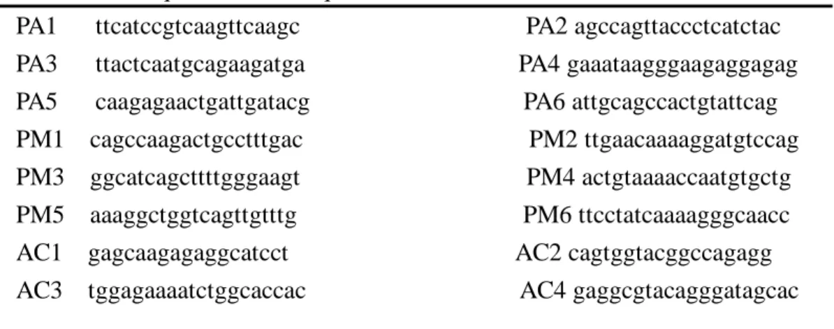
Table 1. Oliqonucleotide Sequence of Primers
PA1 ttcatccgtcaagttcaagc PA2 agccagttaccctcatctac PA3 ttactcaatgcagaagatga PA4 gaaataagggaagaggagag PA5 caagagaactgattgatacg PA6 attgcagccactgtattcag PM1 cagccaagactgcctttgac PM2 ttgaacaaaaggatgtccag PM3 ggcatcagcttttgggaagt PM4 actgtaaaaccaatgtgctg PM5 aaaggctggtcagttgtttg PM6 ttcctatcaaaagggcaacc AC1 gagcaagagaggcatcct AC2 cagtggtacggccagagg AC3 tggagaaaatctggcaccac AC4 gaggcgtacagggatagcac PA1–6, AC1, and AC3, sense primers; PM1–6, AC2, and AC4, antisense primers
Table 2. Size (bp) of the Second-Round Multiplex PCR Products
Second round API2 breakpoint MALT1 breakpoint
Multiplex PCR M541 M814 M1123 M1150
Second PCR-A A1203 122bp 147bp 135bp 108bp Second PCR-B A1446 94bp 119bp 107bp 80bp Second PCR-C A1701 112bp 137bp 125bp 98bp
DNA analysis: PCR, SSCP and direct sequence
1. PCR for primer:
1.1 Repair error phenotypes (RER)
1.2 Microsatellite primer and sequence are retrieved from Genomic Database (http: gdbwww.gdb.org)
2. t(1:14) (Bcl-10):
2.1 The full coding sequence of the Bcl-10 gene is amplified by 5 overlapping PCR, with a single reaction spanning the coding exon and with 2 reactions spanning each of remaining 2 coding exons.40
3. p5339:
GT strand, AGGGATACTATTCAGCCCGAGGTG AC strand, ACTGCCACTCCTTGCCCCATTC 4. c-Myc44: exon-1 and intro-1
5′-CGACTGGAACTTACAACACC-3′ 5′-CTGGCTCACACAGGCGAT-3′ 5. p1642:
Primer sequences is according to previous report. 35 6. FAS/CD9545:
The full coding region, comprising exon-intro junction, is amplified by PCR from genomic DNA using published primers.30
7. Bcl-6. The exon 1 and exon1-intron 1 boundary region sequence of the Bcl-6 gene (the primer sets are synthesized according to Migliazza, et al.46)
結果與討論 (RESULTS and DISCUSSION):
Part I: Identification of molecular and cellular markers, as well as gene expression profiles which may help predict HP-dependence state of high-grade gastric MALToma
Part Ia: Molecular and cellular markers
We have already identified 2 molecular markers and 2 immunologic cellular markers that are associated with HP-dependence state of HG gastric MALToma.
Part Ia1: Nuclear Expression of BCL10 or NF-B is Predictive of
HP-independent Status of Early-Stage HG Gastric MALToma
BCL10 was identified by its direct involvement in t(1;14)(p22;q32) of
MALToma. It normally resides in the cytoplasm to relay antigen-receptor-mediated signals to activate NF-B of T cells. Upregulation of BCL10 may trigger a
constitutive NF-B signal from the antigen receptor, and thereby contribute to
antigen-independent growth and progression of gastric MALToma. We have found that aberrant nuclear expression of BCL10 was detected in seven (87.5%) of 8
HP-independent high-grade gastric MALTomas and in none of 14 HP-dependent
cases (P<0.001). All seven patients with nuclear BCL10 expression had nuclear expression of NF-B, while only two of 15 patients without nuclear BCL10 expression did so (P=0.002) (Table 3 and Fig. 1). Furthermore, the nuclear
co-localization of RelA and BCL10 was confirmed by confocal immunofluorescence microscopy (Fig. 2). As a single variable, the frequency of nuclear expression of NF-B was also significantly higher in the HP-independent tumors than in the
HP-dependent tumors [7 of 8 (87.5%) versus 2 of 15(12.3%), P=0.002]. Nuclear
expression of BCL10 and NF-B, both have a sensitivity of 85.7%, and specificity of 100% and 86.4%, respectively, in predicting HP-independence of HG gastric
MALToma. Manuscripts describing these findings have been submitted for publication.
In LG gastric MALToma, BCl-10 nuclear localization is closely linked to the co-existent chromosomal aberration, t(11;18)(q21;q21). However, this latter genetic change has not been found in HG gastric MALToma. The biologic significance of BCL-10 nuclear localization will be further explored. In order to confirm the discrepant expression of t(11;18)(q21;q21) in HG and LG gastric MALToma, we have established a multiple RT-PCR assay to detect various API2-MALT1 fusion transcript using archival formalin-fixed, paraffin-embedded lymphoma tissues. Using this rapid and simple method, we could detect API2-MALT1 fusion transcript in 4 (50%)
of 8 HP-independent LG gastric MALToma cases. The API2-MALT1 fusion transcript was detected in only one (12.5%) of eight HP-independent cases, and in none of all HP-dependent cases (Fig. 3). We have confirmed that BCL10 nuclear translocation was independent of t(11;18)(q21;q21) in the majority of HG gastric MALToma cases.. Nuclear expression of BCL10 may also be detected in some LG gastric MALTomas without t(11;18)(q21;q21).47,48 These findings suggest that the direct interaction between BCL10 and API2-MALT1 fusion protein may not exist in most MALT lymphomas, and the molecular interaction and biologic consequence of nuclear translocation of BCL10 in gastric MALToma should be further investigated.
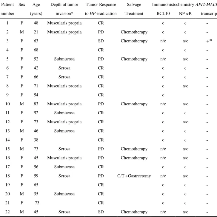
Table 3: Clinicopathologic Features of the Patients and Tumor Expression of BCL10, NF-B, and API2-MALT1
Patient Sex Age Depth of tumor Tumor Response Salvage Immunohistochemistry API2-MALT1 number (years) invasion* to HP eradication Treatment BCL10 NF-B transcript
1 F 48 Muscularis propria CR c c -
2 M 21 Muscularis propria PD Chemotherapy c c -
3 F 63 SD Chemotherapy n/c n/c +* 4 F 68 CR c c - 5 F 52 Submucosa PD Chemotherapy n/c n/c - 6 F 42 Serosa CR c c - 7 F 66 Serosa CR c c - 8 F 71 Muscularis propria CR c n/c - 9 F 54 CR c -
10 M 83 Muscularis propria PD Chemotherapy n/c n/c -
11 F 52 Submucosa CR c c -
12 F 73 Muscularis propria CR c n/c -
13 M 46 Submucosa CR c c -
14 F 38 CR c c -
15 M 73 Serosa PD Chemotherapy n/c n/c -
16 F 45 Muscularis propria PD Chemotherapy n/c n/c -
17 F 56 Submucosa CR c c - 18 F 59 Serosa PD C/T +Gastrectomy n/c n/c - 19 F 65 CR c c - 20 M 35 Submucosa CR c c - 21 F 73 CR c c - 22 M 45 Serosa SD Chemotherapy n/c n/c -
Evaluated by EUS (15 cases) and histologic examination of surgical specimens (1 case).
c, cytoplasmic. n/c, both nuclear and cytoplasmic. CR, complete remission; SD, stable disease; PD, progressive disease. * API2 breakpoint (A1146); MALT1 breakpoint (M1150).
A D
B E
C F
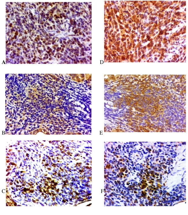
Fig. 1. BCL10 and NF-B protein expression in high-grade gastric MALT lymphoma. (A,B,C): BCL10. (D,E,F): NF-B. (A, D): Nuclear BCL10 and
NF-B expression in the tumor cells of H pylori-independent cases. (B,E):
Cytoplasmic BCL10 and NF-B expression in the tumor cells of H pylori-dependent cases. (C,F): Cytoplasmic BCL10 expression and nuclear NF-B expression in the tumor cells of the two H pylori-dependent cases. Original magnification x 400
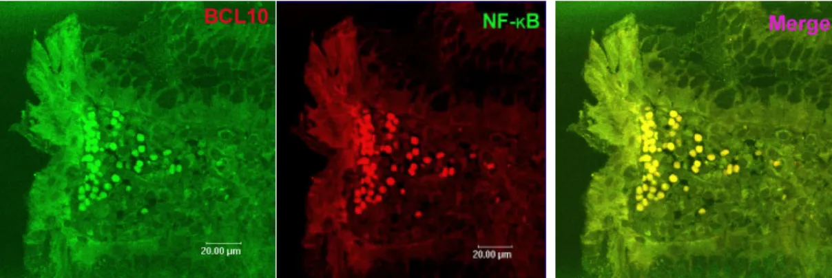
Fig. 2: Co-localization of BCL10 and NF-kB. The nuclear expression of BCL10
(green) and NF-B (red) in the tumor cells of HPi-independent cases analyzed by confocal microscopy. Original magnification x 400
-actin
Fig. 3: Detection of API2-MALT1 fusion transcript by multiplex RT-PCR. B:
Second PCR-B, C: Second PCR-C; lane N, negative control (normal lymph node);
lanes 1, HP-independent HG gastric MALToma positive for API2-MALT1 fusion
transcript; lanes 2-5, HP-independent low-grade gastric MALToma positive for API2-MALT1 fusion transcript; lanes 6-9, HP-independent HG gastric MALToma negative for API2-MALT1 fusion transcript; -actin mRNA is amplified in all cases.
160 137 119 80 160 137 119 80 N 1 2 3 4 5 6 7 8 9 B C
Part Ia2: Non-expression of CD86 on lymphoma cells is associated with
HP-independent state of HG gastric MALToma
Proliferation of neoplastic B-cells of MALToma depends at least partly on the stimulation of HP-specific intratumoral T-cells. The presence of
co-stimulatory marker CD86 (B7.2) on lymphoma cells may promote functional T-cell-mediated neoplastic B cell proliferation. The expression of CD86 was detected in 9 (64%) of 14 HP-dependent HG gastric MALTomas but in none of 6
HP -independent cases (p=0.010). Non-expression of CD86 on MALToma cells
predicts HP-independence of HG gastric MALToma. We speculate that loss of co-stimulatory markers might preclude the interaction between MALToma cells and reactive T-cells and thereby facilitate the transition into the HP -independent status of HG gastric MALToma (Fig. 4).
A B
Figure 4: Immunohistochemical analysis of co-stimulatory markers (CD86 ) (A)HP-dependent HG gastric MALToma; (B) Follicular cell
lymphoma (positive control). Original magnification x 400.
Part Ia3: Decreased infiltration of NK Cells in tumor tissues is associated with
HP-independent state of HG gastric MALToma
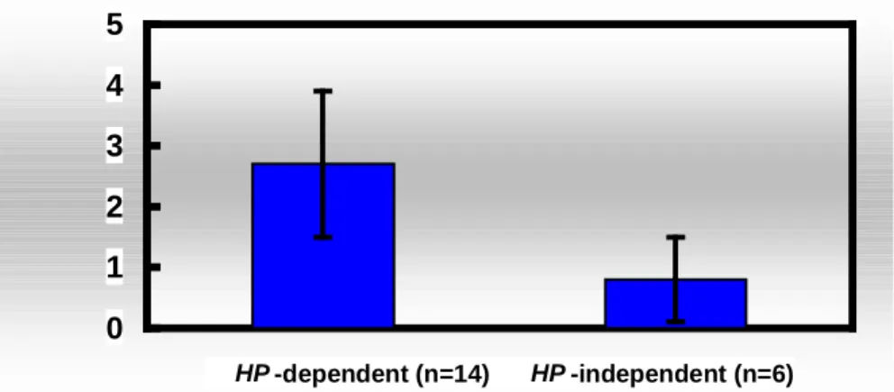
In vitro experiments have suggested that reactive T cell subpopulations in tumor tissues may provide survival and/or growth-promoting signals to the LG gastric MALToma. CD56 (+) natural killer (NK) cells are thought to limit the autoreactive T cell-dependent B cell response elicited by HP, and therefore may contribute to the remission of gastric MALToma after eradication of H. pylori infection. We found that HP -dependent HG gastric MALToma contained significantly higher numbers of CD56 (+) NK cells than HP -independent cases (2.7 ±1.2% versus 0.80±0.70%; p=0.005, nonparametric Mann- Whitney U test) (Figure 5).
Part II: Identification of novel H. pylori–dependence-relevant genes and clarification of their functions
Since BCL10 appears to be one most important determinant of
HP-independence of HG gastric MALToma, we are currently exploring the
mechanism and biologic significance of nuclear translocation of BCL10 in malignant B cells. We are on the way to establish a model system by which BCL10 can be localized to individual cellular compartments. By utilization of Western Blot analysis and IP-MASS analysis, we will address the mechanism that leads to BCL10 nuclear translocation.
Since BCL10 self-associates and interacts with other cytoplasmic
CARD-containing proteins such as MALT-1, CARMA, TRAF-2, and pro-caspase 9, nuclear localization of BCL10 might simply due to the interruption of its ability to self-associate or interact with these cytoplasmic partners. These possibilities will be clarified as well. We showed that NF-B activation was closely associated with aberrant BCL10 nuclear expression. A high degree of co-expression of NF-B and BCL10 in the nuclei was demonstrated in our patients with
HP-independent HG gastric MALTomas. We are testing the hypothesis that
activated NF-B elicits a signal that mediates the translocation of BCL10.
Expression vectors for BCL10 -GFP fusion proteins have been constructed. In the presence or absence of NF-B activators or inhibitors, the localization of
BCL10-GFP fusion protein will be examined by immunofluorescence microscope.
0 1 2 3 4 5 HP -dependent (n=14) HP -independent (n=6)
Figure 5. Histogram demonstrates the mean percentage of cells
Part III: Comparisons of the gene expression profiles of the co-existing HG and LG gastric MALToma, and clarification of their significance in
HP-dependence
Among 20 freshly frozen tissue samples of high-grade gastric MALToma, of which the HP-dependence or –independence state have already been verified by previous prospective clinical study, 16 samples containing microdissectable HG and LG are identified. Microarray approaches to identify expression profiles of the co-existing LG and HG counterpart of the same gastric MALToma patient are ongoing. The results will be interpreted on a background of their individual response to HP eradication. This will help clarify if the genetic changes responsible for HG transformation and HP-independence are different.
計畫成果自評部份
In the first year of this 3-year project, we have successfully identified 2 molecular markers and 2 immunologic cellular markers that highly useful in predicitng HP-independence state of HG gastric MALToma. Since HG gastric MALToma may progress rapidly if unresponsive to HP eradication therapy, thes lines of information are invaluable adjunct for the clinicians to select the right modality of first-line treatment. Manuscripts of these findings have been submitted for publication. We have accomplished what we had proposed for the first year of study. We have also prepared all necessary materials for the second and third year of studies.
參考文獻 (REFERENCES)
1. Isaacson PG: The MALT lymphoma concept updated. Ann Oncol 6: 319-320, 1995
2. Cogliatti S, Schmid U, Schumacher U, et al: Primary B-cell gastric lymphoma: A clinicopathological study of 145 patients. Gastroenterology 101: 1159-1170, 1991
3. Wotherspoon AC, Ortiz-Hidalgo C, Falzon MR, et al: Helicobacter
pylori-associated gastritis and primary B-cell gastric lymphoma. Lancet 338:
1175-1176, 1991
4. Doglioni C, Wotherspoon AC, Moschini A, De Boni M, Isaacson PG: High incidence of primary gastric lymphoma in northeastern Italy. Lancet 339: 834-835, 1992
5. Savio A, Franzin G, Wotherspoon AC, et al: Diagnosis and posttreatment follow-up of Helicobacter pylori-positive gastric lymphoma of
mucosa-associated lymphoid tissue: Histology, polymerase chain reaction or both? Blood 87: 1255-1260, 1996
6. Stolte M: Helicobacter pylori and gastric MALT lymphoma. Lancet 339: 745-746, 1992
7. Isaacson PG, Spencer J: Malignant lymphoma of mucosa-associated lymphoid tissue. Histopathology 11: 445-462, 1987
8. Chan JKC, Ng CS, Isaacson PG: Relationship between high-grade lymphoma and low-grade B-cell lymphoma of mucosa-associated lymphoid tissue (MALToma) of the stomach. Am J Pathol 136: 1153-1164, 1990 9. Harris NL, Jaffe ES, Stein H, et al: A revised European-American
classification of lymphoid neoplasms: A proposal from the International Lymphoma Study Group. Blood 84: 1361-1392, 1994
10. De Jong D, Boot J, Van Heerde P, et al: Histological grading in gastric
lymphoma: Pretreatment criteria and clinical relevance. Gastroenterology 112: 1466-1474, 1997
11. Hussell T, Isaacson PG, Crabtree JE, et al: The response of cells from
low-grade B-cell gastric lymphomas of mucosa-associated lymphoid tissue to
Helicobacter pylori. Lancet 342: 571-574, 1993
12. Hussell T, Isaacson PG, Crabtree JE, et al: Helicobacter pylori specific tumour-infiltrating T-cells provide contact dependent help for the growth of malignant B cells in low-grade gastric lymphoma of mucosa-associated lymphoid tissue. J Pathol 178: 122-127, 1996
infection in primary low-grade gastric lymphoma of mucosa-associated lymphoid tissue. Ann Intern Med 122: 767-769, 1995
14. Wotherspoon AC, Doglioni C, Diss TC, et al: Regression of primary low-grade B-cell gastric lymphoma of mucosa-associated lymphoid tissue type after eradication of Helicobacter pylori. Lancet 342: 575-577, 1993 15. Savio A, Franzin G, Wotherspoon AC, et al: Diagnosis and posttreatment
follow-up of Helicobacter pylori-positive gastric lymphoma of
mucosa-associated lymphoid tissue: Histology, polymerase chain reaction or both? Blood 87: 1255-1260, 1996
16. Bayerdörffer E, Neubauer A, Rudolph B, et al: Regression of primary gastric lymphoma of mucosa-associated lymphoid tissue type after cure of
Helicobacter pylori infection. Lancet 345: 1591-1594, 1995
17. Neubauer A, Thiede C, Morgner A, et al: Cure of Helicobacter pylori infection and duration of remission of low-grade gastric mucosa-associated lymphoid tissue lymphoma. J Natl Cancer Inst 89: 1350-1355, 1997
18. Morgner A, Miehlke S, Fishbach W, et al: Complete remission of primary high-grade B cell gastric lymphoma after cure of Helicobacter pylori infection. J clin Oncol 19:2041-48, 2001.
19. Nakamura S, Matumoto T, Suekane H, et al: Predictive value of endoscopic ultrasonography for regression of gastric low grade and high-grade MALT lymphomas after eradication of Helicobacter pylori. Gut 48:454-460, 2001 20. Chen LT, Lin JW, Shyu RY, et al: Prospective study of Helicobacter pylori
eradication therapy in stage IE high-grade mucosa-associated lymphoid tissue
lymphoma of the stomach. J Clin Oncol 19:4245-51, 2001
21. Isaacson PG. Gastric MALT lymphoma:from concept to cure. Ann Oncol 10:637-645, 1999
22. Koulis A, Diss T, Isaacson PG, Dogan A. Characterization of
tumour-infiltrating T-lymphocytes in B-cell lymphomas of mucosa-associated lymphoid tissue. Am J Pathol 151:1353-1360, 1997
23. Hauer AC, Finn TM, MacDonald TT, Spencer J, Isaacson PG. Analysis of TH1 and TH2 cytokine production in low-grade B-cell gastric MALT-type lymphomas stimulated in vitro with Helicobacter pylori. J Clin Pathol 50: 957-959, 1997
24. Greiner A, Knorr C, Qin Y, et al. Low-grade B-cell lymphomas of mucosa-associated lymphoid tissue (MALT-type) require CD40-mediated signalling and Th2-type cytokines for in vitro growth and differentiation. Am J Pathol 150: 1583-1593, 1997
and antigen-specificity repertoire in Helicobacter pylori specific T cell clones from the antrum of chronic gastritis patients with or without peptic ulcer. Eur J Immunol 27: 1751–55, 1997.
26. Bousiottis VA, Freeman GJ, Gibben JG, Nadler LM. The role of B7-1/B7-2 : CD28/CTLA4 pathways in the prevention of anergy, induction of productive immunity and down-regulation of the immune response. Immunol Rev 153: 5-26, 1996
27. De Jong, Vyth-Dresse F, Dellemijn T, et al: Histological and immunological parameters to predict treatment outcome of Helicobacter pylori eradication in low-grade gastric MALT lymphoma. J of Pathol 193;318-324, 2001.
28. Guidoboni M, Doglioni C, Laurino L, et al: Activation of infiltrating cytotoxic T lymphocytes and lymphoma cell apoptotic rates in gastric MALT
lymphomas: Differences between high-grade and low-grade cases. Am J Pathol 155: 823-829, 1999
29. Shirai M, Arichi T, Nakazawa T, et al: Persistent infection by Helicobacter pylori down-modulates virus-specific CD8+ cytotoxic T cells response and prolongs viral infection. J Infect Dis 177:72-80, 1998.
30. Kos FJ, Engleman EG: Immune regulation: a critical link between NK cells and CTLS. Immunol Today 17: 174-176, 1996
31. Horwitz DA, Gray JD, Ohtsuka K, et al: The immunoregulatory effects of NK cells: the role of TGF-beta and implication for autoimmunity. Immunol Today 18: 538-542, 1997
32. Rosenwald A, Ott G, Stilgenbauer S, et al: Exclusive deletion of the
t(11;18)(q21;q21) in extranodal marginal zone B cell lymphomas (MZBL) of MALT type in contrast to other MZBL and extranodal large B cell lymphomas. Am J Pathol 155:1817-1821, 1999
33. Baens M, maes B, Steyls A, et al: The product of the t(11;18), an API2-MLT fusion, marks nearly half of gastric MALT type lymphomas without large cell proliferation. Am J Pathol 156:1433–1439, 2000
34. Inagaki H, Okabe M, Seto M, Nakamura S, Ueda R, Eimoto T: API2-MALT1 fusion transcripts involved in mucosa-associated lymphoid tissue lymphoma: multiplex RT-PCR detection using formalin-fixed paraffin-embedded
specimens. Am J Pathol 158:699–706, 2001
35. Willis TG, Jadayel DM, Du MQ, et al: Bcl10 is involved in t(1;14)(p22; q32) of MALT B cell lymphoma and mutated in multiple tumor types. Cell 96:35–45, 1999
36. Ye H, Dogan A, Karran L, et al: BCL10 expression in normal and neoplastic lymphoid tissue: nuclear localization in MALT lymphoma. Am J Pathol
157:1147–1154, 2000
37. Liu H, Ye H, Dogan A, et al: T(11;18)(q21;q21) is associated with advanced i. mucosa associated lymphoid tissue lymphoma that expresses nuclear
BCL10. Blood 98:1182–1187, 2001
38. Wang D, You Y, Case SM, et al: A requirement for CARMA1 in TCR-induced
i. NF-kappa B activation. Nat Immunol 3:830-5, 2002
39. Du M, Peng H, Singh N, Isaacson PG, Pan L. The accumulation of p53 abnormalities is associated with progression of mucosa associated lymphoid tissue lymphoma. Blood 86:4587-4593, 1995
40. Ohashi S, Segwa K, Okamura S, et al: A clinicopathologic study of gastric mucosa-associated lymphoid tissue lymphoma. Cancer 88: 2210-2219, 2000 41. Omonishi K, Yoshino T, Sakuma I, et al: Bcl-6 protein is identified in
high-grade but not low-grade mucosa-associated lymphoid tissue lymphomas of the stomach. Mod Pathol 11: 181-185, 1998
42. Martinez-Delgado B, Fernandez-Piqueras J, Garcia MJ, et al:
Hypermethylation of a 5' CpG island of p16 is a frequent event in non- Hodgkin’s lymphoma. Leukemia 11:425-428, 1997
43. Ferreri AJ, Ponzoni M, Pruneri G, et al: Immunoreactivity for p27(KIP1) and cyclin E is an independent predictor of survival in primary gastric
non-Hodgkin's lymphoma. Int J Cancer 15:599-604, 2001
44. Peng H, Diss T, Isaacson PG, Pan L. c-myc gene abnormalities in mucosa-associated lymphoid tissue (MALT) lymphomas. J Pathol 181:381-386, 1997
45. Gronbaek K, Straten PT, Ralfkiaer E, et al: Somatic Fas mutations in non-Hodgkin's lymphoma: association with extranodal disease and autoimmunity. Blood 92:3018-3024, 1998
46. Migliazza A, Martinotti S, Chen W. et al: Frequent somatic hypermutation of the 5′ noncoding region of the BCL6 gene in B-cell lymphoma. Proc Nalt Acad Sci USA 92:12520-4, 1995
47. Okabe M, Inagaki H, Ohshima, et al: API2-MALT1 fusion defines a
distinctive clinicopathologic subtypes of pulmonary extranodal marginal zone B-cell lymphoma of mucosa-associated lymphoid tissue. Am J Pathol
162:1113-1122, 2003
48. Ye H, Liu H, Raderer M, et al: High incidence of t(11;18)(q21;q21) in
Helicobacter pylori-negative gastric MALT lymphoma. Blood 101:2547-2550,
附錄 (出席國際學術會議心得報告及發表之論文)
*: corresponding author Part I.
Expression of nuclear BCL10 is highly correlated with the expression of nuclear NF-B and is predictive of Helicobacter pylori-independent status in early-stage high-grade gastric MALT lymphoma.
Sung-Hsin Kuo, Li-Tzong Chen, Pei-Yen Yeh, Chi-Long Chen, Ih-Jen Su , Jaw-Town Lin2, and Ann-Lii Cheng*. Eur J Cancer 2002: 38, Suppl 7; S161 (abstr. 540) poster presentation.
Manuscripts have been submitted for publication. Abstract content:
Background:
We have recently demonstrated that around 60% of early-stage high-grade gastric mucosa-associated lymphoid tissue (MALT) lymphomas are rendered durable tumor remission by eradication of H pylori (J Clin Oncol 2001; 19:4245 -51). However, the histologic and molecular features that help predict the H pylori -dependent state of these tumors remains elusive. The intracellular signal protein BCL10 was identified by its direct involvement in t(1;14)(p22;q32) of MALT lymphomas, and is a putative regulator of antigen-receptor-mediated NF-B activation. Upregulation of BCL10 may trigger a constitutive NF-B signal from the antigen receptor, and therefore may contribute to antigen-independent growth and progression of gastric MALT lymphoma.
Purpose:
The present study sought to investigate the correlation between nuclear BCL10 expression and nuclear NF-B expression, and the correlation of their expression with the tumor resistance to H pylori eradication therapy in patients with stage IE
high-grade gastric MALT lymphoma.
Materials and Methods:
Lymphoma biopsies of all patients, who had participated in a prospective study of
H pylori-eradication for stage IE high-grade gastric MALT lymphoma, were collected.
The H pylori-dependent status was verified by the results of the prospective clinical trials. There were 16 patients with H pylori-dependent, and 7 patients with H
pylori-independent high-grade gastric MALT lymphoma. The expression of BCL10
and NF-B in pre-treatment paraffin-embedded lymphoma tissues was determined by immunohistochemistry with anti- BCL10 antibody (polyclonal; 1:10; Santa Cruz Biotechnology) and anti-NF-B RelA (p65; 1:150; Santa Cruz Biotechnology). A semi-quantitative method was used to determine the level of expression of RelA.
Reactive spleen and lymph nodes tissue sections were used as the control. The co-expression of nuclear BCL10 and NF-B activity was further analyzed by a confocal immunofluorescence microscopy.
Results:
The aberrant nuclear BCL10 expression was detected in six (86%) of 7 H
pylori-independent high-grade gastric MALT lymphomas but none in 13 H
pylori-dependent cases (P<0.001). All six patients with nuclear BCL10 expression
had co-expression of nuclear NF-B, while only two of 16 patients without nuclear BCL10 expression did so (P=0.002). Interestingly, the latter two patients were found to have tumor invasion of the gastric muscularis propria. There was a significant correlation between nuclear expression of BCL10 and NF-B activation (P<0.001). Furthermore, the nuclear co-localization of RelA and BCL10 was confirmed by confocal immunofluorescence microscopy. The frequency of nuclear translocation of RelA was also significantly higher in H pylori-independent tumors than those H pylori-dependent tumors (6 of 7=86% versus 2 of 16=12.5%, P=0.002).
Conclusion:
The results of this study suggest that nuclear BCL10 expression is closely
associated with the nuclear NF-B expression and support the hypothesis that nuclear BCL10 may activate NF-B. Detection of the nuclear expression of either BCL10 or NF-B is highly useful in the prediction of H pylori-dependent state of early-stage high-grade gastric MALT lymphoma. (This work was supported by grants from NSC91-3112-B-002-009, NHRI-91A1-CANT-1 and NTUH 91-N007)
Part II.
Increased Infiltration of NK Cells in Tumor Tissues and Increased Expression of CD86 on lymphoma cells are Associated with Helicobacter pylori-dependent state of High-Grade Gastric MALToma
Sung-Hsin Kuo 1,4, Li-Tzong Chen5, Hui-Chen Hsu1,4, Pei-Yen Yeh1,4, Chi-Long Chen6, Ih-Jen Su 3, Jaw-Town Lin 2, and Ann-Lii Cheng1, 2,4, *
1
Departments of Oncology, 2Internal Medicine, and 3Pathology, National Taiwan University Hospital; 4Cancer Research Center, National Taiwan University College of Medicine; 5Division of Cancer Research, National Health Research Institutes;
6
Department of Pathology, China Medical College Hospital
Proc Am Assoc Cancer Research 2003, abstract:2329, poster presentation
Abstract content:
Background:
We have recently demonstrated that around 60% of early-stage high-grade gastric mucosa-associated lymphoid tissue lymphoma (MALToma) remains Helicobacter
pylori (HP)-dependent and can be cured by HP eradication (J Clin Oncol 2001;
19:4245-51). However, the histologic and molecular features that help predict the
HP -dependent state of these tumors remains elusive. Proliferation of neoplastic
B-cells of MALToma depends at least partly on the stimulation of HP-specific intratumoral T-cells. CD56 (+) nature killer (NK) cells are thought to limit the
autoreactive T cell-dependent B cell proliferation. Further, the presence of co-stimulatory marker CD86 (B7.2) on lymphoma cells may promote functional T-cell-mediated neoplastic B cell proliferation.
Purpose:
To investigate whether the infiltration of NK cells in tumor tissues and the
expression of CD86 on lymphoma cells may be associated with HP-dependent state of stage IE high-grade gastric MALToma.
Materials and Methods:
Lymphoma biopsies of all patients, who had participated in a prospective study of HP -eradication for stage IE high-grade gastric MALT lymphoma, were collected. There were 14 patients with HP -dependent, and 6 patients with HP -independent high-grade gastric MALToma. The infiltration of CD56 (+) NK cells and the expression of CD86 in pre-treatment paraffin-embedded lymphoma tissues were determined by immunohistochemistry with anti-CD56 antibody (NCL-CD56-1B6,
1:100, Novacastra) and anti-CD86 antibody (AF-141-NA, R and D Systems,
Abingdon, UK). A minimum of 1000 cells (normal and neoplastic) were counted for each single determination and reported as the percentage of CD56 (+) NK cells in total mononuclear cells (as described by Guidoboni et al, Am J Pathol
1999;155:823-29). Staining was considered as positive for CD86 when the protein was detected in more than 10% of tumor cells.
Results:
HP -dependent high-grade gastric MALToma contained significantly higher numbers of CD56 (+) NK cells than HP -independent cases (2.7 ±1.2% versus 0.80±0.70%; p=0.005, nonparametric Mann- Whitney U test ). The expression of CD86 was detected in 9 (64%) of 14 HP-dependent high-grade gastric MALTomas but in none of 6 HP -independent cases (p=0.010).
Conclusion:
Increased infiltration of NK cells in tumor tissues and the expression of co-stimulatory marker CD86 on lymphoma cells are associated with HP-dependent state of early-stage high-grade gastric MALToma. Our data support the hypotheses that increased infiltration of NK cells may suppress the growth of HP- related
autoreactive and neoplastic B lymphoid cells of the stomach, and loss of co-stimulatory markers may preclude functional tumor-B-cell/reactive T-cell interaction and therefore contribute to the transition into the antigen-independent status of high-grade gastric MALToma. (This work was supported by grants from NSC91-3112-B-002-009, NHRI-91A1-CANT-1 and NTUH 91-N007)
Part III.
BCL-2 Expression is Associated with Poor Response to Chemotherapy and Poor Prognosis of Primary High-Grade Gastric Lymphoma
Sung-Hsin Kuo 1,3, Chiun Hsu1,3, Li-Tzong Chen 4, Han-Ting Liu1, Chi-Long Chen5, Chang-Ming Jan6, and Ann-Lii Cheng1, 2,3,*
1Departments of Oncology, and 2Internal Medicine, National Taiwan University Hospital; 3Cancer Research Center, National Taiwan University College of Medicine; 4Division of Cancer Research, National Health Research Institutes; 5Department of Pathology, China Medical College Hospital; Department of Pathology, 6Kaohsiung Medical College Hospital
Proc Am Soc Clin Oncol 2003, abstract:2320, poster discussion
Abstract content:
Background:
Recent data suggest that systemic chemotherapy alone is a highly effective treatment for localized primary large cell lymphoma of stomach. The oncoprotein BCL-2 is a potent suppressor of apoptotic cell death and represses cell death triggered by a variety of anticancer agents, and contributes to chemoresistance of nodal
lymphomas and several other malignancies.
Purpose:
The present study sought to clarify the role of BCL-2 protein expression in predicting chemosensitivity and prognosis of patients with primary high-grade gastric lymphoma.
Materials and methods:
We reviewed the pathologic materials of all patients who had a diagnosis of primary large cell lymphoma of the stomach and who had been treated primarily by systemic chemotherapy in our institutions between January 1, 1988 through
December 31,1999. The expression of BCL-2 in pre-treatment paraffin-embedded lymphoma tissues was determined by immunohistochemistry with anti-BCL-2 antibody (1:25; Dako, Glostrup,Denmark). Staining was considered as positive for BCL-2 when the protein was detected in more than 10% of tumor cells. Tumors were considered chemosensitive if complete remission was documented after chemotherapy.
Results:
anthracyclines or anthracenedione, mostly CHOP (40 cases) and CEpiOP (3 cases). BCL-2 was expressed in 10 (71.4%) of 14 chemoresistant patients but in only 7(21.9%) of 32 chemosensitive patients (P=0.002). The 3-year failure-free survival for tumors with versus without BCL-2 expression was 76% versus 29% (P<0.001, by log-rank test). The median survival time was 16 months in the 17 patients with BCL-2 expression, but it had not yet been reached in the 29 patients without BCL-2
expression. The 3-year overall survival rate was 35 % for patients with BCL-2 expression and 82% for those without (P<0.001, by log-rank test).
Conclusion:
BCL-2 expression of tumor cells is associated with a poor response to systemic chemotherapy and a poor survival of the patients with primary high-grade gastric lymphoma. This work was supported by grants from NSC91-3112-B-002-009, NHRI-91A1-CANT-1 and NTUH 91-N007.