行政院國家科學委員會專題研究計畫 期中進度報告
子計畫七:奈米結構氧化物光子晶體材料製程與光電特性研
究(1/3)
計畫類別: 整合型計畫
計畫編號: NSC91-2216-E-009-029-
執行期間: 91 年 08 月 01 日至 92 年 07 月 31 日
執行單位: 國立交通大學材料科學與工程學系
計畫主持人: 陳三元
報告類型: 精簡報告
處理方式: 本計畫涉及專利或其他智慧財產權,1 年後可公開查詢
中 華 民 國 92 年 5 月 26 日
2
奈米結構氧化物光子晶體材料製程與光電特性研究(1/3)
Low-temperature Growth of Zn-ZnO Polygon Prismatic Nanocrystals by Thermal
Vapor Transport for Photonic Crystals
計畫編號:NSC91-2216-E-009-029
執行時間:91/08/01 ~ 92/07/31
主持人:陳三元 教授
交通大學材料科學與工程學系
中文摘要
本研究利用熱蒸發與沉積的製程,成功
地在矽基板上生長出六方結構的氧化鋅特殊
多面體奈米結構。一般而言,結晶相與成核
的種子的控制,對於奈米晶體在成長開始時
的本質形狀是很重要的,將含有金屬鋅離子
的有機溶液先塗佈在基板上,並以此作為成
核的種子。再利用熱蒸氣傳輸製程以氧化鋅
粉末為成長源,並通入純氬氣,而在基板溫
度為 250℃下即可成長。結果發現,未作任
何後段處理的氧化鋅多面體奈米結構其實是
由,六方結構的金屬鋅與外層氧化後的氧化
鋅薄膜所組成。我們也設計了許多不同的熱
處理來觀察此氧化鋅多面體奈米結構的形貌
與結晶性。並可以確定其成長機制應該是由
金屬鋅與氧化鋅間無機的架橋機制而來。而
這些六方結構的金屬鋅與氧化後的氧化鋅將
進一步作為後續光子晶體結構的研究。
關鍵詞: 氧化鋅
、奈米晶體
、光子晶體
、螢光、熱蒸發與沉積
Abstract
A novel hier ar chical p olygon pr is matic nano-structure of hexagonal ZnO and Zn has been successfully grown on silicon by thermal vapor transport and condensation method. Thus, in this work, the orga nic solvent with zinc chemica l compound was first coated and used as seeds on the silicon substrate. Subsequently, it was grown by thermal vapor transport withZnO powder at 250°C (substrate) in Ar atmosphere. The samples were characterized using X-ray diffraction, scanning and
t r a n s m i s s i o n e l e c t r o n m i c r o s c o p y, a n d p h o t o l u m i n e s c e n c e s p e c t r o s c o p y. T h e as-s y n t h es i z e d Z n O p ol y g o n p r is ma t i c
nano-structure consisted of hexagonal metallic nuclei (Zn) covered with an oxidation outer thin
film (ZnO). Depending on the different annealing temperature and reaction atmosphere, Zn polygon prismatic structure having various morphologies can be developed. The dimension and crystal phase can be controlled by temperature, time, kinetic sur fa ce ener gy, a nd ca pping molecules. The possible formation mechanism for the Zn-ZnO polygon prismatic crystal structure is identified and proposed as the Zn-ZnO mineral bridge mechanism. Although the present non-prefect Zn-ZnO polygon prismatic structure shows weakly UV emission and strongly deep-level emission, the PL properties and crystallization o f Z n-ZnO prismatic nanocrystals could be improved by suitable post-treatment
Keywords: ZnO、nanocrystals 、photonic
crystal、photoluminescence、
thermal vaportransport and condensation
I. Introduction
Zinc oxide possesses unique optical, electrical and structural properties that make it useful for a wide range of technological applications such as photo-detector [1], solar cells [2,3], nanolasers [4], and other highly functional device[5]. Recently,
much effort has been invest ed in fabricating quasi-one-d i m e n s io na l Z nO na n os t r u c t u r es , because the size a nd sha pes of nanocr ystal repres ent key elements t ha t d et er min e t h e ir electrical and optical properties [6]. Currently, both gas and liquid phase methods are used to synthesize and control the nanocrystal. The vapor-liquid-solid (VLS) method is the common approach to grow the nanostructure and many researchers have tried to control the process parameter to produce various shape nanocrystal, such as nanorod [7], nanonail [8], nanoblets [9], etc. On the other hand, the liquid phase synthesis of anisotropic nanocrystals has been far more limit ed, and only the rod-based system has been studied in depth. The liquid phase growth mechanism can sketchy d ivide into two steps. In the first step, spherical seed nanocrystals
are homogeneously nucleated. Precursors with materials for the desired one-dimensional structures are chemically reduced in the presence of capping molecules and seed nanocrystals that serve as nucleation sites. The gas phase process has been explored to produce 1D nanostructure of different cross sections as well as some other exotic shapes
[10]. However, the gas phase and liquid phase syntheses s e e m t o b e ha r d t o r ea c h u ni f or m morphologies at comparatively mild synthesizing conditions.
The photonic band gap crystal is simply a spatially periodic structure consisting of high and low dielectric regions. A promising technique to fabricate 3D photonic crystals with a photonic band ga p in op t ical wa velengths is self-assembling g r o w t h o f -dimensiona l t h r e e inorga nic nano-crystals. In contrast to conventional process, we present both gas and liquid method toward the growth of well-proportional and crystallized Zn and ZnO polygon prismatic nanocrystals. In order to recognize the growth mechanism of Zn polygon prismatic structure, different heat-treatment and heating profiles wer e designed to obser ve the morphology and crystallization of Zn polygon prismatic structure. T h e c r y s t a l s i z e f r o m micrometer to nanometer and phase control of the ZnO polygon pr is ma tic na nocr ysta ls ca n be a c hi e v e d b y va r yi n g t h e gr o wt h t i m e a n d temperature.
II. Experimental
ZnO a nd Zn polygon prismatic nanocrystals were synthesized using a colloidal Zn particles and physical vapor transport system. An alumina tube wa s mou nt ed i ns i d e a hi gh-temperature tube furnace. Preparation of colloidal Zn in methanol: 50 ml of a 1x10⁻² M Zn (ClO4)2 solution is added to 442 ml methanol plus 8 ml 5M NaOH. The solution is vigorously shaken for 10 min and left overnight. After about 24 hours a transparent solution of stable colloidal is obtained. ZnO powder and graphite mixtur e wer e us ed as Zn s ource. T he s ource material was placed on an alumina boat. After placing the boat at the center of the tube and the substrate locating downstream of the carrier gas flow, the tube was sealed and evacuated by a mechanical rotary pump to a pressure of 80 mTorr. As carrier gas of 100% Ar was used, with a flow rate of 10 sccm, reaction was carried out at 1100˚C and kept at that temperature for 0.5-5 hour to form Zn-ZnO polygon prismatic nanocrystals at 250˚C (substrate temperature). The Zn polygon prismatic nanocrystals were a n n ea l ed a t 25 0˚C in p u r e oxygen atmosphere (5N) for 30 minutes to form the ZnO layer on t he Zn crystals. Aft er tha t , the Zn-ZnO polygon prismatic nanocrystals were
thermally annealed at 550˚C for over 3 hours to s t u d y t h e c ha n g es of mor phology and crystallization.
The deposited product was characterized and a n a l y z e d b y s c a n n i n g e l e c t r o n croscopy m i (FE-SEM, S-4100), and the crystal structure was a na lyz ed using XRD (Siemens D5000). T EM studies of the crystal were carried out a Philips CM20 and Hitacit-600 operated at 200KeV and 100 KeV, a nd ener gy-dispersive X-ray spectroscopy (EDS) attached to SEM and TEM, respectively.
III. Results and discussion
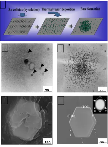
Figur e 1 shows typical scanning electron microcop y (SEM) and T EM ima ge of the Z n polygon prismatic crystals. The diameter of the crystals can be tuned from 30 n m t o 2m by increasing the concentration o f t h e Z n ( C l O4)2 solution. As the concentration of the Zn(ClO4)2 solution increases, the amount of Zn particles would increase. The Zn particles behave as the nucleus sites of the larger Zn crystals. The amount of Zn nucleus sites would be proportional to the concentration of the Zn(ClO4)2 solution. In the highly concentration solution, the large and sharp Zn polygon pr is ma tic cr ysta ls (~2m) wer e synthesized on the substrate by ther mal vapor process (Fig. 1A). On the contrary, as the amount of nucleus sites decr eases, the crystal size of Zn polygon prismatic structure shrinks to ~30 nm scale (Fig. 1C). Figure 2A illustrates the schematic mechanism of such a process. In this case, the initial nucleus sites were prepared by Zn(ClO4)2 solution. When Zn sources were transported b y thermal evaporation process, these nucleus sites would cap these Zn molecules to develop th e nanocrystals. In the following step, some of the fine Zn nanocrystals would get together to for m the large crystals. According to above statement, it was found that the concentration of the Zn(ClO4)2 solution could determine t h e n u c l eu s s i t es . Therefore, the a mount of nucleus sit es wou ld determine t he size of the Zn polygon prismatic crystals. It was found that the initial nuclei of Zn polygon prismatic crystals were emerged into the small Zn polygon prismatic crystals by themselves and its diameter is less than 10 nm, as seen in Fig. 2B. Figure 2C shows the special distribution pattern of Zn polygon prismatic nanocrystals (~30 nm) formed in the initial stage of crystal growth. The spreading region o f t h e s e distributed Z n nanocrystals has diameter around 150 nm that is close to the composing-crystals obtained in our experiments (Fig. 2D). It suggests that the spatial distribution region of Z n nanocrystals would develop the base for the larger size crystals. After the suitable growth time, the Zn polygon prismatic
4
crystals will be formed on that base (Fig. 2E).
The s ha p e of t h e Z n p o l y g o n p r is ma t ic nanocrystals would vary with the growth time. Figure 3 presents the growth of the Zn polygon prismatic nanocrystals as a function of time. The prefect Zn nanocrystals are observed for the growth time of 2 hours. When the growth time exceeds 2 hours, the shape of the Zn polygon prismatic nanocrystals would begin to disintegrate (Fig. 3B). It is well known that metal state will become unstable when the temperature reaches around its melting point. The apparent melting point (Zn: mp 410˚C) would be reduced for nanocrystal with size sma ll enough. The balance between t h e thermodynamic and kinetics would be influenced by the growth time. It implies that if the growth time exceeds the critical time, t he equilibrium status would be destroyed. It could be due to the unstable lattice-site of zinc atom in the Zn polygon prismatic crystals, long time thermal heating will make the crystal structure destructible. To keep the stability of grown crystals, the oxidation treatment to modif y the sur fa ce status of Z n p o l y g o n prismatic crystal is applied. Both Zn and ZnO have the hexagonal (hcp) structure. From the growth kinetics point of view, when ZnO is for med, rearrangement of the sublattices of zinc from hcp (Zn) to hcp (ZnO) has to occur only at Zn reaction front. Therefore, the nucleating ZnO grain has a template to follow the orientation of Zn exactly. Figur e 4 shows the surface morphology of Zn polygon prismatic nanocrystals after ther ma l treatment at 550?C for 3 hours. It is obvious that Zn polygon prismatic crystals with surface treatment could retain the complete crystals structure (Figure 4B). It is due to the ZnO layer formed by surface treatment that prohibits the zinc atoms leaving away lattice and prevents the crystals from being destroyed.
Figure 5 shows the X-ray diffraction (XRD) spectrum of the nanostructures, where the intensity of zinc crystalline phase is different for different treatment. In contrast with crystallization of Z n polygon prismatic nanocrystals, the sample that undergoes the oxidation treatment presents t h e stronger XRD peak intensity than the other. For the Zn polygon prismatic nanocrystals subjected to ox ida tion tr ea tment on Zn sur fa ce, the ZnO crystalline phase can be developed as identified
from the XRD (Fig. 5B). Chemical composition microanalysis by energy-dispersive spectrometry (EDS) reveals t ha t t he Z n pol ygon pr is ma ti c nanocrystals with surface oxidation are composed of more content of zinc compared to those without surface treatment (with an atomic ratio of = 15:1). This is in a good agreement with the XRD analysis result.
The photoluminescence (PL) measurements of the synthesized Zn polygon prismatic nanocrystals are performed at room temperature, using a He-Cd laser line of 325 nm as the excitation source. As shown in Figure 6, UV and green emission with peaks at 380 nm and 530 nm are observed for the Zn polygon prismatic nanocrystals with surface treatment. The PL spectrum is different from that of Zn. Zn polygon prismatic nanocrystals without oxidation treatment. No emission peak is found because the Zn polygon prismatic nanocrystal is only composed by pure zinc metal in this case. On the contrary, as the Zn nanocrystals have undergone oxidation treatment, the surface will form a thin oxide layer. According to the XRD analysis, the oxide layer is basical zinc oxide. ZnO usually displays two major PL peaks, UV (near-band-edge) emission peak and green (or red) emission peak (deep-level). The deep-level e m i s s i o n s a r e generally associated with defects in ZnO lattice. Besides, the near-band-edge transition could not effectively exist i n t h e n o n-crystalline ZnO. Although the developed Zn polygon prismatic nanocrystals are well crystalline, the interface between Zn and ZnO surface layer usually results in large lattice mismatch (17%). The kind of interface becomes the defect generator to support the deep-level emissions. In addition to lattice mismatch, the incomplet e oxidation of zi nc produces the defects inside surface layer and gives deep-level emission too.
IV. Conclusion
Single-cr yst a ll ine Z n p ol ygon pr is ma t ic nanocrystals are synthesized by liquid solution seed nucleation a n d va p or-gas growth method. The cation concentration of liquid solution determines the dimension of Zn crystals from the nanometer ( 3 0 n m) t o micrometer scale (2 m). The Zn polygon prismatic nanocrystals a r e gr own up through a thermal evaporation process. The Zn nanocrystals undergone surface treatment will form the ZnO surface layer. The ZnO surface layer could ma intain t he polygon pris matic structure and prevent the zinc crystal from being destroyed at higher temperature. In addition, the ZnO surface layer improves the photoluminescence property. Considering their shape, crystal dimension and oxidation behavior, as expected, the crystals having good optical property could be formed within different microcavities. Th e s e n a n o- a n d microcrystals can be further used as building blocks to assemble two- or three-dimensional assemblies.
Acknowledgments
This work was financially supported by the National Science Council of the Republic of China,
5
T a i w a n u n d e r C o n t r a c t N o . NSC89-2216-E-009-034.
References
[1] J. A. R odr igu ez, T. Jir sa k, J. Dvor a k, S . Sambasivan, D. J. Fischer, Phys. Chem. B 2000, 104,
319.
[2] K. Hara, et al. Sol. Energy Mater. Sol. Cel ls 2000, 64, 115.
[ 3 ] H . R ens mo, K . K eis , H . L in ds t r o m, S . S oder gr en, A. S olbr a nd, A. Ha gf el dt , S . E. Lindquist, L. N. Wang, M. Muhammed, J. Phys.
Chem. B 1997, 101, 2598.
[4] M. H. Huang, S. Mao, H. Feick, H. Q., Y. Y. Yan, H. Kind, E. Weber, R. Russo, P.D. Yang, Science
2001, 292, 1897.
[5] H.-J. Muhr, F. Krumeich, U. P. Schonholzer, F. Bieri, M. Niederberger, L. J. Gauckler, R. Nesper, Adv. Mater. 2000, 12, 231.
[6] A. P. Alivisatos, Science 1996, 271, 933.
[7] H. M. Kim, T. W. Kang, and K. S. Chung, Adv. Mater. 2003, 15, 567.
[8] J. Y. Lao, J. Y. Huang, D. Z. Wang, and Z. F. Ren, Nano Lett. 2003, 3(2), 235.
[9] Z. W. Pan, Z. R. Dai, Z. L. Wang, Science 2001, 291, 1947.
[10] P. Yang, Y. Wu, R. Fan, Int. J. Nanosci. 2002, 1,1
Fig. 1. SEM images of the Zn polygon prismatic crystals synthesized by liquid solution and vapor transport method. A) Zn polygon prismatic crystals of 2-μm diameter. B) Zn polygon prismatic crystals of 300-nm diameter. C) TEM images of the Zn polygon prismatic nanocrystals of 30-nm diameter.
F ig. 2. (A) Schematic i l lu s t r a t ion of gr o wt h mechanism of Zn polygon prismatic nanocrystals. (B) T EM image of the nuclei of Zn polygon prismatic nanocrystals (~7 nm) (C) TEM image of Zn polygon prismatic nanocrystals (30 nm) formed by thermal vapor process. (D) Growing Zn polygon prismatic nanocrystals (0.6μm). (E) SEM image of Zn polygon prismatic crystals (0.7 μm) formed by the collection of the ultra-fine Zn nanocrystals.
A 1 m B 200 nm C 150 15 30 20 11 100 010 010 11 (100) (010) ( ( ( ( A
6 350 4 00 45 0 5 00 550 6 00 65 0 P L int en s ity ( a. u .) 380 W a v eleng th ( nm ) Z n n ano crysta ls Z nO /Z n na noc r y s tals F ig. 3. S EM ima ges of Zn polygon pr isma tic
nanocrystals growing as a function of time. (A) 2 hours. (B) more than 3 hours.
Fig. 4. SEM images of Zn p olygon pris matic nanocrystals were thermally heated at 550?C and kept for 3 hours (A) without and (B) with oxidation treatment.
Fig. 5. X-ray diffraction (XRD) spectrum of Zn polygon prismatic nanocrystals were thermally heated at 550?C and kept for 3 hours (A) without and (B) with oxidation treatment.
F Fig. 6. Room-temperature photoluminescence spectr a r ec or ded fr om Zn polygon prismatic
nanocrystals w i t h a n d w i t h o u t o x i d a t i o n 1 m B 500 nm A 500 nm B 2 0 3 0 4 0 5 0 6 0 Z n (0 0 2 ) Z n (1 0 2 ) Z n (1 0 1 ) Z n (1 0 0) 2 C oun t A 2 5 3 0 3 5 4 0 4 5 5 0 5 5 6 0 Z n (1 0 2 ) Z n (1 0 1 ) Z n (1 0 0 ) Z n (0 0 2 ) Z n O (1 0 2 ) Z n O (1 1 0 ) Z n O (1 0 1 ) Z n O (1 0 0 ) Z n O (0 0 2 ) C o u n t 2 B 1 m B