國立臺灣大學理學院心理學研究所 碩士論文
Graduate Institute of Psychology College of Science
National Taiwan University Master Thesis
正常老年人與阿茲海默病及其臨床前期病人對臉部情 緒辨認之同年齡效應研究
An Exploration of the Own-Age Effect on Facial Emotion Recognition in Normal Elderly People and
Individuals with the Preclinical and Demented Alzheimer’s Disease
莊祐蓁
Yu-Chen Chuang
指導教授:花茂棽 博士、張玉玲 博士
Advisors: Mau-Sun Hua, Ph.D., Yu-Ling Chang, Ph.D.
中華民國 109 年 1 月
January 2020
摘要
背景:同年齡效應之探討於近年逐漸獲得重視,然而,因方法學上的限制,
過去研究同年齡效應是否存在於臉部情緒辨認能力之結果並不一致。除健康年長 者,阿茲海默型失智症之病人具臉孔情緒辨認能力之受損,然尚未有研究探討同 年齡效應是否存在於病人之臉孔情緒辨識能力。故本研究先解決過去文獻於方法 學上之限制,再探討同年齡效應是否存在於健康年長者及阿茲海默型失智症病 人。方法:本研究共納入 138 位受試者。實驗一納入 27 位健康老年受試者及 31 位健康年輕受試者;於實驗二納入 27 位健康老年受試者及 80 位記憶抱怨受試者 (分為主觀認知衰退組、記憶型輕度認知障礙組及阿茲海默型失智症組)。每位受 試者接受臉孔情緒辨認作業以測得其臉部情緒辨認能力。結果:實驗一,除年輕 人組在看年輕臉孔之中性表情,在健康老年人組、健康年輕人組未呈現顯著之同 年齡效應。除生氣之情緒辨認,本研究未發現顯著組間差異。不同年紀之臉孔依 不同情緒具不同影響結果:在難過、悲傷之情緒辨認上,年輕人臉孔比老年人臉 孔好辨認,而在快樂的情緒辨認上相反。實驗二,僅記憶型輕度認知障礙組、阿 茲海默型失智症組於難過情緒辨認時呈同年齡效應之傾向,並易將年輕人的難過 情緒誤認為生氣、將老年人的難過情緒誤認為中性。結論:僅在記憶型輕度認知 障礙組、阿茲海默型失智症組發現同年齡效應之傾向,反映因病程進展而導致臉 部情緒辨認能力之受損。本研究之低強度情緒—難過之臉部情緒辨認作業,可視 為偵測早期阿茲海默症之指標。
關鍵字:同年齡效應、臉部情緒辨認、主觀記憶衰退、記憶型輕度認知障礙、阿 茲海默型失智症、臉部情緒辨認作業
An Exploration of the Own-Age Effect on Facial Emotion Recognition in Normal Elderly People and Individuals with the Preclinical and Demented Alzheimer’s
Disease
Yu-Chen Chuang
Abstract
Background: The own-age effect, which may affect the accuracy of facial emotion
recognition (FER), has been investigated over the last decade. However, due to
methodologic limitations and differences, the results were inconsistent. Patients with
Alzheimer’s disease (AD) have been reported to show deficits in FER even in early
phases. Nevertheless, no study has examined the own-age effect in AD patients. The
present study, minimizing prior methodologic drawbacks, thus was to examine this
issue in normal adults, and patients with subjective cognitive decline (SCD), amnestic
mild cognitive impairment (aMCI) and very mild AD. Methods: The total of 138
participants was recruited in the present study. In experiment 1, 27 healthy older
adults and 31 healthy young adults were recruited. In experiment 2, 27 healthy control
(HC) and 80 patients with memory complaints, among 3 groups, SCD, MCI, and AD,
were recruited. The facial emotion recognition function of all participants was
evaluated through our Facial Emotion Recognition Task (FER Task) with Taiwanese
facial emotion stimuli. Results: In experiment 1, the own-age effect was not observed
in the older adults, but was found in younger adults when decoding neutral photos. No
group difference in performing the FER Task was found, except for anger. The photo
age effect of the FER on distinct emotions was significant. Younger faces are more
accurate than older faces to decode difficult emotions in both younger and older
adults. In experiment 2, a tendency of the own-age effect occurred in MCI and AD
groups, who showed significant deficits when decoding sadness, and tended to
mislabel sadness as anger in younger-face photos, neutral in older-face photos.
Conclusions: A tendency of the own-age effect occurred only in MCI and AD groups,
but not in normal individuals and SCD groups can reflect the FER deficits in the
progression of AD. The results displayed that our FER Task, especially for those items
of low-intensity emotion (i.e., sadness), can be a sensitive index for early detection of
early dementia.
Keywords: own-age effect, facial emotion recognition, subjective memory
decline, amnestic mild cognitive impairment, Alzheimer’s disease,
Facial Emotion Recognition Task
Contents
1. Introduction ... 1
2. Methods ... 13
2.1. Participants ... 13
2.2. Measurements ... 14
2.3. Procedure ... 18
2.4. Statistical Analysis ... 19
3. Results ... 21
3.1. Experiment 1: ... 21
3.1.1. Demographics and Clinical Characteristics ... 21
3.1.2. Does the own-age effect of FER exist in normal aging? ... 21
3.2. Experiment 2: ... 23
3.2.1. Demographics and Clinical Characteristics ... 23
3.2.2. Is the own-age effect evident in the patients while performing the FER? . 24 3.2.3. Clinical Utilities ... 27
4. Discussion ... 28
4.1. Does the own-age effect of FER exist in healthy adults? ... 28
4.2. Is the own-age effect evident in the patients while performing the FER? ... 32
5. References ... 36
6. Tables ... 50
7. Figures ... 56
1. Introduction
Facial emotion recognition (FER), one of the essential components of social
cognition (Adolphs, 2001), represents the ability to recognize facial emotional
expressions. It enables individuals to sense their social environment and modify their
behavior accordingly (McCade, Savage, Guastella, Lewis, & Naismith, 2013); it also
contributes to more efficient social interactions (Sze, Goodkind, Gyurak, & Levenson,
2012). Thus, this ability is undoubtedly crucial for social behavior (Hargrave,
Maddock, & Stone, 2002); furthermore, engaging in satisfying social interactions and
avoiding social isolation are important to our health and well-being throughout life
(Cacioppo, Berntson, Bechara, Tranel, & Hawkley, 2011). Consequently, deficits in
this ability may contribute to difficulties in social communication, damage
self-esteem, and even diminish the quality of life (Ciarrochi, Chan, & Caputi, 2000).
FER has drawn considerable attention in clinical and functional imaging studies
recently. Studies have demonstrated that dissociable neural substrates are associated
with the facial recognition of basic emotions (Hennenlotter & Schroeder, 2006;
Schroeder et al., 2004). The occipital and posterior temporal cortices are responsible
for the perceptual analysis of facial expressive features (Haxby, Hoffman, & Gobbini,
2000; 2002), and the extraction of emotional meaning from faces is linked to the
orbitofrontal, ventral prefrontal cortex-related, and somatosensory regions (Adolphs,
2002). However, these emotional circuits, including the hippocampus, amygdala, and
frontal regions, were reported to show age-related neurological changes (Greenwood,
2000). In addition, certain types of neurodegenerative diseases, such as
frontotemporal dementia (FTD) and Alzheimer’s disease (AD), can also damage these
brain regions (Keane, Calder, Hodges, & Young, 2002; Pietschnig et al., 2016). Thus,
deficits in decoding specific emotions have been reported in normal aging as well as
in patients with neurodegenerative diseases (Keane et al., 2002; Torres et al., 2015).
To help families realize the patients’ difficulties and improve their life quality,
choosing an appropriate clinical assessment for early detection of deficits in FER is
undoubtedly crucial.
Furthermore, studies have indicated that several characteristics of emotional
stimuli could affect the accuracy and memory of FER, including cultural, gender-, and
age-based factors (Bäckman, 1991; Hess, Blairy, & Kleck, 1997; Malpass & Kravitz,
1969; Wells, Gillespie, & Rotshtein, 2016). Indeed, the own-race bias refers to the
tendency of recognizing and memorizing one’s own race or ethnicity relatively more
accurately than another race or ethnicity (Malpass, & Kravitz, 1969). Gender has also
been reported to have different effects depending on the type of expressions (Wells et
al., 2016); for example, female faces are reported to be easier to recognize with regard
to expressions of happiness (Hess et al., 1997), while male faces are better recognized
in expressions of disgust, sadness (Hess et al., 1997), and anger (Becker, Kenrick,
Neuberg, Blackwell, & Smith, 2007). To our knowledge, very few studies have
examined the effects of photo age on FER. This is why studies conducted so far in the
domain of age group differences in processing emotional expressions have mostly
used younger faces (some included middle-aged faces) but did not systematically vary
the age of the presented faces. However, the study by Lamont, Stewart-Williams, and
Podd (2005) using neutral faces as stimuli found that observers of different ages
recognize faces of their own age more accurately and rapidly as opposed to those of
other ages (referred to as the own-age bias; Bäckman, 1991). Such findings suggest
that the age of a face constitutes an important factor that influences how we attend to,
encode, and remember faces. Evidence of the own-age bias challenges any
interpretation of observed age group differences in FER, as older observers may have
been at a disadvantage relative to younger observers when the stimuli consisted only
of faces of young individuals.
The own-age effect (in most studies called own-age bias or own-age advantage, while we used the term “own-age effect” because we did not want to emphasize it as
good or bad) is explained by two main theories: experience (or expertise) accounts
(Rhodes & Anastasi, 2012) and social-cognitive accounts (Sporer, 2001). The former
means that more experience and contact with own-age groups increases the
individual’s familiarity with the expressive style of own-age faces, and thus, decoding
of own-age faces is more efficient (Rhodes & Anastasi, 2012). The latter means that
there is a greater motivation to process and attend to the characteristics of own-age
faces (Sporer, 2001); thus, individuals who identify with an ethnic or social group will
exert more effort when decoding the emotional expressions of the own-group
(Thibault, Bourgeois, & Hess, 2006). The own-age effect was initially proposed in
facial recognition memory studies, indicated that facial recognition memory is
superior for own-age relative to other-age faces (Bäckman, 1991; Lamont et al., 2005;
Wright & Stroud, 2002). Further studies have also observed the own-age phenomenon
in tasks that involve recognizing facial emotional expressions across different fields.
For example, participants tended to look longer at own-age faces, and this was
thought to predict more accurate FER in own-age faces (Ebner, He, & Johnson, 2011).
Functional magnetic resonance imaging studies also reported different activities for
own-age and other-age faces regarding neutral and happy expressions (Ebner et al.,
2013). In addition, studies that used electroencephalography reported partly own-age
and own-race effects on the event-related potentials for neutral expressions (Melinder,
Gredebäck, Westerlund, & Nelson, 2010). Based on these empirical evidence and
theories, it may be assumed that own-age photos can enhance the accuracy of FER for
observers. That is, the own-age effect might appear in FER.
Indeed, this hypothesis has been proposed and investigated in several studies
over the last decade, and some have confirmed this effect in older observers. For
example, Riediger, Voelkle, Ebner, and Lindenberger (2011) used posed expression
with multi-dimensional response format and found that middle-aged and older
observers performed well in their target ratings of happiness and anger by the age of
the own-age photos than did young observers. Another study by Riediger, Studtmann,
Westphal, Rauers, & Weber (2014) which only used spontaneous and posed smile as
the test material also supported that older participants could better identify older
rather than younger faces.
However, contrary to the results of the above studies, most research that was
carried out by modifying the age of the photographed or videoed individuals indicated
that there was no own-age effect or that it was observed only for younger observers.
For example, Borod et al. (2004) presented younger, middle-aged, and older female
observers as stimuli, and the results showed that the expressions of older posers were
rated significantly less accurately than those of younger posers for all groups. Further
studies by Ebner and Johnson (2009), Murphy, Lehrfeld, and Isaacowitz (2010), and
Hühnel, Fölster, Werheid, and Hess (2014) also reported similar patterns. In addition,
Malatesta, Izard, Culver, and Nicolich (1987) found that this effect exists only in
younger observers. Older observers were better at rating older faces than they were at
rating younger faces, while the difference was not significant. A study by Richter,
Dietzel, and Kunzmann (2010) also supported this finding in younger observers.
Nevertheless, the results of these studies were inconsistent, and it should be
noted that some methodologic limitations existed in all these studies. First, the gender
of the stimuli and observers in some studies was exclusively female (Borod et al.,
2004; Hühnel et al., 2014; Murphy et al., 2010), even though it is known that gender
can influence the accuracy of the results based on the type of emotion (Wells et al.,
2016). Second, the numbers of photos and observers in some studies were too small
(Borod et al., 2004; Ebner & Johnson, 2009; Hühnel et al., 2014). Third, the target
emotions in these studies were inconsistent; besides, some examined the own-age
effect by averaging the accuracy of emotions (Malatesta et al., 1987). These factors
not only make it difficult to conclude the type of emotion which was reported
consistently enough to show the own-age effect, but also make it hard to analyze the
different effects of distinct emotions based on the finding that different types of
expressed emotions have different effects on accuracy (Wells et al., 2016). Therefore,
it is necessary to assess enough types of emotions and to examine their effects
separately rather than as averages. In conclusion, gender imbalance, small stimuli and
observer sample sizes, selecting incomparable types of emotions, and ignoring the
effect of different emotions were existing methodologic problems in prior studies, and
these might have resulted in inconsistent results. The present study sought to address
these methodological limitations of earlier investigations.
Apart from the problems we have mentioned above, other methodologic
differences existed might also cause inconsistent results, namely, the types of
emotional expressions presented (dynamic or static and posed or spontaneous),
measured approaches of response (the forced-choice approach and the
multi-dimensional response format), and the stimuli database. First, dynamic
spontaneous stimuli were reported to show more ecological validity; thus, they could
increase accuracy (Bartlett et al., 2006; Murphy et al., 2010), while the results of
examining the own-age effect were still inconsistent after controlling it (Murphy et al.,
2010; Riediger, 2014) due to other methodologic problems. Besides, the dynamic
spontaneous stimuli established so far did not include enough stimuli, and most were
female faces only (Murphy et al., 2010; Richter et al., 2010). Thus, there are no
appropriate stimuli that can be selected yet, even if we do not consider including the
East Asian faces. Second, the multi-dimensional response format, the way that
participants rate the percentage across all emotions within a photograph. And the
responses were considered as accurate if the percentage of the target emotion was
higher than the percentage on the remaining scales (Gunes & Pantic, 2010). It was
developed based on the theory that emotional experiences are often multi-faceted
(Hemenover & Schimmack, 2007), and so it was thought to be more appropriate
(Kreibig, Samson, & Gross, 2013). However, some studies that used the forced-choice
approach still confirmed the own-age effect successfully; thus, it seems that different
types of rating formats did not play an important role in the inconsistency of the
results. In addition, Hühnel et al. (2014) indicated that the hit rates in their study were
relatively low because of using the multi-dimensional response format. Although the
multi-dimensional response format was reported to show more ecological validity, the
forced-choice approach might be more appropriate for developing our task to a
clinical measurement. Finally, most studies used the static posed expressions of the
FACES database (Ebner, Riediger, & Lindenberger, 2010) as materials, while the
remaining studies used stimuli (including photos and videos) developed by their
respective laboratories (Borod et al., 2004; Hühnel et al., 2014; Malatesta et al., 1987;
Richter et al., 2010; Riediger, 2014); thus, the stimuli in those studies are
heterogeneous in nature. To control the influence of race on FER (Young &
Hugenberg, 2012), we chose the stimuli from Taiwanese individuals (Tu, Lin, Suzuki,
& Goh, 2018) and included a large number of static posed photos. In conclusion, we
determined to use the forced-choice rating as our response measurement, emotion
stimuli from Taiwanese individuals with static posed photos as stimuli.
Apart from the changes in the brain in normal aging, abnormal cerebral atrophy
and neuropathological changes occur in patients with AD, resulting in damage to the
circuits related to emotions (McLellan, Johnston, Dalrymple‐Alford, & Porter, 2008;
Spoletini et al., 2008). Thus, AD has been reported to result in deficits in FER, with
gradually increasing impairment, especially in specific emotions, as the disease
progresses (Pietschnig et al., 2016), and changes may be evident even in the early
phases (Virtanen et al., 2017). In addition, the onset of AD mostly begins at an age of
over 65 years. If the own-age effect exists in AD or the preclinical and prodromal of
AD patients, the clinical utility of the assessment protocol which uses younger faces
only would decline and underestimate the ability of older patients. Therefore, in
addition to healthy older adults, it is important to examine whether the own-age effect
exists in those with AD, and moreover, in the preclinical and prodromal AD patients.
However, no study has investigated whether the own-age effect exists in FER in the
preclinical and prodromal of AD, and AD patients. Therefore, most studies that
examined the FER performances in AD and the preclinical or prodromal AD patients
used stimuli either without varied age of photos or did not provide exact information
about the age and number.
As the preclinical and prodromal stages of AD respectively, subjective cognitive
decline (SCD) and mild cognitive impairment (MCI) have recently received attention.
The literature on the related neuropathological locations remains heterogeneous in
individuals with SCD. However, the recent study found that people with SCD had
higher amounts of neurotic amyloid plaques evident in the medial temporal lobes and
neocortex regions (Studart Neto & Nitrini, 2016). Accordingly, it might be possible
that the underlying neuropathologic changes have partially influenced the FER
performances in individuals with SCD. However, only one study has investigated
FER performance between adults with SCD and healthy adults, and the results
showed no difference (Pietschnig et al., 2016). The study used the Vienna Emotion
Recognition Tasks (36 pictures, including 6 individuals with anger, disgust, fear,
happiness, sadness, and neutral facial expressions) (Derntl, Kryspin-Exner, Fernbach,
Moser, & Habel, 2008; Gur et al., 2002) with an equal number of photos of both
genders as stimuli but younger faces only.
Several studies have indicated emotion-specific deficits in patients with MCI;
different stimuli were used in these studies. For example, Fujie et al. (2008) found
that patients with MCI showed deficits in decoding sadness and anger, while Spoletini
et al. (2008) indicated an impairment only in decoding low-intensity stimuli,
especially in fearful faces. The former study used the Facial Expressions of Emotion:
Stimuli and Tests (FEEST) (60 pictures, including 6 females and 4 males for six basic
and neutral emotions) (Young et al., 2002) as stimuli. The latter used the Penn
Emotion Recognition Test (ER40) (40 pictures, including 4 female faces and 4 male
facial expressions of happiness, sadness, anger, fear, and neutral) (Gur et al., 2002) as
stimuli. Both the FEEST and the ER40 have mentioned that their photos were
controlled for the photo age, while no further information was presented. Moreover,
Weiss et al. (2008) also used ER40 as stimuli and indicated that patients with single
domain (sd)-MCI did not have significantly altered emotion recognition abilities, and
only multiple domains (md)-MCI patients showed impairments in recognizing sad,
fearful, and neutral faces. This observation of deficits only in md-MCI and not
sd-MCI was also supported by Teng, Lu, and Cummings (2007) and Varjassyová et al.
(2013), but the results of these studies did not examine distinct types of emotion;
therefore, we do not know which types of emotions showed deficits. The stimuli used
by Teng et al. (2007) was the Florida Affect Battery (FAB; 20 pictures, including 4
females of happy, sad, anger, fear, neutral) (Bowers, Blonder, & Heilman, 1998), and
the stimuli used by Varjassyová et al. (2013) were only 4 faces (gender was not
mentioned) for six basic and neutral emotions from FEEST; both studies did not
mention the age in their photos.
From the above data, we find that these studies did not put much emphasis on the
effect of photo age. As it cannot be said that the effect of photo ages was controlled in
these studies, we can assume that the inconsistent results might be partly due to not
considering the own-age effect. Besides, as we have mentioned that no research has
investigated whether the own-age effect in FER exists in patients with SCD, aMCI,
and very mild AD. Therefore, it is necessary to examine whether the own-age effect
exists in these patients before investigating their performances.
The first aim of our study was to control the prior methodologic differences and
limitations and then to investigate whether the own-age effect in FER exists in healthy
elderly adults when considering the different effects of distinct emotions. The second
aim extended to patients with SCD, aMCI, and very mild AD; we first questioned
whether the own-age effect in FER exists in patients and then investigated their
performances in FER in case of different types of emotions. Finally, we used the
emotion stimuli from Taiwanese (Tu et al., 2018) individuals. As it is the first face
emotion database from the Taiwanese population, we collected participants to rate the
intensities and accuracies of these photos, and explored the clinical utility of this test
for further study to develop a FER assessment.
2.
Methods
2.1. Participants
A total of 138 participants (20 to 85 years old) were recruited for the present
study. In experiment 1, 27 older participants, ranging from 55 to 85 years old, were
enlisted through notices advertising our study in their communities, and 31 younger
participants with a range from 20 to 35 years old, who were either college students or
working individuals, were recruited through notices advertising the study on the
internet. In experiment 2, 27 older participants in experiment 1 were also used as
control subjects, and 80 patients, ranging from 55 to 85 years old, with memory
complaints were invited from the Neurology Clinic of the National Taiwan University
Hospital.
Patients were interviewed, screened at the clinic, and diagnosed by neurologists
and a clinical neuropsychologist. Individuals who performed normally in the clinical
neuropsychological assessment with a reported subjective decline in memory within
five years (Jessen et al., 2014) were classified into the SCD group. Individuals whose
performances on the episodic-memory task was 1.5 standard deviation (SD) or more
below the normative data with normal performance on other neuropsychological
assessments were classified into the MCI group (Albert et al., 2011). Individuals who met the established criteria of the National Institute on Aging and Alzheimer’s
Association and had a clinical dementia rating (CDR) of 0.5 points were divided into
the very mild dementia due to AD group. Twenty-seven community-dwelling
volunteers without memory complaints were recruited into the healthy control (HC)
group. Thirty-one younger volunteers were recruited into the younger group.
Exclusion criteria included a current or past history of alcohol or substance abuse,
intellectual disability, brain injury, stroke, endocrine dysfunction, neurological
disorders, or psychiatric disorders. All participants had normal or corrected vision and
hearing abilities. Patients with cardiovascular disease and its risk factors were
excluded if their cardiovascular disease status exceeded 4 points on the Hachinski
Ischemic Score (HIS) (Hachinski et al., 1975).
2.2. Measurements
FER Task. To assess the FER ability, we designed the FER Task. The stimuli
were taken from the database of the East Asian face expression stimuli (Tu et al.,
2018). The database consisted of 628 photos, including seven basic face emotion
expression categories (happiness, sadness, anger, surprise, fear, disgust, and neutral).
Forty-eight young (age range: 18–51 years; 23/25 males/females) and 42 older (age
range: 58–86 years; 21/21 males/females) adults were included in this database.
However, among these, 29 young individuals (15 males, 14 females) from Cheng,
Chen, Chan, Su, and Tseng’s (2013) database were actors; besides, the background
and brightness of their photos were different from those of the Tu et al. (2018)
database. Thus, we excluded these photos and others that were incomplete or
inappropriate. Finally, 406 photos (58 individuals with seven expressions each) were
selected as the emotion stimuli in our pilot study. All selected individuals are
Taiwanese and lived around Taipei; none of them are actors. They were instructed to
move their facial muscles to produce prototypical expressions based on the Facial
Action Coding System (Ekman and Friesen, 1978; Ekman, Friesen, & Hager, 2002).
All photographs were colored, front-view head shots on white backgrounds.
Our Task used the multiple forced-choice rating, and the 5-point Likert scale to
measure the accuracy and the intensity of each photo (ranging from 1: very slightly or
not at all to 5: extremely), respectively. The response options appeared in black on a
white background below the faces and were always presented in the same order. For
reducing the practice effect, the presentation order of emotional faces was identical
for each participant; besides, the lists were pseudo-randomized with the constraint
that no more than two faces of the same face presenter or the same facial expression
were repeated in a row. Stimulus presentation and response collection (accuracy and
intensity) were controlled using E-Prime (Schneider, Eschman, & Zuccolotto, 2002)
and were displayed on a 14-inch notebook.
During the FER Task, participants saw one face at a time. They were asked to
indicate the emotion of the face as soon as possible by pressing one of the response
buttons on a button box. The photos and the response options (emotional category)
were always presented for reducing the need of memory. After participants choose the
emotion of the photo, they were asked to rate the intensity of the selected emotion
presented in the photo. The instruction was, taking a happy expression for example,
“how intense does this image look in terms of happiness?”).
A pilot test was designed to establish the applicability of the tools. An additional
20 younger adults and 20 healthy older adults were recruited to rate the accuracy and
the intensity of the 406 emotional faces. The procedure and design were the same as
in the normal experiment. After the pilot test, we found that disgust was highly
mislabeled as anger thus showed lower accuracy. This pattern was similar to the
previous results by Widen, Russell, and Brooks (2004); besides, they indicated that
the categories of anger and disgust are overlapping, and the prototypical ‘disgust’ face
may tend to be seen as a subtype of anger. As stated above, disgust was removed from
our emotion category. In addition, we found that fear was highly mislabeled as
surprise. However, fear has been reported to be the most difficult emotion to decode
(Derntl et al., 2008; Wells et al., 2016). It is worthy for us to retain fear rather than
surprise in our final emotion categories to examine the performances in both healthy
individuals and in patients. Therefore, we removed surprise from category. Moreover,
the photos from 21 individuals were also screened out because the accuracy of these
photos was lower than 50% of the overall score. One hundred and fifty-five pictures,
in which there are 9 old female and male, 6 young female, and 7 young male photos
for each of the 5 emotion types and neutral, were finally selected as stimulating
materials for the FER Task. The age of older pictures ranges from 55-80 years old; the
age of younger pictures ranges from 20-30 years old.
Neuropsychological assessment. All the younger, HC, SCD, MCI, and AD
participants underwent a neuropsychological assessment conducted by a
neuropsychologist or a project coordinator. Mini mental status examination (MMSE)
and screening for cognitive impairments were performed initially. To rule out the possibility that the intellectual ability might interfere with participants’ FER ability,
participants’ intellectual quotient (IQ) performances on the Wechsler Adult
Intelligence Scale-Third Edition (WAIS-III) or WAIS-IV were collected through the
record of their recent neuropsychological examination. The Logical Memory Subtests
I and II of the Wechsler Memory Scale-III (WMS-III) (Hua et al., 2005; Petersen &
Morris, 2005) were performed to obtain the scores for episodic memory. For those
who did not have previous record, full-scale IQ estimated by performances on the
Similarities, the Arithmetic, the Matrix Reasoning, and the Digit Symbol Substitution
subtests from the WAIS-III (Chen, Hua, Zhu, & Chen, 2008) and the Logical Memory
Subtests I and II of the WMS-III were conducted by the project coordinator. To
control for perceptually based face processing deficits, the Short Form Benton Facial
Recognition Test (BFRT; Benton et al., 1994) was administrated. All older
participants underwent the Taiwan Geriatric Depression Scale (TGDS) (Liao et al.,
2004) test for emotional status evaluation. For patients with SCD, MCI, and AD, a
neuropsychologist also interviewed their informant to complete the CDR.
2.3. Procedure
All participants were explained the purpose of the research and signed an
informed consent form, which was approved by the institutional review board of the
National Taiwan University Hospital. Detailed demographic data are shown in Table
1. Information regarding participants’ age, education, medical history, current health
status, and medication regimen was obtained through a semi-structured interview. For
older participants, the TGDS and the HIS were presented to screen mood and
cardiovascular disease status. BFRT was administrated to screen the ability of
perceptually based faces, then the FER Task was presented. Following, the MMSE
or/and neuropsychological assessment were administered to participants as the
cognitive function screening instrument. At the end of the session, participants were
debriefed about their general performances.
2.4. Statistical Analysis
All statistical tests were performed using the Statistical Package for Social
Sciences (SPSS version 22.0). Demographics and clinical characteristics were
compared using a one-way/two-way analysis of variance (ANOVA) or chi-square
tests. As the results of ANOVA revealed significant between groups, Scheffe’s
pairwise-comparison analysis was used for post-hoc pairwise-comparison analysis. To
test whether the own-age effect existed in older adults in distinct emotions
(experiment 1), and whether the effect exists in SCD, MCI, and AD patients in
different emotions (experiment 2), two mixed-effects analysis of covariance
(ANCOVAs) with three factors were utilized. To control any demographic and/or
neuropsychological performance variables found to be significantly associated with
individual emotion recognition measures, the factor of education was controlled in
experiment 1, and age, education, and IQ were controlled in experiment 2.
Dunn-Bonferroni pairwise comparisons were set for the post-hoc analysis following
ANCOVAs, and the level of significance was fixed at < .05.
Effect sizes were analyzed with partial eta squared (ηp2) reflecting the proportion
of the total variance attributable to the effect. The value ranging from 0.01 to 0.06
indicates a small effect size, 0.06 to 0.14 medium, and above .14 large. Moreover, to
identify different performances to discriminate individuals who showed deficit in
FER from the healthy elderly, we used the receiver operator characteristic (ROC)
curve analysis with the Youden index (Youden, 1950) to determine the cutoff values.
The point on the ROC curve closest to point (0, 1) was chosen to discriminate
impaired from normal FER performances.
3. Results
3.1. Experiment 1:
3.1.1. Demographics and Clinical Characteristics
Table 1 presents group comparisons of demographics and clinical characteristics
in experiment 1. A significant difference was observed with regard to education and
age, which indicated that the education levels of younger groups were higher than
those of the older groups (F(1, 56) = 18.61, p < .001, ηp2 = .249). No statistically
significant differences were found in gender ratio or IQ scores between groups.
3.1.2. Does the own-age effect of FER exist in normal aging?
To examine whether the own-age effect affects the accuracy of FER in different
emotions, we conducted a mixed-effects ANCOVA with three factors: group age
(between-subjects), photo age (within-subjects), and stimulus emotion
(within-subjects). To control the possible effects of education, we included education
as a covariate. The dependent variable was the proportion of correct classifications for
each stimulus emotion in different photo ages (i.e., younger and older faces). The
results are shown in Table 2.
No main effect of group age, photo age, and emotion was revealed. However, the
results showed a significant two-way interaction of photo age ✕ emotion (F(4, 220) =
5.086, p = .001, ηp2= .85) and a significant three-way interaction of photo age ✕
emotion ✕ observer age (F(4, 220) = 2.532, p = .041, ηp2 = .044). Thus, the effect of
photo ages in FER appear to depend not only on different emotions but also on
different age of observers.
To examine the three-way interaction in more detail, we further performed a
simple interaction analysis, and the results revealed a significant simple interaction
effect of photo age ✕ emotion for both younger and older observers. We further
conducted a simple simple main effect analysis. In decoding happiness, sad, and fear,
there is a significant simple simple main effect of photo age for both younger and
older observers. The younger faces were more accurate than the older faces for both
groups to decode in fear and sadness expressions, while the older faces were more
accurate than the younger faces for both groups in decoding happiness. In decoding
anger, no significant simple simple main effect of photo age was observed for both
younger and older observers. However, we found that the older observers generally
seemed to perform better than the younger observers. We assumed that observer-age
did not show the effect was due to analyzing in terms of total accuracy; thus, the
effect of anger might be eliminated by other emotions. To confirm this suggestion, we
conducted separate ANCOVAs for each emotion with education as a covariate. As we
expected, the results in Table 3 showed that a significant main effect of observer age
appeared in decoding anger only (F(1, 55) = 5.604, p = .021, ηp2 = .092).
In summary, the results showed no own-age effect in older and younger
observers in distinct emotions. No group difference appeared while the older
observers perform significantly better than the younger observers in decoding anger.
Moreover, different photo ages showed different effects in different
emotions—younger faces were significantly more accurate than older faces for both
younger and older observers in decoding fear and sad expressions—however, the
reverse condition happened in decoding happiness. The degree of discrimination in
five emotions were presented in Table 3—for the older observers, from easy to hard
was happiness, anger, neutral, fear, and sadness; for younger observers the order was
happiness, neutral, anger, fear, and sadness.
3.2. Experiment 2:
3.2.1. Demographics and Clinical Characteristics
Table 4 presents group comparisons of demographics and clinical characteristics.
The results showed main effects of age (F(3, 103) = 12.83, p < .001), education (F(3,
103) = 3.19, p = .027), IQ (F(3, 103) = 4.64, p = .004), and MMSE score (F(3, 103) =
10.41, p < .001) across four groups. Post hoc pairwise-comparison analyses using
Scheffe’s method indicated that the age of the HC group was younger than that of the
MCI and AD groups, and the age of SCD group was younger than that of AD group.
The education level in MCI group was significantly lower than that in the SCD.
Individuals in the HC and SCD groups showed higher IQ scores than did individuals
in AD group, whereas MCI group did not differ significantly from other groups. HC,
SCD, and MCI groups had higher scores on the MMSE than AD groups. No
differences in terms of other demographics, clinical characteristics, or
neuropsychological performance were found between HC and SCD groups.
3.2.2. Is the own-age effect evident in the patients while performing the FER?
To investigate whether the own-age effect exists in SCD, MCI, and AD groups,
and to evaluate the FER abilities in these groups, a mixed-effects ANCOVA with three
factors: group (between-subjects), photo age (within-subjects), and stimulus emotion
(within-subjects), was conducted. To control the possible effects of age, education,
and IQ, we included these factors as covariates. The dependent variable was the
proportion of correct classifications for each stimulus emotion in different photo ages
(i.e., younger and older faces). The results are shown in Table 5.
The results revealed a significant main effect of group (F(3, 100) = 7.34, p < .001,
ηp2 = .180) and significant two-way interactions of photo age ✕ group (F(3, 100) =
3.04, p = .033, ηp2 = .083) and emotion ✕ group (F(12, 400) = 2.12, p = .015, ηp2
= .060). A post hoc comparison using Scheffe’s method revealed that both HC and
SCD groups did significantly better than MCI and AD groups, while there was no
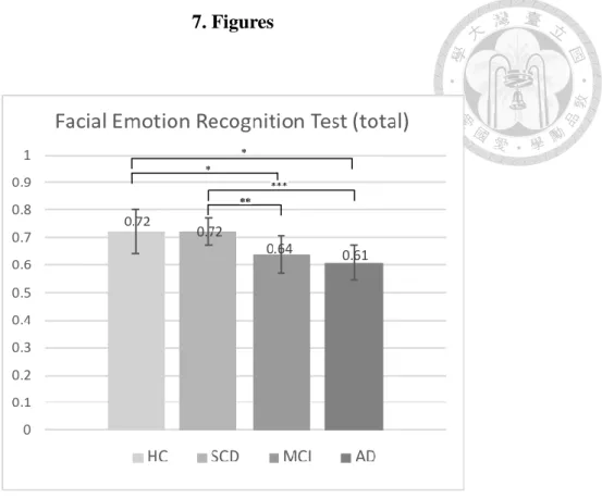
difference between HC and SCD as well as MCI and AD groups (see Figure 1). For
the interaction of photo age and group, further tests of simple main effect of photo age
showed that SCD group performed significantly better in decoding younger face than
in older face (F(1, 100) = 6.89, p = .001, ηp2 = .065). For the interaction of emotion
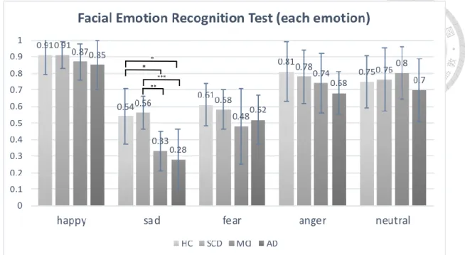
and group, further tests of simple main effect of group in decoding sadness indicated
that HC and SCD groups performed significantly better than MCI and AD groups (F(3,
403) = 20.86, p < .001, ηp2 = .134) in sadness expressions (see Figure 2), suggesting
that the accuracy difference between groups was mainly from the discrepancy in
decoding this category of expressions.
As the scores of sadness between groups could discriminate MCI and AD groups
from HC and SCD groups, it means that sadness recognition presents a remarkable
opportunity to discriminate patients who showed deficits in FER. Although the photo
ages did not show the main effect or interaction with the group in ANCOVA, we still
checked whether the own-age effect exists in sadness across groups to account for the
possibility that the own-age effect in sadness might be diminished by averaging total
emotion. We conducted a mixed-effects ANCOVA in sadness with two factors: photo
age (within-subjects) and group age (between-subjects). No significant interaction
between photo age and group (F(3, 100) = 1.261, p = .292) was found. However, the
results showed a trend that HC and SCD groups performed more accurately in
decoding younger faces compared to older faces (this trend also appeared for older
and younger observers in experiment 1), while MCI and AD groups performed more
accurately in decoding older faces than younger faces (see Figure 3). That is, it
seemed to show that the own-age trend existed in MCI and AD, but not HC and SCD.
To further explore which emotions tended to be mislabeled as from sadness by MCI
and AD groups, two-way mixed ANOVAs were conducted in these two groups.
Post-hoc analysis using the Bonferroni method found that the scores of judging
sadness to sadness were not significantly different from the scores of judging sadness
to anger and sadness to neutral. This means that MCI and AD groups tend to mislabel
sadness to either anger or neutral. To further examine whether there were differences
between mislabeling sadness as anger and as neutral under different photo ages
between the four groups, we conducted separate one-way ANCOVAs between the
four groups. The dependent variable was the proportion of wrong classifications from
sad to anger and neutral in younger and older faces, respectively. The results showed a
significant difference (F(3, 100) = 4.692, p = .004, ηp2 = .123) in mislabeling sadness
as anger in younger faces and a significant difference (F(3, 100) = 3.141, p = .029, ηp2
= .086) in mislabeling sadness as neutral in older faces across four groups. Post-hoc
analysis using the Bonferroni method confirmed that MCI and AD groups got higher
mislabeling scores compared to HC and SCD groups.
3.2.3. Clinical Utilities
From the above results, we thought sadness recognition presents a remarkable
opportunity to discriminate patients who showed deficits in FER. Thus, we conducted
the ROC curve analysis in sadness scores between SCD and MCI. The results
indicated that the sadness accuracies in younger and older faces were different
between SCD and MCI groups (area under the curve [AUC] of younger faces = 80%;
AUC of old faces = 77%). According to the Youden index (Youden, 1950), the data
showed that using a cutoff score of 0.35 for the accuracy in younger faces and a cutoff
score of 0.36 for the accuracy in older faces yielded the most desirable combination of
sensitivity (91%) and specificity (39%) in younger faces and sensitivity (81%) and
specificity (39%) in older faces respectively for identifying significant differences
between the SCD and MCI groups on the FER.
4. Discussion
The present study examined whether the own-age effect exists in healthy adults,
and patients with SCD, aMCI, and very mild AD when performing the FER Task. Our
discussion could be divided into two parts: the issue in healthy adults and patients,
respectively.
4.1. Does the own-age effect of FER exist in healthy adults?
The own-age effect means that individuals showed better performances in
recognizing the own-aged emotional expressions. Methodologically, studies on this
issue need to involve in presenting different ages of photos to different ages of groups.
Some studies included the young-aged, middle-aged, and old-aged faces and
observers, others included the young-aged and the old-aged faces and observers. In
other words, the own-age effect consists of two age-related factors, the cohort-effect
and the photo-age effect.
Considering the cohort-effect, the present study did not find the group-age effect
in terms of average accuracy of emotion recognition. Besides, having analyzed
different emotion stimuli, we also did not find the group difference in decoding
happiness, sadness, and fear. However, the old observers performed better than
younger observers in decoding anger. Our results were inconsistent with those
findings of previous studies that performances of older adults were inferior to those of
their younger counterparts in decoding sadness, fear, and anger (Calder et al., 2003;
Ruffman, Henry, Livingstone, & Phillips 2008; Isaacowitz and Stanley, 2011). The
study discrepancy might be due to higher educational level in our healthy older
participants. Although the effects of higher education on preventing MCI and AD
remain equivocal, favorable study findings indicated that people with higher
education not only performed cognitive tests better than those with lower-educated
ones, but also delayed the onset of cognitive impairment (Lenehan, Summers,
Saunders, Summers, & Vickers, 2015). Besides, Pietschnig et al. (2016) also reported
that higher-educated individuals did have better performance on the emotion
recognition task. Accordingly, it appears that higher education facilitates protective
effects not only on the decline of cognitive function (Matyas et al., 2017), but also of
emotional recognition.
In respect to the issue of the photo-age effect, the present study found a
significant interaction effect between the photo ages and the emotion types. The
younger-face was significantly easier than the older-face recognition for both younger
and older observers in decoding fear and sad expressions; however, the reverse was
observed in decoding happiness. For fear and sadness recognitions, our results were
consistent with those findings of previous studies indicating that healthy adults
decoding younger faces were more accurate than older faces (Ebner et al., 2010,
2011). Such an outcome, as suggested by researchers (Albert, Ricanek, & Patterson,
2007; Porcheron, Mauger, & Russell, 2013) may be attributed to age-related changes
of older faces (e.g., wrinkles and folds) that were more dissimulated, mixed, and
fragmental than their younger counterparts’ ones. However, for the happiness stimuli,
our results were inconsistent with those findings of prior studies (Ebner & Johnson,
2009; Ebner et al., 2010, 2011, 2012; Richter et al., 2010; Riediger et al., 2011;
Hühnel et al., 2014). The discrepancy might be due to two methodologic limitations
in the present study: 1. our posed photo stimuli were less spontaneous in nature; 2. the
intensities of our happy photos in younger faces were relatively low (the intensity was
measured by the 5-point Likert scale, for more details see Methods). The posed
photos were reported to be less ecological validity than the spontaneous photos
(Bartlett et al., 2006; Murphy et al., 2010). Additionally, our results revealed that the
intensities of our younger faces were significantly lower than those of the older faces
in happiness (see Table 6). That is, the younger performers in our photos tended to
present low-intense happy expressions than the older performers. Thus, in the younger
faces, both of our younger and older observers tended to misrecognize happy
expressions to neutral expression.
Taken together, the present study did not find the own-age effect on
emotional-expression recognition in older observers. Likewise, it was also the case for
younger observers, with the exception in decoding neutral photos. Given the present
results consistent with most studies (Borod et al., 2004; Ebner and Johnson, 2009;
Murphy et al., 2010; Hühnel et al., 2014), the own-age effect on the facial emotional
recognition appeared not remarkable, irrespective of younger or older healthy people.
However, the present results were inconsistent with other prior studies displaying the
own-age effect evident in older adults when performing happiness and anger
recognitions (Riediger et al., 2011; 2014), and in younger adults (Malatesta et al.,
1987; Richter et al., 2010) performing the happiness, anger, and sadness ones. Three
methodologic factors might attribute the inconsistent results. One factor was the
unrepresentative sample in those studies (Malatesta et al., 1987; Richter et al., 2010).
The other factor was limited stimuli (Riediger et al., 2014). The last possible
contributor was the discrepancy of the emotional rating format. The forced-choice
approach in our study has generally been used in most studies; however, the
multi-dimensional response format (measuring the percentage across all emotions for
every individual photo, for more details, see Introduction) was used to measure each
of the photos stimuli in Riediger and coworkers (2011). In fact, the type of emotional
experiences in real life is always multi-faceted (Hemenover & Schimmack, 2007);
thus, this response format was more ecologically valid in nature. Nevertheless,
whether this methodologic discrepancy can fully attribute to the inconsistent results
remains further investigation.
4.2. Is the own-age effect evident in the patients while performing the FER?
The present study examined the FER performances in SCD, aMCI and very mild
AD patients after minimizing the methodologic problems. The present study found
that the MCI and AD groups showed FER deficits as compared to the HC and SCD
groups in decoding sadness (see Figure1, Figure 2). Furthermore, the patients tended
to mislabel sadness for anger and for neutral expression when perceiving younger and
old faces respectively. Our results supported the previous findings indicating that FER
deficits occurred in MCI and early-stage AD patients (McCade, Savage, & Naismith,
2011; Varjassyová et al., 2013; Torres et al., 2015), but not in SCD patients compared
to healthy older adults (Pietschnig et al., 2016).
Davidson, Putnam, and Larson (2000) proposed that a neural network
responsible for emotion involving the orbital prefrontal cortex, ventromedial
prefrontal cortex, dorsolateral prefrontal cortex, amygdala, hippocampus,
hypothalamus, anterior cingulate cortex (ACC), insular cortex, and ventral striatum.
Based on the locationist hypothesis, each of the distinct emotions has its own
underlying neural substrate (Barrett, 2006). In fact, a recent study found that the ACC
plays an essential role in processing sadness-related information (Lindquist, Wager,
Kober, Bliss-Moreau & Barrett, 2012). Accordingly, it appears feasible to speculate
such sad recognition problems possibly due to the ACC dysfunction which might
indirectly result from pathological changes of the hippocampal cortices and related
regions commonly evident in early AD and aMCI (Hyman, Van Hoesen, Damasio &
Barnes, 1984). Nevertheless, given that a small group of the patients (particularly
early AD) was sampled in the present study, and the mechanism for the results of
defective recognition of sadness remains unclear, further investigation on a large scale
is necessary.
Regarding the issue of the own-age effect, the present study found that aMCI and
very mild AD patients tended to have the own-age effect on the FER compared with
the healthy compartments (see Figure 3). However, the effect was not significant, and
the accuracy of FER in decoding the own-aged photos remained low.
In short, the present study found that aMCI and very mild AD patients showed
defective FER in sadness with a tendency to mislabel it to anger and neutral in
younger and older faces, respectively. Accordingly, it appears that a measure with
low-intensity of FER (i.e., sadness) can be sensitive to detect patients with very mild
AD and aMCI. Our results also revealed that a tendency of the own-age effect
occurred in patients.
To our knowledge, our study is the first one to investigate several issues,
including whether the own-age effect on FER exists in patients with aMCI and very
mild AD, and is also one or two studies examining FER functioning in individuals
with SCD. However, there were some limitations in the present study: 1. The
educational level of all participants in this study was relatively high, especially in
SCD group. 2. The measuring format (i.e., the forced-choice approach) and the type
of photos (posed and static photos) were less ecological validity. 3. The aMCI
participants were not classified into single or multiple domains. 4. Given the doctrine
of “ZhongYong” responding style to the odd-level rating scale in most
Taiwanese/Chinese people (吳毓瑩,1996; 黃金蘭、林以正、楊中芳,2012; 廖培
珊,2010; Chen, Lee, & Stevenson, 1995), our participants might have a biased rather
than a true rating on the 5-point Likert scale for the intensity of each emotional photo
stimulus. 5. The confounding effect due to incomparable intensity of facial-emotion
stimuli between younger- and older- individual photos in the present study might
influence the results though currently adequate matching means remains unavailable.
Further studies on these issues are thus requisite.
In summary, the present finding indicated that younger faces are easier than older
faces to decode fear and sadness for both younger and older adults. Although the
own-age effect was not evident in healthy adults, the tendency of such an effect
appeared in patients with aMCI and very mild AD when decoding sadness. The
present study elucidated the potential pathophysiological mechanism underlying the
relationships between AD and the sad recognition problems. Nevertheless, due to
small sampling in our study and still lacking neuroimaging evidence, future research
with a larger sample size and regarding the neuroimaging confirmation is needed.
Besides, even though different ages of photos did not affect the FER, using older
faces as a clinical stimuli might increase the medical relationship and the mental
caring of patients. Nevertheless, the hit rates of certain expressions in our stimuli
database were low. Further research using the multi-dimensional response format and
the dynamic and spontaneous photos might eliminate the problem.
5. References
Chinese References
(Each reference in this part was translated to English from the original Chinese
language listed right below).
吳毓瑩 (1996):〈量表奇偶點數的效度議題〉。 《調查研究: 方法與應用》,2,
5-34。
廖培珊 (2010):〈態度量表之選項標示語: 調查資料之潛藏類別分析〉。 《調查
研究-方法與應用》,24,91-134。
黃金蘭、林以正、楊中芳 (2012) :〈中庸信念-價值量表之修訂〉。 《本土心
理學研究》,38,3-41。
English References
Adolphs, R. (2001). The neurobiology of social cognition. Current Opinion in
Neurobiology, 11, 231-239.
Adolphs, R. (2002). Neural systems for recognizing emotion. Current Opinion in
Neurobiology, 12, 169-177.
Albert, A. M., Ricanek Jr, K., & Patterson, E. (2007). A review of the literature on the
aging adult skull and face: Implications for forensic science research and
applications. Forensic Science International, 172, 1-9.
Albert, M. S., DeKosky, S. T., Dickson, D., Dubois, B., Feldman, H. H., Fox, N. C., ...
Snyder, P. J. (2011). The diagnosis of mild cognitive impairment due to Alzheimer’s disease: Recommendations from the National Institute on
Aging-Alzheimer’s Association workgroups on diagnostic guidelines for
Alzheimer's disease. Alzheimer's & Dementia, 7, 270-279.
Bäckman, L. (1991). Recognition memory across the adult life span: The role of prior
knowledge. Memory & Cognition, 19, 63-71.
Barrett, L. F. (2006). Are emotions natural kinds?. Perspectives on Psychological
Science, 1, 28-58.
Bartlett, M. S., Littlewort, G., Frank, M. G., Lainscsek, C., Fasel, I. R., & Movellan, J.
R. (2006). Automatic recognition of facial actions in spontaneous expressions.
Journal of Multimedia, 1, 22-35.
Becker, D. V., Kenrick, D. T., Neuberg, S. L., Blackwell, K. C., & Smith, D. M.
(2007). The confounded nature of angry men and happy women. Journal of
Personality and Social Psychology, 92, 179.
Benton, A. L., Abigail, B., Sivan, A. B., Hamsher, K. D., Varney, N. R., & Spreen, O.
(1994). Contributions to neuropsychological assessment: A clinical manual.
New York: Oxford University Press.
Bowers, D., Blonder, L. X., & Heilman, K. M. (1998). Florida affect battery.
Gainesville: University of Florida.
Borod, J. C., Yecker, S. A., Brickman, A. M., Moreno, C. R., Sliwinski, M., Foldi, N.
S., ... Welkowitz, J. (2004). Changes in posed facial expression of emotion
across the adult life span. Experimental Aging Research, 30, 305-331.
Calder, A. J., Keane, J., Manly, T., Sprengelmeyer, R., & Scott, S. Nimmo-Smith, I.
(2003). Facial expression recognition across the adult life span.
Neuropsychologia, 41, 195-202.
Cacioppo, J. T., Berntson, G. G., Bechara, A., Tranel, D., & Hawkley, L. C. (2011).
Could an aging brain contribute to subjective well-being? The value added by
a social neuroscience perspective. In A. Tadorov, S. T. Fiske & D. Prentice
(Eds.), Social neuroscience: Toward understanding the underpinnings of the
social mind (pp. 249-262). New York: Oxford University Press.
Cheng, C. M., Chen, H. C., Chan, Y. C., Su, Y. C., & Tseng, C. C. (2013). Taiwan
corpora of Chinese emotions and relevant psychophysiological
data—Normative Data for Chinese Jokes. Chinese Journal of Psychology, 55,
555–569.
Chen, C., Lee, S. Y., & Stevenson, H. W. (1995). Response style and cross-cultural
comparisons of rating scales among East Asian and North American students.
Psychological Science, 6, 170-175.
Chen, H. Y., Hua, M. S., Zhu, J., & Chen, Y. H. (2008). Selection of factor-based
WAIS-III tetrads in the Taiwan standardization sample: A guide to clinical
practice. Chinese Journal of Psychology, 50, 91-109.
Ciarrochi, J. V., Chan, A. Y., & Caputi, P. (2000). A critical evaluation of the
emotional intelligence construct. Personality and Individual Differences, 28,
539-561.
Davidson, R. J., Putnam, K. M., & Larson, C. L. (2000). Dysfunction in the neural
circuitry of emotion regulation--a possible prelude to violence. Science, 289,
591-594.
Derntl, B., Kryspin-Exner, I., Fernbach, E., Moser, E., & Habel, U. (2008). Emotion
recognition accuracy in healthy young females is associated with cycle phase.
Hormones and Behavior, 53, 90-95.
Ebner, N. C., & Johnson, M. K. (2009). Young and older emotional faces: Are there
age group differences in expression identification and memory?. Emotion, 9,
329.
Ebner, N. C., Riediger, M., & Lindenberger, U. (2010). FACES—A database of facial
expressions in young, middle-aged, and older women and men: Development
and validation. Behavior Research Methods, 42, 351-362.
Ebner, N. C., He, Y. I., & Johnson, M. K. (2011). Age and emotion affect how we
look at a face: Visual scan patterns differ for own-age versus other-age
emotional faces. Cognition & Emotion, 25, 983-997.
Ebner, N. C., Johnson, M. R., Rieckmann, A., Durbin, K. A., Johnson, M. K., &
Fischer, H. (2013). Processing own-age vs. other-age faces: Neuro-behavioral
correlates and effects of emotion. Neuroimage, 78, 363-371.
Ekman, P. & Friesen, W.V. (1978). Facial action coding system: A technique for the
Measurement of Facial Movement. Palo Alto, CA: Consulting Psychologists
Press.
Fujie, S., Namiki, C., Nishi, H., Yamada, M., Miyata, J., Sakata, D., ... Murai, T.
(2008). The role of the uncinate fasciculus in memory and emotional
recognition in amnestic mild cognitive impairment. Dementia and Geriatric
Cognitive Disorders, 26, 432-439.
Greenwood, P. M. (2000). The frontal aging hypothesis evaluated. Journal of the
International Neuropsychological Society, 6, 705-726.
Gur, R. C., Sara, R., Hagendoorn, M., Marom, O., Hughett, P., Macy, L., ... Gur, R. E.
(2002). A method for obtaining 3-dimensional facial expressions and its
standardization for use in neurocognitive studies. Journal of Neuroscience
Methods, 115, 137-143.
Gunes, H., & Pantic, M. (2010). Automatic, dimensional and continuous emotion
recognition. International Journal of Synthetic Emotions (IJSE), 1, 68-99.
Hachinski, V. C., Iliff, L. D., Zilhka, E., Du Boulay, G. H., McAllister, V. L.,
Marshall, J., ... Symon, L. (1975). Cerebral blood flow in dementia. Archives
of Neurology, 32, 632-637.
Hargrave, R., Maddock, R. J., & Stone, V. (2002). Impaired recognition of facial
expressions of emotion in Alzheimer's disease. The Journal of
Neuropsychiatry and Clinical Neurosciences, 14, 64-71.
Haxby, J. V., Hoffman, E. A., & Gobbini, M. I. (2000). The distributed human neural
system for face perception. Trends in Cognitive Sciences, 4, 223-233.
Haxby, J. V., Hoffman, E. A., & Gobbini, M. I. (2002). Human neural systems for
face recognition and social communication. Biological Psychiatry, 51, 59-67.
Hess, U., Blairy, S., & Kleck, R. E. (1997). The intensity of emotional facial
expressions and decoding accuracy. Journal of Nonverbal Behavior, 21,
241-257.
Hennenlotter, A., & Schroeder, U. (2006). Partly dissociable neural substrates for
recognizing basic emotions: A critical review. Progress in Brain Research,
156, 443-456.
Hemenover, S. H., & Schimmack, U. (2007). That's disgusting!…, but very amusing:
Mixed feelings of amusement and disgust. Cognition and Emotion, 21,
1102-1113.
Hühnel, I., Fölster, M., Werheid, K., & Hess, U. (2014). Empathic reactions of
younger and older adults: No age related decline in affective responding.
Journal of Experimental Social Psychology, 50, 136-143.
Hua, M., Chang, B., Lin, K., Yang, C., & Chen, H. (2005). Wechsler Memory Scale
Third Edition (WMS-III) Manual for Taiwan. Taipei, Taiwan: The Chinese
Behavioral Science Corporation.
Hyman, B. T., Van Hoesen, G. W., Damasio, A. R., & Barnes, C. L. (1984).
Alzheimer's disease: Cell-specific pathology isolates the hippocampal
formation. Science, 225, 1168-1170.
Isaacowitz, D. M., & Stanley, J. T. (2011). Bringing an ecological perspective to the
study of aging and recognition of emotional facial expressions: Past, current,
and future methods. Journal of Nonverbal Behavior, 35, 261.
Jessen, F., Amariglio, R. E., van Boxtel, M., Breteler, M., Ceccaldi, M., Chetelat,
G., . . . Subjective Cognitive Decline Initiative Working, G. (2014). A
conceptual framework for research on subjective cognitive decline in
preclinical Alzheimer's disease. Alzheimer’s & Dementia, 10, 844-852.
doi:10.1016/j.jalz.2014.01.001
Keane, J., Calder, A. J., Hodges, J. R., & Young, A. W. (2002). Face and emotion
processing in frontal variant frontotemporal dementia. Neuropsychologia, 40,
655– 665. doi:10.1016/S0028-3932(01)00156-7
Kreibig, S. D., Samson, A. C., & Gross, J. J. (2013). The psychophysiology of mixed
emotional states. Psychophysiology, 50, 799-811.
Lamont, A. C., Stewart-Williams, S., & Podd, J. (2005). Face recognition and aging:
Effects of target age and memory load. Memory & Cognition, 33, 1017-1024.
Lenehan, M. E., Summers, M. J., Saunders, N. L., Summers, J. J., & Vickers, J. C.
(2015). Relationship between education and age‐related cognitive decline: A
review of recent research. Psychogeriatrics, 15, 154-162.
Liao, Y. C., Yeh, T. L., Yang, Y. K., Lu, F. H., Chang, C. J., Ko, H. C., & Lo, C. M.
(2004). Reliability and validation of the Taiwan geriatric depression scale.
Taiwanese Journal of Psychiatry, 18, 30-41.
Lindquist, K. A., Wager, T. D., Kober, H., Bliss-Moreau, E., & Barrett, L. F. (2012).
The brain basis of emotion: A meta-analytic review. The Behavioral and
Brain Sciences, 35, 121.
Schneider, W., Eschman, A., & Zuccolotto, A. (2002). E-Prime: User's guide.
Pittsburgh, PA: Psychology Software Tools.
Schroeder, U., Hennenlotter, A., Erhard, P., Haslinger, B., Stahl, R., Lange, K. W., &
Ceballos‐Baumann, A. O. (2004). Functional neuroanatomy of perceiving
surprised faces. Human Brain Mapping, 23, 181-187.
Spoletini, I., Marra, C., Di Iulio, F., Gianni, W., Sancesario, G., Giubilei, F., ...
Spalletta, G. (2008). Facial emotion recognition deficit in amnestic mild
cognitive impairment and Alzheimer disease. The American Journal of
Geriatric Psychiatry, 16, 389-398.
Sporer, S. L. (2001). Recognizing faces of other ethnic groups: An integration of
theories. Psychology, Public Policy, and Law, 7, 36.
Studart Neto, A., & Nitrini, R. (2016). Subjective cognitive decline: The first clinical
manifestation of Alzheimer's disease?. Dementia & Neuropsychologia, 10,
170-177.
Sze, J. A., Goodkind, M. S., Gyurak, A., & Levenson, R. W. (2012). Aging and
emotion recognition: Not just a losing matter. Psychology and Aging, 27, 940.