光交聯型乙二醇幾丁聚醣水凝膠用於組織黏合劑和具 備抗菌功能之止血材料
81
0
0
全文
(2) 致謝 感謝蔡偉博老師兩年來的悉心指導,讓我學習如何獨自完成專題研究,同時 也給予我許多實驗技巧、數據分析和論文表達上的各種建議,更讓我習得正面積 極的研究態度。在共同定期會議中游佳欣老師也時常給予我不同的觀點和實驗建 議,讓我在研究歷程中獲益良多。特別感謝游佳欣老師和貴實驗室林哲緯學長協 助操作動物實驗,感謝王孟菊老師與貴實驗室同學協助借用廻轉式動態流變儀, 以及台灣大學高分子所協助代測流變測試,感謝中央大學黃俊仁、黃俊銘教授與 黃詠琪同學協助操作抗菌實驗。同時感謝實驗室的張睿學長、劉藝學長、黃宗駿 學長、徐靖峰學長、陳政宏學長、陳玟璇學姐、蔡品妏學姐、廖子瑩學姐、張甯 筑學姐、伍世昌同學、林詠皓同學和黃楷文同學,傳授實驗技巧和與我討論實驗 細節。尤其感謝家人與朋友對我的支持與鼓勵。. I.
(3) 摘要 本研究期望製備出以乙二醇幾丁聚醣為主要材料和透過光交聯機制快速成 膠之水凝膠,本研究將探討其材料性質、組織黏合、抵抗細菌感染和止血的能力, 驗證此水凝膠能有效做為手術、創傷組織黏合劑以及止血和抗菌功能之材料。乙 二醇幾丁聚醣除保有幾丁聚醣本身優秀的生物相容性、生物降解性、止血和抗菌 能力外,良好的水溶性克服原本幾丁聚醣需溶於酸性環境下的限制,使得此水凝 膠製備和使用更加容易。本研究所使用之光交聯機制是以銣錯合物和過硫酸鈉為 交聯劑,混入經酚基團修飾之乙二醇幾丁聚醣溶液後,僅需以藍光波段照射數秒, 即可快速成膠,此交聯方式產生之共價鍵能提供水凝膠本身良好的機械強度,同 時能與凝膠接觸之動物組織表面形成鍵結達到組織黏合的功效,此外,本交聯方 法除所需成膠時間少,能賦予此水凝膠及時注射於不規則傷口和控制成膠時機等 優勢。本水凝膠亦可作為藥物載體和釋放系統,本研究透過混入抗生素並以光交 聯方式使抗生素包覆於水凝膠中來達到適當的釋放效果,確保傷口於癒合期間能 有效抵抗細菌感染。本水凝膠除本身具備強勁的組織黏合力來黏合傷口出血點作 為止血的物理性阻隔外,幾丁聚醣因其本身帶電基團的效應能進一步達到凝血與 止血效果。本研究藉由核磁共振光譜和可見光紫外光分光光譜檢驗材料合成,以 壓力測試機和雞蛋內膜測試動物組織黏合力,水合和降解實驗測試材料穩定性, 以細胞存活率測試方法檢驗水凝膠可能造成的細胞毒性,利用藥物釋放實驗記錄 水凝膠釋放效果,並以抑菌圈實驗確認水凝膠包覆抗生素的抗菌能力,最後以小 鼠肝臟出血實驗觀察水凝膠止血效果與實際動物應用之成效,由以上實驗與數據 來驗證本水凝膠確實能作為理想的組織黏合劑、抗菌和止血材料。. 關鍵字: 乙二醇幾丁聚醣 ; 水凝膠 ; 光交聯 ; 止血 ; 組織黏合劑 ; 抗菌材 料 ; 藥物釋放. II.
(4) Abstract Tissue adhesives commonly serve as wound dressing and sealant used in surgery. In this study, injectable glycol chitosan based hydrogels were produced by covalent crosslinking initiated by blue light, ruthenium complex, and persulfate. By this photochemical crosslinking method, hydrogels can be made rapidly less than 10 seconds and have strong bond with animal tissue. With these advantages, this hydrogel is able to seal irregular wound and stop wound from bleeding simultaneously. Egg membrane was utilized as animal tissue to test tissue adhesion of hydrogel, and the result demonstrates that it has outstanding ability to adhere to animal tissue. In MTS test, this hydrogel also show its low cytotoxicity and great biocompatibility. In vivo hemostasis, applying hydrogel solution on the wound of mice live and crosslinking the gel by blue light, the hydrogel successfully adhere to the wound and arrested bleeding within 20 seconds. Moreover, mixing antibiotics in hydrogel and releasing antibiotics properly, the hydrogel can initially prevent wound from bacterial infection. These rapidly photocrosslinked glycol chitosan hydrogels are expected to be used as injectable tissue adhesives, drug delivery system, antibacterial wound dressing, and hemostatic materials in its promising future. Key words: glycol chitosan, hydrogel, photochemical crosslink, hemostasis, tissue adhesive, antibacterial material, drug release. III.
(5) Content 致謝................................................................................................................................ I 摘要............................................................................................................................... II Abstract ...................................................................................................................... III Content ....................................................................................................................... IV List of Figures ......................................................................................................... VIII List of Tables.............................................................................................................. XI Chapter 1 Introduction ................................................................................................ 1 1.1 Tissue Adhesives ................................................................................................. 1 1.1.1 Background of tissue adhesives .................................................................... 1 1.1.2 Problem-solving ability of tissue adhesives .................................................. 2 1.1.3 Developed tissue adhesives and hemostats ................................................... 3 1.2 Crosslinking Mechanism of Fabricating Tissue Adhesives ............................ 6 1.2.1 Commonly used crosslinking mechanism .................................................... 7 1.2.2 A ruthenium-based photochemical crosslinking ........................................... 7 1.3 Polymers for Producing Tissue Adhesives ....................................................... 8 1.3.1 Previously used polymers ............................................................................. 8 1.3.2 Chitosan and its derivatives .......................................................................... 9 IV.
(6) 1.3.3 Glycol chitosan based tissue adhesive ........................................................ 10 1.4 Functionalized With Hemostatic and Antibacterial Ability ......................... 10 1.4.1 Hemostatic ability of this new tissue adhesive ........................................... 10 1.4.2 Introducing antibiotics in this new tissue adhesive..................................... 10 1.5 Motivation and Aims........................................................................................ 11 1.6 Research Framework ....................................................................................... 12 Chapter 2 Materials and Methods............................................................................ 14 2.1 Chemicals .......................................................................................................... 14 2.1.1 Synthesis of HPP-GC.................................................................................. 14 2.1.2 Preparation of photo-crosslinked adhesives................................................ 14 2.1.3 Swelling and degradation ............................................................................ 14 2.1.4 MTS assay................................................................................................... 15 2.1.5 In vitro drug release .................................................................................... 15 2.1.6 Antibacterial ability..................................................................................... 15 2.2 Experimental Instruments and Materials ..................................................... 16 2.2.1 Experimental instruments ........................................................................... 16 2.2.2 Experimental materials ............................................................................... 17 2.3 Solution Formula ............................................................................................. 17 2.4 Methods of Material Evaluation ..................................................................... 18. V.
(7) 2.4.1 Synthesis of HPP-GC.................................................................................. 18 2.4.2 Characterization of HPP-GC....................................................................... 19 2.4.3 Preparation of photo-crosslinked HPP-GC adhesives ................................ 19 2.4.4 Tissue adhesion ........................................................................................... 20 2.4.5 Rheological analysis ................................................................................... 20 2.4.6 Hydration .................................................................................................... 21 2.4.7 Degradation ................................................................................................. 21 2.4.8 MTS assay................................................................................................... 22 2.5 Methods of Functional Evaluation ................................................................. 23 2.5.1 In vitro drug release .................................................................................... 23 2.5.2 Antibacterial ability..................................................................................... 23 2.5.3 In vivo hemostatic ability............................................................................ 24 2.6 Statistic Analysis............................................................................................... 25 Chapter 3 Results and Discussion ............................................................................ 30 3.1 Synthesis and Characterization of HPP-GC ................................................. 30 3.2 Fabrication of Photocrosslinked HPP-GC Hydrogel .................................... 31 3.3 Strong Tissue Adhesion.................................................................................... 32 3.4 Rheological Analysis and Mechanical Property ............................................ 33 3.5 Hydration of Different DS HPP-GC Hydrogels ............................................ 34. VI.
(8) 3.6 Degradation and Stability of HPP-GC Hydrogel .......................................... 35 3.7 Cell Viability and Cytotoxicity ........................................................................ 36 3.8 In Vitro Drug Release ...................................................................................... 36 3.9 Measurement of Antibacterial Ability ............................................................ 37 3.10 In Vivo Hemostatic Ability ............................................................................ 38 Chapter 4 Conclusion ................................................................................................ 58 References ................................................................................................................... 59. VII.
(9) List of Figures Figure 1-1 Experimental scheme of this research ........................................................ 13 Figure 2-1 Synthesis of phenol groups modified glycol chitosan via EDC & NHS method. ............................................................................................................. 26 Figure 2-2 Fabrication of HPP-GC hydrogel. .............................................................. 27 Figure 2-3 Mechanism of photochemical crosslinking ................................................ 28 Figure 2-4 (A) Digital force gauge. (B) Procedure of testing the detachment stress of hydrogel by using egg membrane. ................................................................... 29 Figure 3-1 1H NMR analysis of glycol chitosan and HPP-GC. ................................... 40 Figure 3-2 Uv-visible analysis of glycol chitosan, HPP and HPP-GC. ....................... 41 Figure 3-3 Gelation of HPP-GC hydrogel. .................................................................. 42 Figure 3-4 Appearance of hydrogel with different DS. (A) low DS (~0.250 µmole/mg HPP-GC) (B) moderate DS (~ 0.370 µmole/mg HPP-GC) (C) high DS (> 0.534 µmole/mg HPP-GC)......................................................................................... 43 Figure 3-5 Detachment stress of hydrogel with different DS. (n = 5, * and*** represent p < 0.05 and < 0.001 in comparison with sample of 0.378 DS) ...................... 44 Figure 3-6 Detachment stress of hydrogel with different concentration. (DS of sample: 0.368 μmole/mg HPP-GC) (n = 5, * represents p < 0.05 in comparison with 3%) .......................................................................................................................... 45 VIII.
(10) Figure 3-7 Storage modulus (G’) and viscous modulus (G’’) of HPP-GC hydrogel as a function of frequency (Hz) at 37 ℃. ............................................................... 46 Figure 3-8 Hydration of dry hydrogel with different DS. (n = 4, * represents p < 0.05 in comparison with sample of moderate DS) ................................................... 47 Figure 3-9 Degradation of hydrogels. (n = 5) .............................................................. 48 Figure 3-10 Degradation of hydrogels after removing the water on surface. (n = 4) .. 49 Figure 3-11 Cell viability of different concentration of extraction form hydrogel. (n = 5) ...................................................................................................................... 50 Figure 3-12 Amoxicillin released from hydrogel. (n = 4) ............................................ 51 Figure 3-13 (A) E coli. and (B) S. epidermidis were the used. (1) 1000, (2) 200 and (3) 50 μg/ml of gentamycin were entrapped in hydrogel. (4) Commercial gauzes were immersed in 1000 μg/ml gentamycin and (5) 10 µl 1000 μg/ml gentamycin as control. ......................................................................................................... 52 Figure 3-14 (A) E coli. and (B) S. epidermidis were the used. (1) 200, (2) 100 and (3) 50 μg/ml of gentamycin were entrapped in hydrogel. (4) HPP-GC hydrogel without gentamycin (5) 10 µl 200 μg/ml gentamycin as control. .................... 53 Figure 3-15 Procedure of in vivo hemostatic experiment. ........................................... 54 Figure 3-16 Blood loss from wound. (n = 3) ............................................................... 55 Figure 3-17 Filter paper, which absorbed the blood from wound (A) Control (B). IX.
(11) Applying HPP-GC without crosslinking (C) Applying hydrogel. ................... 56. X.
(12) List of Tables Table 3-1 Inhibition of zone of different concentration of gentamycin against E coli. and S. epidermidis. (n = 4) ............................................................................... 57. XI.
(13) Chapter 1 Introduction 1.1 Tissue Adhesives 1.1.1 Background of tissue adhesives Traditional treatment of wound closure and healing such as sutures has some weakness and is recognized ineffective today. Sutures often cause additional damage and inflammation to tissue, and cannot arrest body fluid of wounds [1, 2]. Tissue adhesives attract much attention nowadays, such materials could be appealing alternatives to sutures and staples since they can be applied more quickly, causes less pain and may require less equipment in certain circumstances. Besides, two main factors are now affecting the modern practice of surgery. One is cost containment and the other is aging population. Because of aging population, increasing need of medical care and surgery, which cause augment of medical cost, become serious problem. In order to overcome these challenges, the skills and materials utilized in surgery should be improved. One solution is the effective use of hemostats, sealants, and adhesives. These materials are useful in surgical procedure and are crucial components of the surgical toolbox. The ideal hemostat, sealant, and adhesive, nevertheless, were not effective enough at present. This lack of a promising product sometimes cause frustration and waste of money and time. Continued research and 1.
(14) development is still required to provide new materials [2]. For the purposes of resolving the challenges, new biomaterials and tissue adhesives are necessary. A desirable surgical tissue adhesive should allow rapid adhesion and maintain strong and close bond with tissue around wound for an amount of time sufficient to allow wound healing [3]. Ideal tissue adhesives cannot only hold tissues together but also serve as barriers to leakage when used for wound closure [3]. Moreover, it should not interfere with body’s natural healing mechanisms and should degrade without producing an excessive localized or generalized inflammatory response. With all these features mentioned above, tissue adhesives could serve as sealant, hemostatic material and non-invasive wound dressing [4, 5]. It is our first priority to develop an ideal tissue adhesive possessing those merits.. 1.1.2 Problem-solving ability of tissue adhesives Looking forward to creating a versatile tissue adhesive, which can handle a wide range of situation in medical treatment, a prominent tissue adhesive should possess additional functions. Tissue adhesive can also utilized as hemostat, and it is expected to solve many problems in surgical procedure. For example, hemostasis for reducing blood loss is significant for patients in clinical application [6]. Biomaterials serve as sealant and hemostat play pivotal role in this field, and some widely used tissue adhesives and hemostats will be. 2.
(15) discussed. Those materials have their own drawbacks and that is the reason why new advanced tissue adhesives and hemostats should be invented. Another challenge has to be overwhelmed is the infection caused by bacteria, which often occurs during the healing process of wound, and sometimes a wound dressing itself can cause infection as well [7]. As a result, wound dressings including tissue adhesives with antibacterial ability have been emphasized recently. For example, hydrogels made from chitosan and silver nitrate or hydrogels fabricated with silver nanoparticles and ZnO nanocomposite hydrogels have been developed [8-10]. Nevertheless, silver nitrate and metal oxides exhibit a cytotoxicity even at low concentrations. In order to make a better tissue adhesive with hemostatic and antibacterial ability, alternative methods and polymers should be considered, and how this research enable new material to perform these functions would be mentioned subsequently.. 1.1.3 Developed tissue adhesives and hemostats There are some developed tissue adhesives including Fibrin glues [11, 12], albuminglutaraldehyde adhesives [13], cyanoacrylates [3] and polyethylene glycol glues [14] that are currently used in many surgical procedures [6]. Fibrin glues and cyanoacrylates, receiving plenty of attractiveness before, would be mainly discuss as following. Through realizing the challenge encountered by these currently used tissue adhesives. 3.
(16) and hemostats, we could find out what problem we should solve, and then an ideal material can be designed. Fibrin tissue glue was first used as an adhesive material in 1940. Fibrin based tissue adhesives are composed of purified fibrinogen and thrombin, and form a bond via the physiological cascade of coagulation [3]. Fibrin glue is very successful as a sealant, especially, utilized with sutures or clips. It has been used clinically in many fields including anal fistulae closure [15], prevention of esophageal leakage and stricture after esophageal reconstruction from caustic injury [16] and so forth. However, with some drawback, fibrin glue have some limitation when utilized in surgery. Fibrin glue need complicate preparation that is time-consuming and costly. While fibrin glues are less toxic than cyanoacrylates, their low adhesive strength limits their applicability in many surgical procedures [17], and it has risks of allergic reactions to patients [18-20]. In addition, since fibrin tissue adhesives are prepared from pooled human blood, there is concern. for. potential. viral. transmission,. especially hepatitis. and. human. immunodeficiency virus [21]. Cyanoacrylates were first synthesized in 1949 [22] and were first reported as tissue adhesives 1959 [23]. There are three FDA-approved cyanoacrylate products. They are 2-octyl cyanoacrylate (Dermabond) and the two commercial forms of n-butyl-2cyanoacrylates (Indermil, and Histoacryl and Histoacryl Blue). They primarily serve as. 4.
(17) surgical closure of skin incisions. All the cyanoacrylate may be used to hold skin edges together. The adhesive polymerizes within about 30 seconds in the presence of local hydroxyl groups [2]. Nevertheless, the toxicities of aldehyde-containing and cyanoacrylates adhesives significantly circumscribe the application [24-26]. The toxicity of cyanoacrylate glues results from many factors including direct toxicity of monomers such as methyl-2-cyanoacrylate or of byproducts such as cyanoacetate and formaldehyde [27], insufficient tissue vascularization [17], and all these products release heat when placed on the skin and may result in discomfort [2]. Another limit of cyanoacrylates is the fact that they are hard and brittle; hence, they are inflexible for the dynamic nature in vivo [28]. There are some developed hemostats, and they can be divided into three main categories: Mechanical agents, active agents and flowables. Porcine gelatins, kind of mechanical agents, are produced as sponges and powders. They serve as mechanical barriers to bleeding by forming a matrix. There are some safety issues such as the placement of a foreign body in the surgical field and swelling of these materials as well. Hence, these hemostats should not be used in infected fields and they may cause a foreign body reaction. Swelling may also cause injury to nearby tissues and cells including nerves. The other products in this category including bovine collagen, oxidized regenerated cellulose and polysaccharide spheres [2].. 5.
(18) Thrombin, kind of active agents, is marketed as bovine human pooled plasma and recombinant. It can actively convert the fibrinogen contained in blood to fibrin and thereby creating a clot. However, there are some safety concerns that thrombin products should never be utilized for intravascular injection since it can reverse the anticoagulation effect of heparin and result in clotting. Other concern is the bovinederived product should not be used in patients with bovine allergies [2]. Human plasma thrombin with bovine gelatin matrix, kind of flowables hemostats, uses a combination of human thrombin and bovine gelatin to make a granular hemostat that uses both active and mechanical components to achieve hemostasis. Safety concerns are like those of the mechanical agents and active agents such as swelling and reversing anticoagulation [2]. Another well-known hemostat and sealant, fibrin glue, was introduced in previous section [20]; however, with the drawback mentioned above, better materials must be invented to overcome the weakness of fibrin glue.. 1.2 Crosslinking Mechanism of Fabricating Tissue Adhesives The ways to fabricate hydrogels and tissue adhesives also have critical influence on their application. In this section, commonly used crosslinking mechanism would be introduced, and the major mechanism utilized in this research would be emphasized as. 6.
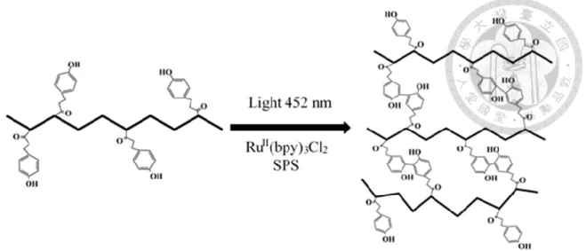
(19) following.. 1.2.1 Commonly used crosslinking mechanism A variety of methods of fabricating modified chitosan hydrogel has been used such as metal-ion mediated [29], enzymatically crosslinked [30, 31], catalytically crosslinked [32] and hybrid crosslinked hydrogel [33]. However, those methods have some disadvantages including time-consuming crosslinking process, high toxicity, low mechanical strength, inability of in-situ formation and so forth [29, 34]. Among these mechanisms, enzymatically crosslinked utilizing HRP and H2O2 is one of the most extensively investigated approaches. The HRP cross-linking allows rapid crosslinking when HRP, H2O2 and phenol-rich polymers react together [30]. Despite the advantages of in-situ gelation and rapidity of crosslinking, HRP and H2O2 are inevitably incorporated within hydrogels. Excessive use of H2O2 may lead to cytotoxicity [34].. 1.2.2 A ruthenium-based photochemical crosslinking A ruthenium-based photochemical crosslinking process was described previously [35]. This method enables the crosslinking of protein such as resilin [36], fibrinogen [37] keratin [38] and gelatin [39, 40] through rapid formation of covalent dityrosine bond. In addition, the covalent bond can be formed by irradiation of blue light with nearby tyrosine, thiol or amino groups on animal tissue surface within a short time that is a. 7.
(20) promising advantage to serve as tissue adhesives [35, 41]. This photochemical crosslinking method can create desirable biomaterials, which are less costly, highly elastic, highly adhesive and would not cause inflammation in vivo [41]. This method also make rapid in-situ gelation and low cytotoxicity possible. In this study, a phenolmodified glycol chitosan hydrogel was also fabricated by this photochemical crosslinking method.. 1.3 Polymers for Producing Tissue Adhesives 1.3.1 Previously used polymers In order to make a desirable tissue adhesive, the choice of polymer is significant. Various polymers were selected to fabricate surgical sealants and hydrogel for their biocompatibility, degradable and mechanical properties. Polyethylene glycol [31, 42], polyvinyl alcohol [43, 44], fibrin [20], keratins [38] and gelatin[39, 45] based materials have been invented for a period of time. Especially, fibrinogen [37, 46], gelatin [40], chitosan [47] and keratins [38] were used as base for fabricating tissue adhesives by the same ruthenium-based photochemical crosslinking method in previous research. Nonetheless, despite the advantages of biocompatibility, degradability and inducing cell adhesion fibrinogen, gelatin and keratin having [1, 40], the poor mechanical property and concern of immune response reduce the applicability of these polymers based tissue adhesives. Take another commercial tissue adhesive as an example, the. 8.
(21) well-known gelatin tissue adhesives is gelatin-resorcinol-aldehyde glue [48]. This glue is produced by crosslinking of the gelatin and resorcinol by the aldehyde takes place in about 30 seconds [49]. Although this glue is less toxic than cyanoacrylate, it is limited by cytotoxicity of aldehyde and byproduct, low adherence to wet surfaces [50]. For improving the quality of tissue adhesive, overcoming the weakness of previous used polymers by selected a more desirable one is necessary.. 1.3.2 Chitosan and its derivatives Among these polymers, chitosan and its derivatives have received great attention. The advantages of polysaccharide-based tissue adhesives are that they are composed of naturally occurring sugar building blocks, which makes it easier to construct materials that are highly biodegradable and biocompatible and would not induce immune responses. In certain cases, polysaccharides possess antibacterial ability, which is helpful to prevent bacterial infections during surgical and healing process [48]. Chitosan has been used in various fields like wound dressing, hydrogel, scaffold and tissue engineering because of its mucoadhesive property, hemostatic ability, low cytotoxicity, biodegradability and antibacterial ability [51-53]. Additionally, chitosan hydrogel also has pH-sensitive swelling ability, drug-release characteristics, and morphology [54] that allow it to be versatile in biomaterial. Therefore, chitosan and its derivatives are ideal objects to be utilized as polymer of new tissue adhesives.. 9.
(22) 1.3.3 Glycol chitosan based tissue adhesive In this study, glycol chitosan, a chitosan derivative with a glycol moiety that makes it greatly water-soluble and preserve the ideal properties of chitosan [30], is used as the polymer to produce hydrogel as tissue adhesives in order to make the fabrication of hydrogel be done faster and easier in neutral water condition. This advantage could conquer the limitation of dissolving chitosan in acidic environment. Better mechanical strength of hydrogels is also expected by utilizing glycol chitosan as backbone.. 1.4 Functionalized With Hemostatic and Antibacterial Ability 1.4.1 Hemostatic ability of this new tissue adhesive The primary function of tissue adhesive is holding tissues together and serve as barriers to leakage when used for wound closure [3]. An ideal tissue adhesive can be mechanical block to prevent wound from bleeding. Furthermore, in previous research, chitosanbased hemostatic materials [55] could promote blood coagulation by charge effect which can attracts negatively charged red blood cells to form blood clot [48, 56]. With this outstanding ability, this strong glycol chitosan based tissue adhesive cannot only form mechanical blocking barrier [2] but accelerate blood coagulation to stop bleeding.. 1.4.2 Introducing antibiotics in this new tissue adhesive Wound dressing with antibiotics have been investigated [57], hydrogels especially are. 10.
(23) regards as promising drug delivery and release system due to their soft tissue biocompatibility, controllable morphology and structure [58], pH-sensitive swelling property [59], biodegradability [60], and capability of delivering drug at constant rate without systematic toxicity [61-63]. This photochemcially crosslinked hydrogel can entrap antibiotics in-situ and then release properly to perform antibacterial activity. With this additional antibacterial activity, this hydrogel is able to serve as tissue adhesive and hemostatic materials without the concerns of infection.. 1.5 Motivation and Aims Wound closure and healing are significant for medical treatment. Since there are plenty of disadvantages of commercial tissue adhesive, sealant and hemostat, a new kind of tissue adhesive should be invented to overcome those weakness. An desirable tissue adhesive needs to be developed to meet some requirements, including biocompatibility, biodegradability, non-toxic, proper mechanical strength, high adhesion, short preparation time, in situ gelation and so forth. In this study, we attempted to develop a phenol modified glycol chitosan hydrogels (HPP-GC) by rapid photochemical crosslinking. By using glycol chitosan, its better solubility enables HPP-GC samples to be dissolved in water easily than using chitosan. Through controlling degree of substitution of phenol group, the most ideal hydrogel properties such as strong tissue adhesion and stable mechanical property could be. 11.
(24) achieved. Rapid crosslinking process enabled hydrogel to be fabricated in-situ, applied to irregular wound, and stop bleeding immediately. Antibiotic was introduced to resist bacterial infection by proper drug release. Lastly, by intrinsic characteristics of glycol chitosan and the mechanical barriers formed, the bleeding from wound could be arrested. The properties of using glycol chitosan as backbone and the functions of hemostasis, antibacterial ability have not been investigated in previous research, and they are considered as our aim in this study. In conclusion, this new tissue adhesive is able to perform various functions that can solve several problem in surgical and healing process.. 1.6 Research Framework In this study, the first task was to successfully synthesize phenol-modified glycol chitosan, and then verified the synthesis by 1H-NMR and UV spectroscopy analysis. Then, the hydrogels were fabricated by photochemical crosslinking method. Secondly, the main characteristic of this tissue adhesive is tissue adhesion with animal tissue; thus, the experiment of tissue adhesion was done first before other experiments. After the experiment of tissue adhesion, the best-modified hydrogels, which performed maximum of tissue adhesion, could be selected to be the research object. The selected hydrogels would be tested via various experiments of material evaluation, including rheological analysis, hydration, degradation and cell viability, to observe material. 12.
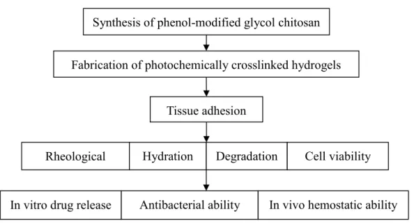
(25) property. Subsequently, the experiments of functional evaluation, such as in vitro drug release, antibacterial activities and in vivo hemostatic ability, would be done to verify whether this hydrogels could serve as tissue adhesive, drug release system, antibacterial material and hemostat, and be efficacious in pragmatic application. Synthesis of phenol-modified glycol chitosan Fabrication of photochemically crosslinked hydrogels. Tissue adhesion. Rheological analysis In vitro drug release. Hydration. Degradation. Antibacterial ability. Cell viability. In vivo hemostatic ability. Figure 1-1 Experimental scheme of this research. 13.
(26) Chapter 2 Materials and Methods 2.1 Chemicals 2.1.1 Synthesis of HPP-GC 1.. Glycol-chitosan (≧60% (titration), crystalline): Cat. # G7753, Sigma-Aldrich, USA. 2.. N-(3-Dimethylaminopropyl)-N′-ethylcarbodiimide hydrochloride (EDC): Cat. # 03450, Fluka, United Kingdom. 3.. N-Hydroxysuccinimide (NHS): Cat. # H7377, Sigma-Aldrich, USA. 4.. 3-(4-Hydroxyphenyl)propionic acid (phloretic acid, PA): Cat. # A14567, Alfa Aesar, Great Britain. 5.. 2-(N-Morpholino)ethanesulfonic acid hydrate (MES hydrate): Cat. # M2933, Sigma, USA. 2.1.2 Preparation of photo-crosslinked adhesives 1.. Tris(2,2′-bipyridyl)dichlororuthenium(II) hexahydrate (Ru(bpy)3Cl2): Cat. # 224758, Aldrich, USA. 2.. Sodium persulphate (SPS): Cat. # 13457, Sigma-Aldrich, USA. 2.1.3 Swelling and degradation 1.. Sodium chloride (NaCl): Cat. #4058-01, J. T. Baker, USA 14.
(27) 2.. Lysozyme from chicken egg white: Cat. # L6876, Sigma-Aldrich, USA. 2.1.4 MTS assay 1.. CellTiter 96® AQueous One Solution Reagent: Cat. # G358A, Promega, USA. 2.. Fetal bovine serum (FBS): Cat. # 04-001-1A, BI, USA. 3.. Gentamicin solution: Cat. # SV30080.01, GE, USA. 4.. Minimum essential medium alpha medium (alpha-MEM): Cat. # 11900-024, Gibco, USA. 5.. 2-Mercaptoehanol: Cat. # M3148, Sigma-Aldrich, USA. 6.. Penicillin Streptomycin (P/S): Cat. # 15140122, Gibco, USA. 7.. Phenol: Cat. # P1037, Sigma-Aldrich, USA. 8.. Sodium bicarbonate: Cat. # S7277, Sigma, USA. 9.. Sodium chloride (NaCl): Cat. #4058-01, J. T. Baker, USA. 10. Tris(hydroxymethyl)aminomethane (Tris): Cat. # 4109-02, J.T.Baker, USA 11. Trypan blue: Cat. # T8154, Sigma, USA 12. Trypsin-EDTA solution: Cat. # T4174, Sigma, USA. 2.1.5 In vitro drug release 1.. Amoxicillin: Cat. # A8523 Sigma-Aldrich, USA. 2.1.6 Antibacterial ability 1.. LB Broth, Miller (Luria-Bertani): Cat. # 244620, Difco, USA. 15.
(28) 2.. Agar Granulated: Cat. # 214530, Difco, USA. 3.. Gentamicin sulfate salt: Cat. # G1264, Sigma-Aldrich, USA. 2.2 Experimental Instruments and Materials 2.2.1 Experimental instruments 1.. Absorbance microplate readers: ELx800, BioTek, USA. 2.. Analytical balances: AB104-S, Mettler Toledo, USA. 3.. Autoclave: TM-326, Tomin, ROC. 4.. Centrifuge: 5804R, Eppendorf, Germany. 5.. Constant temperature water bath: WB212-B2, Kansin, Taiwan. 6.. Digital force gauge: FGP-5, NIDEC-SHIMPO, Japan. 7.. Incubator: Class-100 HEPA, Thermo Scientific, USA. 8.. Laminar flow hood. 9.. LED high power lamp (~8 W, 440–460 nm): PAR20, VITALUX, ROC. 10. Motorized test stand: FGS-50VB-H, NIDEC-SHIMPO, Japan 11. Nuclear magnetic resonance (NMR): AVIII-500MHz FT-NMR, Bruker, USA 12. Orbital Shaker Incubator, DENG YNG, ROC 13. Oven, HSIANGTAI, ROC 14. pH electrodes, TN-TXW600-GB, TNI, ROC 15. Phase contrast optical microscopy: TS-100, Nikon, Japan. 16.
(29) 16. Modular compact rheometer: MCR-102, Anton Paar, Canada 17. Stirrer/hot plate: Model PC-420, Corning, USA 18. UV-visible spectrophotometer: Cary 300, Agilent, USA 19. Vortex-genie 2: G560, Scientific Industries, Inc., USA 20. Vacuum dry oven, DOV40, DENG YNG, ROC. 2.2.2 Experimental materials 1.. Dialysis membranes (MWCO 12,000~14,000), Cat. # 1230-45, Cellu Sep, USA. 2.. 75 mm bottle top filter, Cat. # 595-3320, Thermo Scientific, USA. 3.. 96-well cell culture microplate, Cat. # 267313, Nunc, Denmark. 4.. 100 mm TC-treated culture dish, Cat. # 430167, Corning, USA. 5.. 15 mL PP centrifuge tube, Cat. # 430791, Corning, USA. 6.. 50 mL PP centrifuge tube, Cat. # 430829, Corning, USA. 7.. 1.5 mL microcentrifuge tubes, Cat. # 1260-00, SSI, USA. 8.. 2 mL microcentrifuge tubes, Cat. # 1310-00, SSI, USA. 2.3 Solution Formula 1. MES buffer, pH 5.0 19.52 g MES hydrate is dissolved in 800 mL of deionized water. The pH is adjusted to 5.0 with NaOH, and deionized water is added to a total volume of 1 L. MES buffer is sterilized by filtration through a 0.2 µm filter and stored at 4 °C.. 17.
(30) 2. Minimum Essential Medium Alpha Medium A bag of alpha-MEM power and 2.2 g sodium bicarbonate are dissolved in 800 mL deionized water. Then, 400 μL 2-Mercaptoehanol, 1 mL gentamycin solution, 10 mL P/S, and 100 mL FBS are added. The pH is adjusted with HCl and NaOH to 7.4, and deionized water is added to a total volume of 1 L. The solution is sterilized by filtration through a 0.2 µm filter and stored at 4 °C. 3. Phosphate buffered saline solution (PBS) PBS consisted of 8 g NaCl, 0.2 g KCl, 1.44 g Na2HPO4, and 0.245 g KH2PO4 in 1 liter deionized water with its pH value of 7.4. The pH value was adjusted to pH 7.4 by 0.1 N HCl and 0.1 N NaOH.. 2.4 Methods of Material Evaluation 2.4.1 Synthesis of HPP-GC The synthesis of HPP-GC was modified from the previous research [30] (Figure 2-1). Briefly, 0.2 g glycol chitosan was dissolved in 50 ml pH 5.0 MES buffer; the solution was put into Erlenmeyer flasks with magnetic stirring bar continuously stirring for 1 hour until glycol chitosan was totally dissolved. Secondly, 0.03 g of HPP were dissolved in 50 ml pH 5.0 MES buffer. HPP solution was avoided from light and then wait for 1 hour until HPP was totally dissolved. Afterwards, two solution mentioned above were mixed, and then 0.22 g EDC and 0.12 g NHS were added into the flask. The solution. 18.
(31) was stirred at room temperature overnight. After reaction completed, the product was purified by dialysis against deionized water through a seamless cellulose tube (MWCO: 12,000~14,000, Cellu Sep, USA) for 2 days. Last, the frozen solution lyophilized for three days to obtain HPP-GC sample.. 2.4.2 Characterization of HPP-GC 1. H-NMR and UV spectroscopy analysis were used to verify the successful. derivatization and determine the degree of substitutions (DS) of phenol groups in HPPGC samples. For 1H-NMR, 10 mg samples were dissolved in 0.5 ml deuterium oxide and analyzed by using Nuclear magnetic resonance (AVIII-500MHz FT-NMR, Bruker, USA). The DS of phenol groups in HPP-GC polymer was quantified using UV-Visible Spectrophotometer (Cary 300, Agilent, USA). The glycol chitosan and HPP-GC samples were dissolved in deionized water and the absorbance was measured at 276 nm. Phenolic substitution was calculated from a standard curve prepared using various concentrations (200, 100, 50, 25, 12.5 µg HPP / ml ultra-pure water) of HPP dissolved in ultra-pure water.. 2.4.3 Preparation of photo-crosslinked HPP-GC adhesives All hydrogel precursor solutions were prepared in deionized water and the concentration of solutions are 3% (wt. %). The solution was mixed with light-activated metal catalyst RuII(bpy)3Cl2 (1mM) and SPS (20 mM). Then, irradiating the solutions. 19.
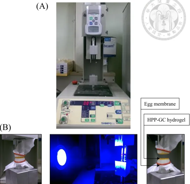
(32) for 30 s with a LED high power lamp (8 W, 440–460 nm: PAR20, VITALUX, ROC) from a distance of 15 cm. The crosslinking mechanism was illustrated in Figure 2-2 and 2-3.. 2.4.4 Tissue adhesion Tissue adhesion was examined using a digital force gauge (FGP-5, NIDEC-SHIMPO, Japan) shown in Figure 2-4 (A) with a 50 N digital force gauge. Two pieces of egg membrane was attached to glass cylindrical vials (diameter: 10 mm) by using O-ring. 90 μl HPP-GC solution was applied on the top of the egg membrane, and then make the upper glass cylinder move downward to attach to the hydrogel. LED high power lamp was turned up, and the hydrogel was exposed to the light for 30 s. After turning off the lamp, wait 30 secs until hydrogel was stable. Afterwards, the detachment stress was tested by making the upper glass cylinder move upward to pull the hydrogel apart. The maximum of detachment stress was recorded. The procedure is shown in Figure 2-4 (B). This experiment mainly aims at testing the tissue adhesion of hydrogel with animal tissue (egg membrane). The reason that using egg membrane is its low cost, easiness to operate and thinness that can let blue light penetrate appropriately.. 2.4.5 Rheological analysis The rheological properties were evaluated by using a Modular compact rheometer (MCR-102, Anton Paar, Canada). Parallel plate geometry (20 mm diameter) was. 20.
(33) performed at 37℃ to monitor the elastic modulus values (G’) and the viscous modulus values (G’’). A constant strain 1 % was used for the measurements. For the frequency sweep measurements, the sweep of the frequency varied from 0.1 Hz to 1 Hz, and each frequency sweep took 10 points to be completed. Through rheological analysis, the mechanical strength and elastic property can be measured to ensure the hydrogel is able to serve as adhesives that would not break or be destroyed easily.. 2.4.6 Hydration After the fabrication of HPP-GC hydrogels, hydrogels were lyophilized and the dry weights of hydrogels (Wdry) were measured. Afterwards, the dry samples were immersed in pH 7.4 PBS. Then, at specific time interval, water on the surface of hydrogels was removed by kimwipes, and the wet weights of hydrogels (Wwet) were recorded. Hydration values of HPP-GC hydrogels with different degree of substitutions were calculated by the following equation. Hydration is highly correlated with morphology and stability of hydrogel, this test can help us analyze other characteristics such as degradation and drug release of this hydrogel. Hydration (%) =. (𝑊𝑤𝑒𝑡 − 𝑊𝑑𝑟𝑦 ) × 100% 𝑊𝑑𝑟𝑦. 2.4.7 Degradation Hydrogel samples (200 mg) were prepared in vials accurately weighted (Wi). Subsequently, 5 mL of PBS solutions containing lysozyme (1 mg/mL) were added into. 21.
(34) the vials to cover the hydrogels and then incubated in 37 ℃ at 50 rpm Orbital Shaker Incubator (DENG YNG, ROC). At specific time intervals (1, 4, 7, 14 days), the buffer solution was removed from the samples and the hydrogels were weighted (W t) after water on the surface of hydrogels was removed by kimwipes. The remaining mass ratios were determined by the following equation. Degradation time can demonstrate that whether the hydrogel can sustain for enough time to serve as adhesives, wound dressing in wound healing process. Remaining mass ratios (%) =. 𝑊𝑡 × 100% 𝑊𝑖. 2.4.8 MTS assay Cytotoxicity of hydrogel was studied using L929 cells according to ISO10993 standard test. The hydrogel was prepared in a 2 mL centrifuge tube from 0.2 mL (3 wt. %) HPPGC solution. Each tube of hydrogels was extracted using 1 mL of serum-free medium and incubated for 48 hours at 37°C shaker (50 rpm). Followed by incubation, the final extract solutions containing 10% fetal bovine serum were diluted using culture medium to 100%, 50%, and 10% for culturing L929 cells. L929 cells were seeded in 96-well plate at a density of 2 × 10⁴ cells per well, and cultured in 100 µL of culture medium for 1 day in 37 ℃ incubator. Afterward, the culture medium was replaced with 100 µL of prepared dilution, and the plate was incubated for 1 day in 37 ℃ incubator. Subsequently, 20μl of CellTiter 96® AQueous One Solution Reagent (MTS) in 100μl. 22.
(35) of culture medium replaced the solution of each well, and the plate was incubated for 3 hours. Finally, absorbance was measured at 490 nm using an Elisa reader (ELx800, BioTek, USA). The results were compared with cells treated using culture medium and 0.64% phenol solution in culture medium. This test is significant for this biomaterial since this hydrogel will directly adhere to human tissue; thus, the cytotoxicity should be seriously considered.. 2.5 Methods of Functional Evaluation 2.5.1 In vitro drug release Amoxicillin solution (0.2 wt. %) was prepared, and then HPP-GC samples were dissolved in this solution (3 wt. %). After crosslinked by irradiation of blue light, the hydrogels were incubated in 10 ml pH 7.4 PBS, and 0.5 ml solution would be taken out from the vials for Uv-visible test; 0.5 ml fresh pH 7.4 PBS was then added to the vial. The solution would be taken out in different time intervals including 10, 20, 30, 45, 60, 90, 120, 180, 300 minutes. Amoxicillin could be detected at 229 nm in Uv-visible spectrophotometer (Cary 300, Agilent, USA), and then the release ratios of antibiotics were also determined from the calibration curves.. 2.5.2 Antibacterial ability Antibacterial activities were tested by inhibition of zone. E. coli as an example of Gramnegative bacteria and Staphylococcus epidermidis as an example of Gram-positive. 23.
(36) bacteria were used as target organisms. Concretely, each bacterium was incubated in sterile Luria-Bertani (LB) liquid culture medium at 37 ℃, with a 200 rpm shaking rate. Afterward, the bacteria solutions were diluted to a 0.01 optical density at 600 nm with fresh LB broth. LB agar medium was prepared and sterilized in an autoclave. Then the agar medium was poured into the sterile petri plates when the medium was in a warm molten state. After the medium solidified, 10 μl of bacterial solution was transferred into sterile petri plates and daubed the surface uniformly. Then, hydrogels (containing 0.005%, 0.01%, 0.02% and 0.1% gentamycin) was tailored into circle with the diameter of 8 mm and placed on the LB agar medium. The agar plates were then placed in an incubating oven at 37 °C and were left for 14 hours to assess the zone of inhibition for each of the hydrogel. The image of zone of inhibition was photographed and percentage of the inhibition was estimated through the following equation. By this antibacterial experiment, the fact that whether the hydrogels can serve as ideal drug release system and whether the gentamycin entrapped hydrogels by photochemically crosslinked method can effectually against bacterial can be verify. %inhibition =. 𝑑𝑖𝑎𝑚𝑒𝑡𝑒𝑟 𝑜𝑓 𝑧𝑜𝑛𝑒 𝑜𝑓 𝑖𝑛ℎ𝑖𝑏𝑖𝑡𝑖𝑜𝑛 (𝑚𝑚) × 100% 𝑑𝑖𝑎𝑚𝑒𝑡𝑒𝑟 𝑜𝑓 ℎ𝑦𝑑𝑟𝑜𝑔𝑒𝑙 (𝑚𝑚). 2.5.3 In vivo hemostatic ability To evaluate the hemostatic potential of the HPP-GC hydrogels, a hemorrhaging liver mice model was employed (22–25 g, 5 weeks, male). All animal studies were performed. 24.
(37) in compliance with guidelines set by national regulations and were approved by the local animal experiments ethical committee. Briefly, a mice was anesthetized using 400~600 μl 2% xylocaine mixed with water (vol.: vol. = 1:3) and fixed on a surgical corkboard. The liver of the mouse was exposed by abdominal incision, and serous fluid around the liver was carefully removed to prevent inaccuracies in the estimation of the blood weight obtained by the filter paper. A pre-weighted filter paper on a paraffin film was placed beneath the liver. Bleeding from the liver was induced using a 28 G needle and 100 μl of hydrogel was immediately applied to the bleeding site and expose the gel to blue light 10s (solution: 3 wt.% of HPP-GC). After bleeding for 2 min, the weight of the filter paper with absorbed blood was measured and compared with a control group.. 2.6 Statistic Analysis The data was presented as means ± standard error of the mean (SEM). The statistical analyses between different groups were determined with Student- Newman-Keuls Multiple Comparisons Test. A value of p ≤ 0.05 was considered statistically significant difference.. 25.
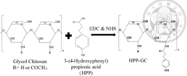
(38) Figure 2-1 Synthesis of phenol groups modified glycol chitosan via EDC & NHS method.. 26.
(39) Figure 2-2 Fabrication of HPP-GC hydrogel.. 27.
(40) Figure 2-3 Mechanism of photochemical crosslinking [35].. 28.
(41) (A). Egg membrane HPP-GC hydrogel. (B). Figure 2-4 (A) Digital force gauge. (B) Procedure of testing the detachment stress of hydrogel by using egg membrane.. 29.
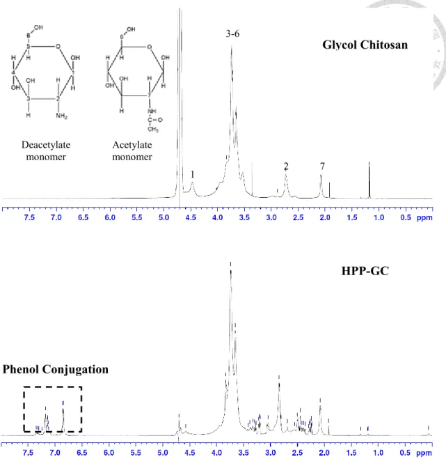
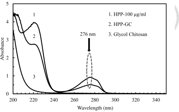
(42) Chapter 3 Results and Discussion 3.1 Synthesis and Characterization of HPP-GC HPP-GC was synthesized by the reaction of glycol chitosan (GC) with 3-(4Hydroxyphenyl) propionic acid (HPP). The successful derivatization was verified by 1. H NMR (Figure 3-1). Quantitative Uv-visible analysis (Figure 3-2) was used to. determine the degree of substitution (DS) of phenol groups in HPP-GC samples. The successful synthesis was verified by 1H NMR. Figure 3-1 shows 1H NMR spectra of glycol chitosan and HPP-GC. By comparison with the spectrum of glycol chitosan, new signals were observed at δ = 6.8 ppm and δ = 7.2 ppm in the spectrum of HPPGC. The signals of new peaks corresponding to aromatic protons confirmed the successful graft of phenol groups in the HPP-GC polymer. The reduction of the signal of hydrogen on amino groups at δ = 2.7 ppm after the reaction confirmed that the reaction completed correctly. The degree of substitution (DS) was determined by Uv-visible spectrophotometer. According to the Figure 3-2, the phenol groups on HPP-GC could be detected at 276 nm wavelength. Among all the samples produced, the DS arranged from 0.200 to 0.600 μmoles of phenol groups per mg of HPP-GC. The DS could be controlled by the amount of HPP. After the actual DS was determined, the following experiments of different 30.
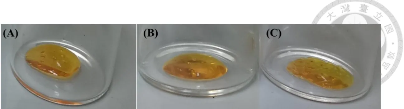
(43) characteristics could be analyzed and explained.. 3.2. Fabrication. of. Photocrosslinked. HPP-GC. Hydrogel After mixed with light-activated metal catalyst RuII(bpy)3Cl2 and SPS, the solution become orange ready to be crosslinked. This injectable solution could be crosslinked whenever the user wants and could be made into any shape (Figure 3-3). In the photo crosslinking process, the solution could be crosslinked rapidly within 5 seconds, yet waiting for 30 seconds to make the hydrogel crosslinked completely in our procedure. These advantages make applying hydrogel to irregular wound possible and the moment for crosslinking could be controlled easily. Compared with other crosslinking methods, widely used enzymatically crosslinked hydrogels always need special device to fabricate hydrogel such as double syringes [30, 34], that is an additional expense and hard to operate. Besides, after mixing with HRP and H2O2, the gelation will occur immediately and the moment is hard to control that would definitely cause inconvenience in certain condition. Moreover, some hybrid crosslinking methods are under restraint like temperature and pH of environment [33]. This photochemically crosslinked hydrogel can conquer those disadvantages with the merits mentioned above. As the Figure 3-4 shown, different DS have significant influence on the mechanical properties, structure and appearance of hydrogels. High DS (> 0.500 µmole/mg HPP-. 31.
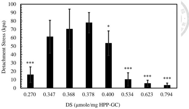
(44) GC) would make the hydrogel shrink, and the high crosslinking density would squeeze out the water of hydrogels. Sometimes the hydrogel may be stiff and crack after crosslinked.. 3.3 Strong Tissue Adhesion Strong tissue adhesion is the main advantage of this photochemically crosslinked HPPGC hydrogel. In previous study, it puts forth the fact that by irradiation of blue light, the phenol groups can not only form intermolecular crosslinking but bind the amino and thiol groups on animals’ tissue as well [35, 41]. Tissue adhesion could be measured by utilizing digital force gauge to measure the detachment stress at the break point of hydrogels when they were pulled apart. The results indicates that different DS would critically affect the tissue adhesion of hydrogels. Figure 3-5 shows that the detachment stress of the least DS (around 0.250 µmole/mg HPP-GC) is 15.99 ± 9.29 kpa, the moderate DS (around 0.370 µmole/mg HPP-GC) is 77.96 ± 12.10 kpa, the higher DS (larger than 0.534 µmole/mg HPP-GC) is lower than 10.40 ± 7.90 kpa. This photochemically crosslinked HPP-GC hydrogel possess approximately same or larger adhesive strength than other crosslinking method or polymer [31, 33, 64]. Especially, compared with the commercial fibrin sealant fabricated by same photochemical method [46], the adhesive strength of this HPP-GC hydrogel is much larger. From Figure 3-5, we could also find out that low DS (< 0.300 µmole/mg HPP-GC). 32.
(45) would not provide enough phenol groups to crosslink that the hydrogel would not have adequate crosslinking density; therefore, it would lose the cohesive ability to become hydrogel and could not form enough bonds with animal tissue for adhesion as well. As for hydrogels with high DS, they tend to have too high crosslinking density and that would cause the hydrogel have less elasticity, shrink and sometimes crack into pieces [41]. This tendency would severely destroy the structure of hydrogel and finally fail to bind animal tissue. After finding out the proper DS of hydrogels, which possess the strongest tissue adhesion, this most promising hydrogel would serve as the major object of this research. In addition, as Figure 3-6 shown, different concentration also have crucial effect on tissue adhesion. Higher concentration (3%) possessed better tissue adhesion and moderate concentration (2%) just slightly lower than the higher one (3%); nevertheless, low concentration (1%) could not become strong hydrogel and it still had a lot of water which had not been crosslinked by irradiation of blue light. Therefore, it is ideal to fabricate hydrogel between 2% to 3%.. 3.4 Rheological Analysis and Mechanical Property The rheological analysis can characterize the elastic and viscous property of this photocrosslinked HPP-GC hydrogel. As Figure 3-7 shown, the storage modulus of 3% HPP-GC hydrogel is approximately 6663 ± 65 Pa, which indicates the fact that this. 33.
(46) hydrogel is highly elastic and it has great mechanical strength [65]. Previous studies have shown that photo cross-linking 2% glycol chitosan solution has storage modulus of 1800 Pa [66]. Similarly, crosslinking 1.5% glycol chitosan solution with various concentrations of benzaldehyde functionalized PEG analogues has storage modulus ranging from 210~1400 Pa [30]. Hence, we can say that this ruthenium-based photochemical crosslinking process make HPP-GC hydrogel have much higher elastic and mechanical strength. The high storage modulus can also verify the fact that this photochemical crosslinking method can make hydrogel form strong intermolecular covalent bond between polymers. The outstanding elastic and viscosity of HPP-GC hydrogels is significant for tissue adhesion, which can better bear load and dissipate elastic energy, and can be applicable to flexible soft tissue [67].. 3.5 Hydration of Different DS HPP-GC Hydrogels Swelling and hydration ability of taking up liquid play important part in wound dressing. Stable swelling and hydration can provide proper absorbency for wound dressing to treat wound exudate. The values of hydration of different DS of hydrogel were measured. As Figure 3-8 shows that the dry hydrogels took up water fast in the first 5 hours. The hydration equilibrium was reached at approximately 24 hours, and the values of hydration of least DS hydrogels (0.250 µmole/mg), moderate DS hydrogels and high DS hydrogels are 2222% ± 33%, 1610% ± 314% and 1288% ± 261%. 34.
(47) respectively. It is obvious that different DS of hydrogel can affect crosslinking density of hydrogel, and that definitely change the morphology and porosity of hydrogel [68]. The higher the crosslinking density is, the smaller the porosity is. Therefore, smaller porosity absorbs less liquid. On the contrary, if the crosslinking density is lower, the higher porosity can absorb liquid more and faster. The appropriate hydration can be controlled by the degree of substitution of phenol groups. Since the hydrogels can also serve as hemostatic material, the hydration of taking up blood is significant as well [69].. 3.6 Degradation and Stability of HPP-GC Hydrogel Degradation is an important factor for the application of this hydrogel. The hydrogel should sustain for sufficient time and have adequate strength for sealing the wound and the curation [69]. Moreover, for the using purpose of being drug delivery system and wound dressing, the hydrogel must have ideal rate of degradation to control the drug release or the degradation at expected time point [58]. According to the Figure 3-9, the hydrogel initially degrade 20% of weight since the hydrogel would shrink and squeeze out some water, and the weight of hydrogel would lose along with the renewal of PBS. The following 13 days, the hydrogel only lost 20% of mass, which indicates that it cannot only sustain for adequate time but degrade with proper rate. In order to observe the tendency of degradation after the initial loss of water squeezed out, another. 35.
(48) experiment was done as Figure 3-10. The low DS cause hydrogels have more loose structure; thus, they would degrade faster than moderate ones. Nevertheless, high DS often results in crack of hydrogel make them lose their weight even faster than low DS hydrogel.. 3.7 Cell Viability and Cytotoxicity In this study, this photocrosslinked hydrogels were designed to be applied and crosslinked directly on the site of wound. Thus, the cytotoxicity of the HPP-GC and this photochemically crosslinking processes were evaluated by cultivating L929 cells with extraction of hydrogel and then quantify via MTS method. By immersing hydrogel into serum-free medium to get extraction, it could be regarded as the condition that when hydrogel adhere to human tissue, how body fluid would change and whether it would cause cytotoxicity. Different concentration of extraction were utilized to cultivate L929 for 1 day. As shown in Figure 3-11, the 100%, 50% and 10% have 53% ± 7%, 77% ± 2% and 97% ± 4% of cell viability respectively. The result demonstrates that most of the toxicity of SPS was consumed after the rapid photocrosslinking process, which has been studied in previous study [39]; hence, the hydrogels have low toxicity at most of concentration except the highest concentration.. 3.8 In Vitro Drug Release Hydrogels are frequently used as drug delivery system and have promising controlled. 36.
(49) release by modifying their structure and porosity [58, 60]. The Figure 3-12 presents an initial fast release, which may be attributed to the diffusion caused by swelling and the water squeezed out toward the surface of hydrogel after crosslinking. Since this photocrosslinked hydrogel expected to serve as wound dressing and sealant of surgical procedure such as sealing incision of gastrointestinal tract surgery, this fast release of amoxicillin is a desirable feature in an acidic condition of the stomach to overcome the limitations of gastric emptying times [59, 70]. In previous study of hydration, the porous structure of hydrogel also strongly correlate with the rate of drug release. For different using purpose, user can design different degree of substation of phenol groups to modify porous structure.. 3.9 Measurement of Antibacterial Ability In this in-vitro antibacterial experiment, E coli. and S. epidermidis were used to test the antimicrobial ability of hydrogel, which entrapped gentamycin by photocrosslinking. Different concentration of gentamycin (1000, 200, 50 μg/ml) were tested in order to observer the efficacy and then user could select proper concentration of antibiotics to use in the future. As Figure 3-13 shown, both antibiotic presented evident inhibition zone, it demonstrates the fact that gentamycin can against wide range of bacteria including gram-positive and gram-negative ones. By comparing with commercial gauze, there is only small inhibition zone around gauze, and it shows that this hydrogels are. 37.
(50) more efficient drug delivery and better release system. Although antibiotics can prevent bacterial infection, user must choose appropriate concentration of antibiotics for not causing damage to cells. Therefore, another groups of different concentration of samples were tested, and the Figure 3-14 shows that different concentrations of gentamycin (200, 100, 50 μg/ml) would affect the inhibition zone. As Table 3-1 shown, when using E coli. as the object of the experiment, 200, 100 and 50 μg/ml of gentamycin-entrapped hydrogel had 306% ± 41%, 304% ± 38% and 249% ± 6% of inhibition respectively. When using S. epidermidis as the object of the experiment, 200, 100 and 50 μg/ml of gentamycin-entrapped hydrogel had 305% ± 42%, 271% ± 32% and 192% ± 22% of inhibition respectively. The result demonstrates that 200 and 100 μg/ml of gentamycin-entrapped hydrogel could efficiently treat against two kinds of bacterial. Nonetheless, 50 μg/ml of gentamycin-entrapped hydrogel had relatively small inhibition zone. As a result, 200 and 100 μg/ml of gentamycin-entrapped hydrogel can serve as promising antibacterial material.. 3.10 In Vivo Hemostatic Ability This rapidly crosslinked hydrogel can utilized as hemostatic material because of outstanding adhesive property and fast gelation. This injectable hydrogel is able to be applied onto the hemorrhaging site and adhere to tissue around directly by irradiation of blue light and then become a hemostatic barrier. Moreover, glycol chitosan, which is. 38.
(51) a kind of derivative of chitosan, have some hemostatic ability. The hemostatic ability was measured by determine the quantity of bleeding on mice liver (Figure 3-15). As shown in Figure 3-16, for the control group, the mice live was bleeding without applying any material and the blood loss was 179.2 ± 95.5 mg. The other group was applying HPP-GC solution and then crosslink it by blue light, the blood loss was 73.0 ± 15.9 mg. According to the observation, the bleeding site on mice wound could be covered and stopped it from bleeding by this hydrogel within 10s, and significantly reduce the blood loss of the wound. Likewise, the filter papers could also indicates that there was obvious different amount of blood was absorbed (Figure 3-17). As a result, with the adhesive and in situ crosslinked ability, this HPP-GC hydrogel can serve as antibleeding barrier to deal with irregular wound and bleeding of wound. Additionally, by these results, the applicability of this hydrogel to animal tissue is verified.. 39.
(52) 3-6. Deacetylate monomer. Acetylate monomer. Glycol Chitosan. 2. 1. 7. HPP-GC. Phenol Conjugation. Figure 3-1 1H NMR analysis of glycol chitosan and HPP-GC.. 40.
(53) 5 1. HPP-100 μg/ml. 1. Absobance. 4. 2. HPP-GC 276 nm. 2. 3. 3. Glycol Chitosan. 2. 3. 1. 0 200. 220. 240. 260. 280. 300. 320. 340. Wavelength (nm) Figure 3-2 Uv-visible analysis of glycol chitosan, HPP and HPP-GC.. 41.
(54) Figure 3-3 Gelation of HPP-GC hydrogel.. 42.
(55) (A). (B). (C). Figure 3-4 Appearance of hydrogel with different DS. (A) low DS (~0.250 µmole/mg HPP-GC) (B) moderate DS (~ 0.370 µmole/mg HPP-GC) (C) high DS (> 0.534 µmole/mg HPP-GC). 43.
(56) 100. Detachment Stress (kpa). 90 80 *. 70 60 50 40 30. ***. ***. 20 10. ***. ***. 0.623. 0.794. 0 0.270. 0.347. 0.368. 0.378. 0.400. 0.534. DS (μmole/mg HPP-GC) Figure 3-5 Detachment stress of hydrogel with different DS. (n = 5, * and*** represent p < 0.05 and < 0.001 in comparison with sample of 0.378 DS). 44.
(57) 100. Detachment Stress (kpa). 90 80 70 60 50 40. *. 30 20 10 0 3%. 2%. 1%. Concentration (wt%) Figure 3-6 Detachment stress of hydrogel with different concentration. (DS of sample: 0.368 μmole/mg HPP-GC) (n = 5, * represents p < 0.05 in comparison with 3%). 45.
(58) Modulus G', G'' (Pa). 10000. 1000. 100 G' 10. G''. 1. 0. 0.2. 0.4. 0.6. 0.8. 1. 1.2. Frequency (Hz) Figure 3-7 Storage modulus (G’) and viscous modulus (G’’) of HPP-GC hydrogel as a function of frequency (Hz) at 37 ℃.. 46.
(59) 2500% * Hydration (%). 2000% 1500% 1000%. Low DS Moderate DS. 500%. Excessive DS. 0% 0. 5. 10. 15. 20. 25. 30. Hours Figure 3-8 Hydration of dry hydrogel with different DS. (n = 4, * represents p < 0.05 in comparison with sample of moderate DS). 47.
(60) Mass Remains (%). 100% 80% 60% 40% 20%. 0% 0. 2. 4. 6 8 Days. 10. 12. Figure 3-9 Degradation of hydrogels. (n = 5). 48. 14. 16.
(61) 100% Low DS. Moderate DS. Excessive DS. Mass Remains (%). 90% 80% 70% 60% 50% 40% 0. 1. 2. 3. 4. 5. 6. 7. Weeks Figure 3-10 Degradation of hydrogels after removing the water on surface. (n = 4). 49.
(62) 120%. Cell Viability (%). 100% 80% 60% 40% 20% 0% 100%. 50%. 10%. blank. control. Exraction concentration & Control Figure 3-11 Cell viability of different concentration of extraction form hydrogel. (n = 5). 50.
(63) Release Ratio (%). 100% 90% 80% 70% 60% 50% 40% 30% 20% 10% 0% 0. 50. 100. 150. 200. 250. Minutes Figure 3-12 Amoxicillin released from hydrogel. (n = 4). 51. 300. 350.
(64) Figure 3-13 (A) E coli. and (B) S. epidermidis were the used. (1) 1000, (2) 200 and (3) 50 μg/ml of gentamycin were entrapped in hydrogel. (4) Commercial gauzes were immersed in 1000 μg/ml gentamycin and (5) 10 µl 1000 μg/ml gentamycin as control.. 52.
(65) (A). (2). (B). (1). (5). (1). (2). (3). (5). (3). (4). (4). Figure 3-14 (A) E coli. and (B) S. epidermidis were the used. (1) 200, (2) 100 and (3) 50 μg/ml of gentamycin were entrapped in hydrogel. (4) HPP-GC hydrogel without gentamycin (5) 10 µl 200 μg/ml gentamycin as control.. 53.
(66) Figure 3-15 Procedure of in vivo hemostatic experiment.. 54.
(67) 300.0. Blood (mg). 250.0 200.0 150.0 100.0 50.0 0.0 Control. Applying HPP-GC hydrogel. Figure 3-16 Blood loss from wound. (n = 3). 55.
(68) (A). (B). (C). Figure 3-17 Filter paper, which absorbed the blood from wound (A) Control (B) Applying HPP-GC without crosslinking (C) Applying hydrogel.. 56.
(69) 200 (µg/ml). 100µg/ml. 50µg/ml. E coli.. 306% ± 41%. 304% ± 38%. 249% ± 6%. S. epidermidis. 305% ± 42%. 271% ± 32%. 192% ± 22%. Table 3-1 Inhibition of zone of different concentration of gentamycin against E coli. and S. epidermidis. (n = 4). 57.
(70) Chapter 4 Conclusion In this study, an injectable phenol conjugated glycol chitosan hydrogel were successfully fabricated by rapid photochemical crosslinking. This hydrogel shows outstanding tissue adhesion and its ability of flexible application to irregular wound for its in-situ gelation. Because of these advantages and stable characteristics, this hydrogel is able to seal wound and sustain for adequate time that make it be a great sealant and wound dressing. With antibiotics, this hydrogel cannot only serve as ideal drug release system but also perform desirable antibacterial ability. In vivo hemostatic experiment, photocrosslinked HPP-GC hydrogels were successfully crosslink on bleeding site of mice liver, adhere to the tissue around wound, and arrested blood within 10 seconds that showed excellent hemostatic ability. This result demonstrates that the hydrogel indeed have ability to be utilized on animal tissue. All the merits mentioned above prove that photochemically crosslinked HPP-GC hydrogels are promising tissue adhesives, drug release system, hemostatic and antibacterial materials.. 58.
(71) References [1] N. Annabi, K. Yue, A. Tamayol, A. Khademhosseini, Elastic sealants for surgical applications, Eur J Pharm Biopharm 95(Pt A) (2015) 27-39. [2] W.D. Spotnitz, S. Burks, Hemostats, sealants, and adhesives: components of the surgical toolbox, Transfusion 48(7) (2008) 1502-16. [3] B. Mizrahi, C. Weldon, D.S. Kohane, Tissue Adhesives as Active Implants, 8 (2010) 39-56. [4] J.C. Wheat, J.S. Wolf, Jr., Advances in bioadhesives, tissue sealants, and hemostatic agents, Urol Clin North Am 36(2) (2009) 265-75, x. [5] T.B. Reece, T.S. Maxey, I.L. Kron, A prospectus on tissue adhesives, Am J Surg 182(2) (2001) 40s-44s. [6] S. Ozawa, Patient blood management: use of topical hemostatic and sealant agents, AORN J 98(5) (2013) 461-78. [7] P.T. Kumar, V.K. Lakshmanan, T.V. Anilkumar, C. Ramya, P. Reshmi, A.G. Unnikrishnan, S.V. Nair, R. Jayakumar, Flexible and microporous chitosan hydrogel/nano ZnO composite bandages for wound dressing: in vitro and in vivo evaluation, ACS Appl Mater Interfaces 4(5) (2012) 2618-29. [8] M. Kozicki, M. Kolodziejczyk, M. Szynkowska, A. Pawlaczyk, E. Lesniewska, A. Matusiak, A. Adamus, A. Karolczak, Hydrogels made from chitosan and silver nitrate, 59.
(72) Carbohydr Polym 140 (2016) 74-87. [9] M. Perez-Diaz, E. Alvarado-Gomez, M. Magana-Aquino, R. Sanchez-Sanchez, C. Velasquillo, C. Gonzalez, A. Ganem-Rondero, G. Martinez-Castanon, N. ZavalaAlonso, F. Martinez-Gutierrez, Anti-biofilm activity of chitosan gels formulated with silver nanoparticles and their cytotoxic effect on human fibroblasts, Mater Sci Eng C Mater Biol Appl 60 (2016) 317-23. [10] F. Wahid, J.J. Yin, D.D. Xue, H. Xue, Y.S. Lu, C. Zhong, L.Q. Chu, Synthesis and characterization of antibacterial carboxymethyl Chitosan/ZnO nanocomposite hydrogels, Int J Biol Macromol 88 (2016) 273-9. [11]. H.A.G.. M.. Radosevich,. T.. Burnoufa,. Fibrin. Sealant. Scientific. Rationale,Production M ethods, Properties, andCurrent Clinical Use, Vox Sang 72 (1997) 133-143. [12] D.J. Meng-G Martin Lee, Applications of Fibrin Sealant in Surgery, Surgical Innovation 12 (2005) 203–213. [13] G. Hidas, A. Kastin, M. Mullerad, J. Shental, B. Moskovitz, O. Nativ, Sutureless nephron-sparing surgery: use of albumin glutaraldehyde tissue adhesive (BioGlue), Urology 67(4) (2006) 697-700; discussion 700. [14] M. Ryou, C.C. Thompson, Tissue Adhesives: A Review, Techniques in Gastrointestinal Endoscopy 8(1) (2006) 33-37.. 60.
(73) [15] R. Cirocchi, E. Farinella, F. La Mura, L. Cattorini, B. Rossetti, D. Milani, P. Ricci, P. Covarelli, M. Coccetta, G. Noya, F. Sciannameo, Fibrin glue in the treatment of anal fistula: a systematic review, Ann Surg Innov Res 3 (2009) 12. [16] J.A. Saldana-Cortes, F. Larios-Arceo, E. Prieto-Diaz-Chavez, E.P. De Buen, S. Gonzalez-Mercado, A.S. Alvarez-Villasenor, M.R. Prieto-Aldape, C. Fuentes-Orozco, A. Gonzalez-Ojeda, Role of fibrin glue in the prevention of cervical leakage and strictures after esophageal reconstruction of caustic injury, World J Surg 33(5) (2009) 986-93. [17] A. Lauto, D. Mawad, L.J.R. Foster, Adhesive biomaterials for tissue reconstruction, Journal of Chemical Technology & Biotechnology 83(4) (2008) 464-472. [18] B.J. Kober, A.M. Scheule, V. Voth, N. Deschner, E. Schmid, G. Ziemer, Anaphylactic reaction after systemic application of aprotinin triggered by aprotinincontaining fibrin sealant, Anesth Analg 107(2) (2008) 406-9. [19] S. Ghosh, J.D. Cabral, L.R. Hanton, S.C. Moratti, Strong poly(ethylene oxide) based gel adhesives via oxime cross-linking, Acta Biomater 29 (2016) 206-14. [20] M.D. Thomas E. MacGillivray, Fibrin Sealants and Glues, J Card Surg 18 (2003) 480-485. [21] L. Durham, D. Willatt, M. Yung, I. Jones, P. Stevenson, M. Ramadan, A method for preparation of fibrin glue, The Journal of Laryngology & Otology 101(11) (1987). 61.
(74) 1182-1186. [22] A.E. Ardis, Preparation of monomeric alkyl alpha-cyano-acrylates, Google Patents, 1949. [23] H. Coover, F. Joyner, N. Shearer, T. Wicker, Chemistry and performance of cyanoacrylate adhesives, J Soc Plast Eng 15 (1959) 413-417. [24] W. Furst, A. Banerjee, Release of glutaraldehyde from an albumin-glutaraldehyde tissue adhesive causes significant in vitro and in vivo toxicity, Ann Thorac Surg 79(5) (2005) 1522-8; discussion 1529. [25] P.A. Leggat, D.R. Smith, U. Kedjarune, Surgical applications of cyanoacrylate adhesives: a review of toxicity, ANZ J Surg 77(4) (2007) 209-13. [26] J.B.H. Pt. Stephen C. Woodward, John L. Cameron, , George Brandes, Edwin J. Pulaski, , Fred Leonard, Histotoxicity of Cyanoacrylate Tissue Adhesive, Annals of Surgery 162 (1964) 114-122. [27] B.J. Vote, M.J. Elder, Cyanoacrylate glue for corneal perforations: a description of a surgical technique and a review of the literature, Clinical & experimental ophthalmology 28(6) (2000) 437-442. [28] K. Kimura, K. Sugiura, Adhesive composition, Google Patents, 1982. [29] J.H. Ryu, S. Hong, H. Lee, Bio-inspired adhesive catechol-conjugated chitosan for biomedical applications: A mini review, Acta Biomater 27 (2015) 101-15.. 62.
(75) [30] S.V. Gohil, S.B. Brittain, H.-M. Kan, H. Drissi, D.W. Rowe, L.S. Nair, Evaluation of enzymatically crosslinked injectable glycol chitosan hydrogel, J. Mater. Chem. B 3(27) (2015) 5511-5522. [31] E. Lih, J.S. Lee, K.M. Park, K.D. Park, Rapidly curable chitosan-PEG hydrogels as tissue adhesives for hemostasis and wound healing, Acta Biomater 8(9) (2012) 32619. [32] J.H. Ryu, Y. Lee, M.J. Do, S.D. Jo, J.S. Kim, B.S. Kim, G.I. Im, T.G. Park, H. Lee, Chitosan-g-hematin: enzyme-mimicking polymeric catalyst for adhesive hydrogels, Acta Biomater 10(1) (2014) 224-33. [33] J.H. Ryu, Y. Lee, W.H. Kong, T.G. Kim, T.G. Park, H. Lee, Catecholfunctionalized chitosan/pluronic hydrogels for tissue adhesives and hemostatic materials, Biomacromolecules 12(7) (2011) 2653-9. [34] J.W. Bae, B.Y. Kim, E. Lih, J.H. Choi, Y. Lee, K.D. Park, In situ formation of enzyme-free hydrogels via ferromagnetic microbead-assisted enzymatic cross-linking, Chem Commun (Camb) 50(89) (2014) 13710-3. [35] T.K. DAVID A. FANCY, Chemistry for the analysis of protein–protein interactions: Rapid and efficient cross-linking triggered by long wavelength light, Proc. Natl. Acad. Sci. 96 (1999) 6020–6024. [36] C.M. Elvin, A.G. Carr, M.G. Huson, J.M. Maxwell, R.D. Pearson, T. Vuocolo, N.E.. 63.
數據
![Figure 2-3 Mechanism of photochemical crosslinking [35].](https://thumb-ap.123doks.com/thumbv2/9libinfo/8665362.195415/40.892.143.785.115.888/figure-mechanism-of-photochemical-crosslinking.webp)
+7

相關文件
展生物医学、生物育种等产业。新材料领域重点发展新型电子
接枝共聚合反應是材料改質的主要技術之ㄧ,已廣泛應用於高分子材料及生
使金屬離子均勻分散在纖維中而具有抗菌作用。抗菌
而在後續甲烷化反應試驗方面,以前段經厭氧醱酵產氫後之出流水為進流基 質。在厭氧光合產氫微生物方面,以光合作用產氫細菌中產氫能力最好的菌株 Rhodopseudomonas palustris
包括三維機械設計的所更的功能(SolidWorks 三維建模軟體)、資料管 理軟體 PDMWorks Client、以及用於設計交流的常用工具:eDrawings 專 業版(基於 e-mail 的設計交流工具),
SF15140A 楊勝舜 利用 JKB-122 評估對於經干擾素(長效 型或短效型)或干擾素和 Ribavirin 組合 治療沒有反應的 C 型肝炎病毒陽性患 者之肝臟功能 (丙胺酸轉胺酶
其他紡織材料製女用或女童用汗衫及其他背心、三角褲、短內褲、便服、浴袍、晨衣及類似品 Women's or girls' singlets and other vest, briefs, panties, negliges, bathrobes,
黑木耳 (Auricularia polytricha) 是台灣普遍的食用 菌之一,為一種低熱量的食品,亦是一種富含食用纖維及
