國立臺灣大學牙醫專業學院口腔生物科學研究所 碩士論文
Graduate Institute of Oral Biology College of Dentistry National Taiwan University
Master Thesis
活細胞分析式微流道晶片之開發及其應用於癌症轉移潛能診斷之評估 暨
SIK2 藉由磷酸化 CAPS2 調控胰島素分泌之探討
Study I: A portable microfluidic device for the rapid diagnosis of cancer metastatic potential
Study II: Identification of CAPS2 as a downstream target of SIK2 in regulating insulin secretion
陳立瑜 (Li-Yu Chen)
指導教授﹕周涵怡 博士 (Han-Yi Chou, Ph.D) 中華民國 104 年 7 月
July, 2015
研究一 活細胞分析式微流道晶片之開發及其應用於癌症轉移潛能診
斷之評估 ... 1
摘要 ... 1
Study I: A portable microfluidic device for the rapid diagnosis of cancer metastatic potential which is programmable for temperature and CO2 ... 2
Abstract ... 2
Background ... 3
Lung cancer, the foremost cause of cancer related deaths ... 3
Routine examinations for lung cancer and its limitations ... 4
Aim ... 6
Design ... 7
Design Concept ... 7
Microfluidic chip as a principle part of device ... 7
Microfluidic cell culture ... 8
Metastatic index ... 9
Analytic platform ... 10
Materials and methods ... 13
Microfluidic fabrication ... 13
Chitosan preparation and coating ... 14
Cell Culture ... 14
Cell migration and proliferation measurement ... 15
Fluorescenct reagent and staining ... 15
Cell seeding and detachment assays on MD-CaMP ... 17
Cell seeding and attachment assays on culture dish ... 17
Western blot analysis ... 18
Results ... 19
Design of MD-CaMP ... 19
Migratory abilities of CL1-0, CL1-1 and CL1-5 Cell model for MD-CaMP testing ... 20
Fibronectin expression of CL1-0, CL1-1,CL1-5 ... 21
Correlation of metastatic potential with rate of detachment on chitosan ... 22
Selective fractionation of cells from a mixed population by their differential rate of detachment ... 24
Altered cell morphology with pH ... 25
Differential attaching properties of CL1-0, CL1-1 and CL1-5 on chitosan surface ... 25
Cell fractionation of co-cultured CL1-0, CL1-1, CL1-5 by differential
attachment properties on chitosan surface ... 27
Conclusion and Discussion ... 29
Reference ... 33
Figures ... 34
Appendix ... 43
研究二 SIK2 藉由磷酸化 CAPS2 調控胰島素分泌之探討 ... 44
摘要 ... 44
Study II: Identification of CAPS2 as a downstream target of SIK2 in regulating insulin secretion ... 45
Abstract ... 45
Introduction ... 47
Definition, classification, cause and prevalence of diabetes mellitus ... 47
Secretory pathway, distinct pools and biphasic secretion of insulin vesicles in pancreatic β cells ... 48
Roles of SIK2 in metabolism and insulin function ... 51
The expression and function of SIK2 in pancreatic β cell in regulating insulin secretion ... 53
CAPS2 as a putative substrate of SIK2 in regulation of insulin secretion ... 55
Aim ... 58
Materials and Methods ... 59
Isolation of pancreatic islets ... 59
Antibodies ... 60
Immunofluorescent staining: ... 60
RNA isolation and RT-PCR ... 62
Cloning of full length CAPS2 ... 62
Site direct mutagenesis of pCMV-myc-CAPS2(1-1260) ... 65
SIK2 constructs ... 66
Transfection ... 66
Immunoprecipitation ... 67
In vitro kinase assay ... 67
Western blot analysis ... 68
Insulin ELISA ... 68
Results ... 70
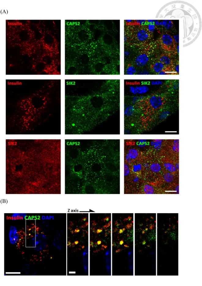
The expression of SIK2 and CAPS2 in mouse pancreatic islets ... 70
Subcellular localization of SIK2 and CAPS2 in mouse pancreatic β cell ... 70
Cloning of β cells expressed CAPS2 isoform ... 71
Physiological relevant proteins interaction between SIK2 and CAPS2 ... 73
Putative SIK2 phosphorylation sites on CAPS2 and CAPS2-MUT-T1016A, T1052A, T1231A construct ... 74
In vitro phosphorylation of SIK2 on CAPS2 ... 75
Transfection and expression of pTimer-phogrin in primarily cultured islet cells ... 76
Insulin secretion of SIK2/CAPS2 overexpressed mouse primary β cells ... 77
Conclusion and Discussion ... 80
References ... 85
Figures ... 89
研究一 活細胞分析式微流道晶片之開發及其應用 於癌症轉移潛能診斷之評估
摘要
肺癌的轉移若及早發現並加以治療,便能顯著提升患者改善病情的機會。
然而,現行臨床癌症檢驗之方式,如胸腔攝影、斷層掃描及組織切片等,均 未能有效判斷肺癌細胞轉移之可能性,以致患者無法獲得適當的治療。本研 究中,吾人設計開發出一新型微流道晶片系統,以期作為快速評估癌症轉移 潛能之臨床檢驗。本系統設計係以恆定環境溫度及酸鹼度為基礎,再以甲殼 素附著之微流道表面偵測目標細胞之貼附性,作為判斷細胞遷移特性之依據。
於實際測試中,吾人將不同轉移潛能之肺癌細胞株,經標定後以本系統進行 細胞貼附性測試。結果顯示本系統確能有效區分不同轉移潛能之肺癌細胞株。
因此,透過本系本研究所開發之微流道系統,僅需少量自患者經活體採樣分 離之細胞樣本,便能迅速取得評估癌細胞轉移潛能之資訊。此微流道系統未 來若能應用於臨床診斷,便能為肺癌及其他癌症患者,於早期研判適合之治 療模式。
Study I: A portable microfluidic device for the rapid diagnosis of cancer metastatic potential which is programmable for temperature and CO
2Abstract
If metastasis of lung cancer can be found and treated early, a patient might have an improved chance to prevail over it, but routine examinations such as chest radiography, computed tomography and biopsy cannot characterize the metastatic potential of lung cancer cells; critical diagnoses to define optimal therapeutic strategies are thus lost. In this study, we designed a portable microfluidic device for the rapid diagnosis of cancer metastatic potential. Featuring a micro system to control temperature and a bicarbonate buffered environment, our device discriminates a rate of surface detachment as an index of the migratory ability of cells cultured on pH-responsive chitosan. We labeled metastatic subpopulations of lung cancer cell lines, and verified that our device is capable of separating cells according to their metastatic ability. As only few cells are needed, a patient's specimen from biopsies, e.g. from fine-needle aspiration, can be processed on site to offer immediate information to physicians. We expect that our design will provide valuable information in pre-operative evaluations to assist the definition of therapeutic plans for lung cancer, as well as for metastatic tumors of other types.
Background
Lung cancer, the foremost cause of cancer related deaths
Nowadays, the biggest challenge in medical is cancer, the leading cause of death worldwide, accounting for 8.2 million deaths in 2012. Around 90% of all cancer deaths are the result of metastases, the spread of cancer from its primary site to other places in the body, rather than of the primary tumors [1].
Among plenty of cancers, lung cancer is the most usually diagnosed malignancy in the world. Meanwhile, lung cancer is the foremost cause of cancer mortality worldwide, accounting for 1.3 million deaths annually and the 5 year survival rate is only about 15% [2]. It is common among men in terms of both incidence and mortality, and among women has the third highest incidence, and is second after breast cancer in mortality [3].
In approximately 40% of patients diagnosed with lung cancer, the diagnosis is made after the disease has advanced because lung cancer may not produce any noticeable symptoms in the early stages. Those early metastasized lung cancers could not be detected before they have become aggravated, causing bad prognosis with a high mortality rate. The only way to decrease mortality of lung cancer patients is to detect it, diagnose it and treat it as early as possible.
Routine examinations for lung cancer and its limitations
Approximately 85% of patients with lung cancer seek medical advice with symptoms, and the remainder is detected by chest radiographic evaluation initiated for other reasons such as general physical examination. Routine diagnostic examinations for patients suspected with lung cancer includes imaging test, sputum cytology test and tissue biopsy test (http://www.cancer.org). Chest x-ray is the most common imaging test for screening any abnormal masses or nodules on the lungs, and may sometimes be combined with CT scans to reveal smaller lesions that would not be detected on an X-ray for detailed information. If patients cough and produce sputum, sputum cytology
test sometimes help them in finding the presence of cancerous cells. Biopsy test is the remove of suspicious cancer tissues or cells for histological, cytological, or molecular analysis by bronchoscopy passed down from throat and into lungs or by image guided needle through chest wall and into the lung [4]. Definite diagnosis of suspected lung cancer will not be made until cancer cells are detected in biopsied tissues obtained from suspicious lump found by imaging tests under microscope.
However, most patients have initiated the metastatic process by the time of diagnosis. Although lung cancer is initially detected by imaging morphological changes inside the lungs and confirmed with biopsy tests, unobvious cancer mass or small
order to prevent early-staged metastasis before spreading continuously, it is important to understand the metastatic potential of cancer cells and plan treatments referring to this characteristic. To do so, it requires technical challenges to functionally assay live cancer cells from patients' specimens which are accessible from biopsy. Nevertheless, patients' specimens are usually fixed for tissue processing or smear tests rather than cultured because handling of living cells clinically is limited by the consumption of time, costs and manpower. Routine examinations such as histological, cytological, or molecular analysis provide us information about cancer classification, expression of tumor markers or tumor subtyping from biopsied tumor samples instead of direct information on the metastatic potential of cancer. Critical diagnoses to define optimal therapeutic strategies are thus lost.
Aim
To make up for limitations of routine examinations for analyzing metastatic potential of living cell samples, we aim to develop a device which utilizes patients' specimen obtained from biopsy to evaluate metastatic characteristics of cancer cells.
Design
Design Concept
According to our demands, this device was designed based on the following four features. Firstly and fundamentally, it provides a bio-mimic environment such as CO2
incubator having temperature and pH value controls to keep cells alive. Secondly, it should be small enough to handle very few cells obtained from the fine needle biopsy.
Thirdly, it directly perform functional assay of patients' living-cell samples on metastatic characteristics. Fourthly, it can be a portable device offering rapid, on site processing and diagnosing.
Microfluidic chip as a principle part of device
Microfluidic device is also known as a lab-on-a-chip (LOC) device or a micro total analysis systems (µTAS), which integrate and scale down laboratory functions and processes to a miniaturized chip format and shrink all analytic steps to micro-level by manipulating small (10−9 – 10−18 L) amounts of fluids in channels of micrometer.
Currently, microfluidic device has been applied to perform all kinds of biomedical uses because it provides various advantages. For example, volumes of fluids within microfluidic channels are generally in the microliter range, it greatly reduces the use of
reagents and analytes. The reacting turnaround time is shorten as whole analytic processes have been condensed to a chip. In addition, the lower fabrication costs of microfluidic devices allow the production of cost-effective disposable chips. With these properties, microfluidic device is beneficial to deal with restricted amount of samples, and is suitable to be applied as a general diagnostic device. Here we explored the advantages offered by microfluidic device to design a 'Microfluidic Device for Cancer Metastatic Potential', MD-CaMP.
Microfluidic cell culture
On the basis of our aim, live cells from biopsied specimen should be maintained alive from the time being collected out of human body and to the functional analysis.
The ability to maintain extracellular environment in a physiological conditions is fundamental for cell culturing. Sometimes when meeting the needs to assay or image live cells for a period of time, most of the laboratory practices utilize microscope incubators to control three important factors – temperature, CO2 and moisture, to attain proper environments for cells to behave physiologically during experimenting. However, a commercial biological microscope incubator currently takes tens of minutes to reach a steady environment; it is also expensive and energy-consuming.
Development in microfluidics enables us to create tools to control the cellular microenvironment. The possibility of microfluidic device to integrate precise and rapid control of their temperature and CO2 can be an effective solution [5]. In the case of temperature control, various configurations of a micro-heater have been extensively discussed [6]. For instance, a microfluidic temperature control device for quick and reversible switch of temperature has been used to study the temperature-sensitized microtubule dynamics of cells [7]. Also, the stable carbon dioxide partial pressure on CO2 chips controlled by the equilibration of pre-equilibrated aqueous flow through the highly gas permeable substrate, poly(dimethylsiloxane) (PDMS) enable long-term culture of mammalian cells. Accordingly, by the incorporation with temperature and CO2 control, our device MD-CaMP is designed as a minimized cell incubator which provides bio-mimic environment for live cell assaying [8].
Metastatic index
Metastasis is a complex process that involves the spread of cancer to distant parts of the body from its original site. There are several steps of metastasis. At first, cancer cells separate from the primary tumor and Invade through around tissues and their basement membranes. Next they penetrate blood or lymphatics vessels and spread into
circulation. Finally they extravasate and colonize the distant area.
The complicated interactions between tumor cells and the extracellular matrix (ECM) play important roles during tumor metastasis. Abnormal extracellular matrix remodeling is related to metastatic disease and promotes cancer progression [9, 10].
Fibronectin, an extracellular matrix glycoprotein, has been reported to be up-regulated in advanced stage lung cancer cells, and the metastatic potential of lung cancer is positively correlated to the amount of fibronectin expression [11, 12]. Higher production of fibronectin is not only a hallmark of cancer cell mesenchymal epithelial transition, it can also relay intracellular signaling pathways that lead to increased aggressiveness, and is an important factor for the “seed and soil” process for distant lung cancer metastasis [13]. Thus, fibronectin can be considered as an index for assaying lung cancer metastatic potential.
Analytic platform
Since fibronectin is a negatively-charged glycoprotein, we attempted to use a biocompatible and electro positive material as an analytic platform to separate cancer cells with different level of fibronectin by electro affinity. Chitosan, is one of many interesting material fitting our requirement. Produced commercially
by de-acetylation of chitin, the structural element in the exoskeleton of crustaceans, chitosan is well-known in biomedical uses due to the characteristics of biodegradable, biocompatible, bio-adhesive, nontoxic, non-antigenic. Structurally, chitosan is a cationic polysaccharide with a variable number of randomly located N-glucosamine and N-acetyl-glucosamine groups. The protonation–deprotonation equilibrium of the primary amine on its glucosamine residues make it a pH-responsive polycation. The isoelectric point (pI) of chitosan occurs at pH7.4 [14], meaning chitosan carries no electrical charge at pH7.4. In other words, if environmental pH value is lower than its pI, chitosan will be positively charged, and conversely if environmental pH value is greater than its pI, chitosan will be negatively charged. It has been reported that by altering its adsorption to fibronectin, chitosan is capable of controlling cell attachment by adjustment of medium pH [15]. While increasing medium pH (to pH 7.65), over 90% of cells would rapidly detached from chitosan surface within 1 h. Coated as a thin film on the surface of tissue culture dish, chitosan can be a pH-responsive substrate to harvest cells without enzymatic treatments (Appendix 1). Furthermore, mixed cells can be fractionated according to their distinguishing detachment capabilities on chitosan surface [16]. Accordingly, chitosan is applicable as an analytic platform of MD-CaMP for fractionating cancer cells by its affinity to fibronectin.
In this study, we propose the design of a microfluidic device for cancer metastatic potential, MD-CaMP, which enables bedside fractionation of minute mass specimens.
MD-CaMP features programmable modules for temperature control, bicarbonate-dependent pH regulation, and a pH-responsive chitosan coating to discriminate the cancer metastatic potential through the adsorption of fibronectin.
Materials and methods
Microfluidic fabrication
The design and fabrication of microfluidic device was made cooperatively with Y.
H. Yu and I. F. Yu, department of Mechanical Engineering, College of Engineering, National Taiwan University.
The microfluidic chambers were fabricated on poly(dimethylsiloxane) (PDMS) using soft lithographic methods. The master mould was fabricated on a polymethylmethacrylate (PMMA) plate using a computer numerical control (CNC) machine (EGX-400, Roland Inc., Japan) equipped with a 0.5 mm drill bit at a feeding speed of 7 mm. s−1 and a rotational rate of 26,000 rpm. PDMS containing the prepolymer and the curing agent at the mass ratio of 10 : 1 was mixed, degassed in vacuum, poured into the PMMA mould and cured at 80 °C. The PDMS structure was treated with an oxygen plasma before bonding and assembly.
A grid of platinum micro-heaters is fabricated on glass to precisely and rapidly control the temperature based on the feedback signal of a designed micro thermal sensor and a control program. The grid-type micro-heater and micro thermal sensor were fabricated by standard lithography and lift-off. The bio-compatible soda-lime glass (thickness 0.7 mm) was first cleaned with piranha solution (H2SO4:H2O2 = 3 : 1). A
positive photoresist (AZ 4620, Clariant Inc., USA) was then spin-coated (3000 rpm, 40 s) and patterned with photolithography to define the micro-heaters and the micro thermal temperature sensor. With E-beam evaporation, we deposited a Pt layer (120 nm) above a Cr layer (30 nm) on the glass substrate (0.7 mm). The Pt layer was then patterned with a standard lift-off process to obtain the micro-heaters and the microthermal sensor.
Chitosan preparation and coating
A chitosan substrate was prepared from 0.5%(w/w) chitosan solution dissolved in 3%(w/v) acetic acid [15]. Chitosan substrate was added (30 µL) into the microchannels or (100 µl/cm2 surface area) into culture dish and dried at 60°C for one day to form a thin film. Then the coating surface needs to be neutralized was by 0.5N NaOH(aq) for 2 hours at room temperature and washed with ddH2O and 1×PBS once. Before cell seeding, these microchannels and culture dishes were exposed to ultraviolet light overnight and washed thoroughly with PBS again.
Cell Culture
(Invitrogen) supplemented with 10% fetal bovine serum at 37°C in a 5% CO2
humidified atmosphere and passage by cell detachment buffer (10 mM EDTA in PBS).
Cell migration and proliferation measurement
For cell migration measurement, wound-healing assay which mimics cell migration during wound healing in vivo was performed.CL1-0, CL1-1 and CL1-5 cells were seeded at same density in 6 well plates until confluent. Making a straight scratch on cultured cells by using a (yellow) pipette tip to create uniform cell-free spaces allows cells to migrate into and "heal" the "wound". Live cell images were captures 24 hours by time lapse microscopy. We measured the original width of the area immediately after the scratch as 100%, and for the subsequent time points, the width were measured again to obtain the remaining percentage of cell-free space.
For cell proliferation measurement, equal amount (1×10⁶) of CL1-0, CL1-1 and CL1-5 cells were seeded in 6 well plates and trypsinazed to count the number of cells after culturing for 24 hours by hemocytometer.
Fluorescenct reagent and staining
MitoTracker Red CM-H2XRos (Invitrogen, M7513), MitoTracker Green FM
(Invitrogen, M7514), Vybrant CFDA SE Cell Tracer (Invitrogen, V12883), Hoechst 34580 (Invrogen, H21486)
For MitoTracker Red CM-H2XRos and MitoTracker Green FM, dilute 1 mM MitoTrackerstock solution to the final working concentration (25–500 nM) in incomplete medium. When cells have reached the desired confluency, media was removed from the dish, then prewarmed (37°C) staining solution containing MitoTracker probe was added and incubate for 15–45 minutes at 37°C. After staining is complete, the staining solution could be replaced with fresh prewarmed media.
For Vybrant CFDA SE Cell Tracer, dilute the 10 mM stock solution in PBS to working concentration (0.5–25 µM). When cells have reached the desired confluency, media was removed from the dish, then prewarmed (37°C) PBS containing Vybrant CFDA probe was added and incubate for 15 minutes at 37°C. After staining is complete, the staining solution could be replaced with fresh prewarmed media and incubate the
cultures for another 30 minutes at 37°C.
For Hoechst 34580, dilute the 10 mg/ml stock solution in PBS to working concentration (0.2 to 5 µg/mL). When cells have reached the desired confluency, media was removed from the dish, then prewarmed (37°C) staining solution containing Hoechst 34580 probe was added and incubate for 20–30 minutes at 37°C. After staining is complete, the staining solution could be replaced with fresh prewarmed media.
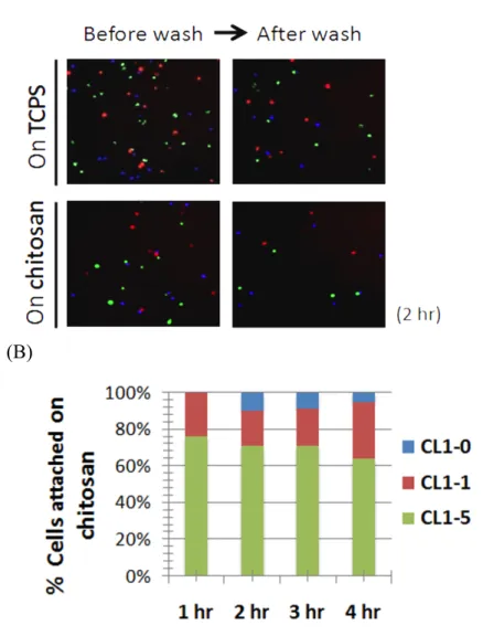
Cell seeding and detachment assays on MD-CaMP
1 × 10⁵ CL1 cells were seeded onto the chip coated with chitosan and cultured at pH 7.4 for 16 h. In co-culture experiments, CL1-1 cells and CL1-5 were pre-stained with 300 nM MitoTracker Red CM-H2XRos (red) and 5µM Vybrant CFDA SE Cell Tracer (blue), respectively, for 15 min at 37 °C and mixed in a ratio of 1 : 1 to seed into the microchannel.
For flushing, a micro-flow syringe pump served to continually infuse PBS (pH7.8) so as to mimic the rate of blood flow in arteries (8 mm s−1).
Cell seeding and attachment assays on culture dish
6 × 10⁵ cells were seeded onto the 35 mm TCPS culture dish coated with chitosan and cultured at pH 7.4 for 0, 1, 2, 3, 4 hr. In co-culture experiments, CL1-0, CL1-1, CL1-5 were pre-stained with MitoTracker Green FM, MitoTracker Red CM-H2XRos and Hoechst 34580 respectively and mixed in a ratio of 1 : 1:1 to seed onto the 35 mm TCPS culture dish coated with and without chitosan and cultured at pH 7.4 for 0, 1, 2, 3, 4 hr. At each point of time, cells were gently washed three times and imaged for calculation of cells still attached on chitosan surface.
Western blot analysis
Whole cell lysates were extracted by lysing cells with RIPA lysis buffer (20mM Tris/HCl, pH 7.4, 0.15M NaCl, 1mM DTT, 1mM EDTA, 1mM EGTA, 5% glycerol, 0.1% Triton X-100, 1X protease inhibitor (Roche) and 1X phosphatase inhibitor (Roche)) at 4 ℃for 15 min followed by centrifugation at 13000g, 4 ℃for 15 min. 30-50 µg of the proteins was separated by 8% SDS-PAGE and eletrophorestically transferred to polyvinylidenedifluoride (PVDF) membranes (PALL) at 300 mA, 2 hr. Membranes were probed with anti-fibronectin antibody [IST-9] (ab6328,abcam) and anti-tubulin antibody (GTX11324, GeneTex) at 4 , O/N. Horseradish peroxidase-conjugated polyclonal goat anti-mouse secondary antibodies were incubated for 1 hour at room temperature. Polypeptide bands were visualized using ECL chemiluminescent substrates (Advansta) and LAS-4000 luminescent image analyzer (Fujifilm).
Results
Design of MD-CaMP
MD-CaMP is designed as a minimized cell incubator which provides a bio-mimic environments — a controlled temperature and partial pressure of CO2 for live cell analysis without the requirement of a general cell-culture incubator.
This device contains two parts; the micro-temperature control system and the main body of this device, microfluidic chip (Figure 1A). Micro-temperature control system, including a micro-heater and a micro thermal sensor, stably provides physiological temperature maintenance at 37°C (Figure 1C). Functionally, the micro-temperature control system was placed underneath the microfluidic chip and controlled temperature from the bottom of the device (Figure 1E). And the main body of this device, microfluidic chip, is composed of outer CO2 chambers and inner cell culture chambers (Figure 1B, D). The outer CO2 chamber is a bicarbonate buffered environment filled with NaHCO3 solution for the adjustment of medium pH value through CO2 penetrating a gas-permeable PDMS wall of chamber ; the inner chamber is a cell culture area with chitosan coated surface on which cancer cells are assayed for their metastatic potential. In addition, the optically clear microfluidic chip made by PDMS permits direct cell observation under microscope. We hope MD-CaMP design
can help to fractionate biopsy or operative samples to provide a rapid index of the cancer metastatic potential.
Migratory abilities of CL1-0, CL1-1 and CL1-5 Cell model for MD-CaMP testing
After the design and fabrication of MD-CaMP, we started to test its function and feasibility. For testing model, we choose three human lung adenocarcinoma sublines CL1-0, CL1-1 and CL1-5, which are subpopulations from a clonal cell line CL1. CL1 was established from a human subject (age 64 years) with a poorly differentiated adenocarcinoma. Subpopulations (sublines CL1-0, CL1-1 and CL1-5) from CL1 cells were selected according to their differing invasiveness through a Boyden chamber, and sibling cell clones were assigned according to the number of rounds through a Boyden chamber selection that each had passed [17].
To verify the authenticity of the cell clones we are using, we performed wound healing assay to reconfirm the differing degree of migratory ability as documented. It is performed by making a restrained scratch on a nearly confluent culture of cells, to create uniform cell-free spaces that allows adjacent cells to migrate into the area. We measured the original width of the area immediately after the scratch as 100%, and for the subsequent time points, the width of the same locations were measured again to
obtain the remaining percentage of cell-free space. The wound closure percentage was measured by subtracting the latter remaining percentage of cell-free space from 100%, the original space immediately after scratching (Figure 2A).
According to our data, there are not so different wound closure percentage in CL1-0 and CL1-1 which presented lower rate of migration. In contrast, the wound closure percentage of CL1-5 is about 4-fold higher than the other two sublines (Figure 2B). Additionally, to ruled out the doubt that the differences between their wound closure percentage were the results of differences in their proliferation rate, we performed a cell proliferation analysis by counting viable cells using Trypan Blue exclusion assay at the end time point for the wound closure assessment (24h post seeding) from 3 independent experiments. In both experiments we verified that the ability of CL1-5 to migrate into spaced regions resulted in significantly faster wound closure, despite it did not display the faster proliferative index (Figure 2C).These results already confirmed the migratory abilities of CL1-0, CL1-1 and CL1-5as originally documented properties. CL1-5 as the representative of high metastatic potential, and CL1-0, CL1-1 as the representative of low metastatic potential, they are ideal tests models for MD-CaMP.
Fibronectin expression of CL1-0, CL1-1,CL1-5
Fibronectin plays a major role in oncogenic transformation, contributing to cancer cell growth and migration. Furthermore, higher level of fibronectin is correlated with cancer metastasis. In MD-CaMP, fibronectin is the indicator for analysis of cancer metastatic potential which depends on the characteristics of chitosan capable of modulating cell attachments through its interaction with fibronectin [15].
With three testing cell model representing differential degree of metastatic potential, we next wanted to check if the metastatic potentialof CL1-0, CL1-1 and CL1-5 were related to their fibronectin expression. Data from western blot probed with fibronectin antibody showed CL1-5 with higher metastatic potentialhad higher fibronectin expression level than CL1-0, CL1-1 cells (Figure 3).
Correlation of metastatic potential with rate of detachment on chitosan
By the adjustment of medium pH value, one can control cell attachment on chitosan depending on the interaction between extracellular fibronectin and chitosan.
When the environmental pH increased which make chitosan become negatively charged, cell attachment on chitosan may decrease because the electro affinity between negatively charged fibronectin and chitosan becoming negatively charged decrease.
Based on the concept, we tested if the highly migratory CL1-5 and lowly migratory
CL1-0, CL1-1 with differential level of fibronectin expression presented differential rate of detachment on chitosan coated MD-CaMP when raising environmental pH value.
CL1-0, CL1-1 and CL1-5 were seeded separately on MD-CaMP to test cell detachment when the culture conditions were altered from pH 7.2 to 7.8, under a simulated blood flow infusion of physiological medium (8 mm s−1 in artery = 64 mL h−1 flow rate in our chip) through a micro-flow syringe pump into the MD-CaMP. Cells were infused by medium at pH 7.2 for 6 min as control, followed by infusion of medium at pH 7.8 for 6 min. After incubated in the environment at pH 7.8 for 35 min, cells were infused again for 6 min (Figure 4A). The time lapse phase contrast microscope was used to image cells remaining in the MD-CaMP at each time point before and after flow infusion. We quantified the number of cells attached per unit area in percentiles to the original number present in the same area (Figure 4B). Notably, results showed that there are no differences in cell detachments at pH 7.2, after the pH changed to 7.8, CL1-0 and CL1-1 still remained attached while the CL1-5 cells displayed substantial detachment from the chitosan surface (Figure 4C).
Since the interaction between chitosan and fibronectin decreased, the more fibronectin-dependent cells such as CL1-5 tended to detach from the chitosan coated surface more rapidly than CL1-0 and CL1-1 when pH value of medium changed from pH7.2 to pH 7.8 (Figure 4D). From this experiment we concluded that the design of
MD-CaMP and our operating scheme can efficiently discriminate cells with different degrees of metastatic ability.
Selective fractionation of cells from a mixed population by their differential rate of detachment
Tumor is a heterogenous tissue containing many different types of cells and cells with different metastatic characteristics. Similarly, cells collected from biopsied specimens are mixed cell populations. If being able to measure the percentages of highly metastatic populations among whole cells, the metastatic potential of cancer can be predicted.
We have tested that MD-CaMP could discriminate cells with different degrees of metastatic ability. In the following experiment, we wanted to test the ability of MD-CaMP to fractionate cells according to their metastatic ability in CL1-1, CL1-5 co-cultured system. We labeled CL1-1 with MitoTracker Red, CL1-5 with CFDA and perform similar experiments as the last one. Labeled CL1-1 cells and CL1-5 cells were mixed and co-seeded on MD-CaMP and introduced buffered culture medium at pH 7.2 and 7.8 under infusion of physiological simulated blood flow through a micro-flow syringe pump. Cells were infused by medium at pH 7.2 for 10 min as control, followed
by 40 min of incubation before infusion of medium at pH 7.8 for 10 min (Figure 5A, B).
Data indicated neither CL1-1 nor CL1-5 cells detached under simulated blood flow at pH 7.2, indicating a stable interaction between chitosan and the cellular fibronectin.
However, after switching to pH 7.8, the percentile of attached CL1-5 cells decreased sharply, from 78% to almost none at the end of the simulated infusion. In contrast, CL1-1 maintained about 90% of the cells attached (Figure 5C). Taken together, our chip design is capable of discriminating between cells with differing metastatic potential, even in mixed cell populations as the clinical specimens are.
Altered cell morphology with pH
To exclude the possibly that the increase in medium pH may cause cellular damage that leads to detachment from the chitosan, we followed the change in cell morphology using DIC imaging. We found that the CL1-5 cells were still completely attached to the chitosan-coated surface after 60 min at pH 7.4. When the pH was switched to 7.8, the cells gathered and began to detach (Figure 6).
Differential attaching properties of CL1-0, CL1-1 and CL1-5 on chitosan surface
We have proven the abilities and advantages of MD-CaMP which is able to
separate cells according to their metastatic ability. In the analysis system of MD-CaMP, cells have to be seeded previously on chitosan for overnight before performing cell detachment assay by infusion of culture medium at pH 7.8 under physiological simulated blood flow velocity. As a result, it takes more than one day for the whole assaying processes. We are thinking if time needed for MD-CaMP analysis can be shorten to improve the testing efficiency. Cell seeded on chitosan surface detach when raising environmental pH (higher than its pI) which makes chitosan become negatively charged because negatively charged extracellular fibronectin repels chitosan with the same electric property. We were wondering in normal cell culturing condition (pH 7.2-7.4) when chitosan is positively charged, if there would be differences in the rate of cell attachment due to differential degree of fibronectin adsorption on chitosan surface.
We hypothesize that the negatively charged fibronectin which composes the extracellular matrices of cancer cells may be more prone to adsorb to positively charged chitosan.
Firstly we tested the attachment rate of CL1-0, CL1-1 and CL1-5 individually on chitosan coated cell culture dish. CL1-0, CL1-1 and CL1-5 were seeded equally on chitosan surface and washed at indicated time point (0, 1, 2, 3, 4, 5 hr after seeding)(Figure 7A). We quantified the number of cells remained after wash in percentiles to the original number of cells before wash. As seen in data, almost all
CL1-5 cells attached while about 60% of CL1-1 and less than 10% of CL1-0 attached after seeding for 4 hours, saying that cells with higher metastatic potential attached faster on chitosan surface (Figure 7B). From this experiment we discovered that cells with differential fibronectin expression could truly led to differential rate of cell attachment on chitosan surface as a result of differential degree of fibronectin adsorption on chitosan surface.
Cell fractionation of co-cultured CL1-0, CL1-1, CL1-5 by differential attachment properties on chitosan surface
Also in co-cultured system where equal amount of CL1-0, CL1-1 and CL1-5 were mixed and seeded on chitosan surface, CL1-5 attached much faster than CL1-1 which attached faster than CL1-0; however, when seeded on general TCPS (tissue culture polystyrene) dish as control, there were no differences in their attachment rate (Figure 8A, B). Data suggested chitosan was able to separate cells with different metastatic potential also by their differential rate of attachment. By means of assaying cell attachment instead of cell detachment on chitosan, we saved a lot of time spending on the whole analysis processes because there was no need for pre-seeding of cells and modulating pH of infused medium. These discoveries gave us a clue that we could
improve our analysis system based on cell attachment rate and reform MD-CaMP according to this concept to provide more efficient analysis for cancer metastatic potential.
Conclusion and Discussion
In this study, we proposed the design of a microfluidic device for detecting cancer metastatic potential with living specimens obtained from biopsy, and we have proved that MD-CaMP can fractionate cells with differential metastatic abilities from a mixed population of cancer cells simulating biopsy specimen. Our design constitutes the first example that integrates the control of both temperature and CO2 for the maintenance and functional assay of live cancer cells in a microfluidic chip. We exploited that fibronectin expression of cancer cells can be an indicator for functional discrimination of cell metastatic ability by its physical interaction with pH-responsive chitosan which as a result alters cell adhesion properties on chitosan surface, to provide a rapid index for cancer metastatic potential. We expect that MD-CaMP can provide valuable information for the planning of optimal treatment clinically.
Microfluidics enables design flexibility, great reproducibility, fluidic control, reduced consumption of reagents and chemical waste, abbreviated duration of sample treatment, selective gas permeability and experimental feasibility. Being the principle part of this device, the microfluidic-based chip can render many benefits.
For examples, microfluidic chip is small enough to handle small specimens even few cells from fine needle biopsy, so the extra specimens is not required from patients for this test. Also, it is a portable device, hence the analytic processes can be easily
performed on bedside for on-site diagnoses. Additionally, because the manufacture of microfluidic chip is inexpensive, it can be massively produced and applied as a screening test for patients suspected with lung cancer and asked to get biopsy.
The outer CO2 chamber surrounding cell culture chamber and the micro-temperature control system put under microfluidic chip serve as a miniaturized microfluidic cell incubator to maintain a basic growth environment that allows both cell culture in the atmosphere and recording cell images directly on this device. The micro-temperature control system offers a uniform and stable distribution of temperature (37 °C) in the area of cell culture. Meanwhile, NaHCO3 solutions filled in the CO2 chamber pass through the gas-permeable chamber wall made from PDMS providing a partial pressure of CO2 that adjusts medium pH to a physiological condition.
At the same time, the humidity of the external environment is maintained and the evaporation of the medium is slowed. This device combines the advantages of both commercial closed and perfusion-type microincubation image-chamber slides: little pollution and the possibility of modifying the medium or the addition of drugs, providing high-resolution cellular imaging more effectively than a plate or microwell imaging more effectively than a plate or microwell.
The pH-responsive chitosan coated on cell culture area discriminates cancer cells having differential metastatic potential through its electro affinity with extracellular
fibronectin that alters cell adhesion properties on chitosan surface. We are able to control cell attachment to and detachment from the surface of pH-sensitive chitosan via varying environmental pH either by bicarbonate buffered CO2 chamber maintaining physiological pH 7.2-7.4 or by the infusion of medium at pH 7.8. On one hand, with a varied rate of detachment of cells on chitosan surface, chitosan is an analytic platform for the discrimination of cancer metastatic potential. On the other hand, cells after analysis can be retrieved by the characteristic of pH responsive chitosan for downstream research, without any additional treatment. The detachment of cells from the surface of the substrate is at present mainly achieved with a conventional proteolytic enzyme treatment that might impair some cell functions and complicate the cell culture. In our case, cells were therefore grown and detached from the chitosan surface by controlling the pH of the environment; protease-based solutions, such as trypsin, are no longer required.
ECMs affect tumor cell behavior including metastasis. Abnormal ECM dynamic is well reported in clinical studies and it is a hallmark of cancer. Fibronectin is one of the extracellular matrix protein and its expression is positively correlated to metastatic potential of lung cancer. Fibronectin can thus be considered as an indicator for assaying lung cancer metastatic potential. In our cell models for testing, three human lung adenocarcinoma sublines CL1-0, CL1-1, CL1-5 with differential migratory abilities
have different degree of fibronectin expression. Besides, higher level of fibronectin also participates in metastasis of oral, breast cancers. Nevertheless, we chose the CL1 series for lung metastatic cancer model to test our device because of their well documented characteristics as a standardized lung cancer sublines representing progressive metastatic abilities. The use of MD-CaMP will not be confined to lung cancer.
Moreover, other ECM proteins have also been reported in the role of cancer progression. For instance, thickening and linearization of collagen fibers are often found in areas where active tissue invasion and tumor vasculature, suggesting that they play an active role in facilitating cancer invasion. Same as fibronectin, collagen or other ECM proteins are as well negatively charged in normal culture condition (pH=7), MD-CaMP can as a result be applied to sort different types of cancer cells with distinct composition and properties of ECMs also by the electro-interaction between negatively charged ECMs and pH-responsive chitosan. The future potential of MD-CaMP can include clinically usage as a quick screening tool widely for many kinds of cancer.
Reference
1. Mehlen, P. and A. Puisieux, Metastasis: a question of life or death. Nat Rev Cancer, 2006. 6(6): p. 449-58.
2. Stewart, B.W., et al., World cancer report 2014. xiv, 630 pages.
3. Jemal, A., et al., Cancer statistics, 2004. CA Cancer J Clin, 2004. 54(1): p. 8-29.
4. Mazzone, P., et al., Bronchoscopy and needle biopsy techniques for diagnosis and staging of lung cancer. Clin Chest Med, 2002. 23(1): p. 137-58, ix.
5. Velve-Casquillas, G., et al., Microfluidic tools for cell biological research. Nano Today, 2010. 5(1): p. 28-47.
6. Velve Casquillas, G., et al., Fast microfluidic temperature control for high resolution live cell imaging. Lab Chip, 2011. 11(3): p. 484-9.
7. Velve-Casquillas, G., et al., A fast microfluidic temperature control device for studying microtubule dynamics in fission yeast. Methods Cell Biol, 2010. 97: p. 185-201.
8. Forry, S.P. and L.E. Locascio, On-chip CO2 control for microfluidic cell culture. Lab Chip, 2011. 11(23): p. 4041-6.
9. Lu, P., V.M. Weaver, and Z. Werb, The extracellular matrix: a dynamic niche in cancer progression. J Cell Biol, 2012. 196(4): p. 395-406.
10. Cox, T.R. and J.T. Erler, Remodeling and homeostasis of the extracellular matrix:
implications for fibrotic diseases and cancer. Dis Model Mech, 2011. 4(2): p. 165-78.
11. Jia, D., et al., Development of a highly metastatic model that reveals a crucial role of fibronectin in lung cancer cell migration and invasion. BMC Cancer, 2010. 10: p. 364.
12. Sun, X., et al., The EDA-containing cellular fibronectin induces epithelial-mesenchymal transition in lung cancer cells through integrin alpha9beta1-mediated activation of PI3-K/AKT and Erk1/2. Carcinogenesis, 2014. 35(1): p.
184-91.
13. Quail, D.F. and J.A. Joyce, Microenvironmental regulation of tumor progression and metastasis. Nat Med, 2013. 19(11): p. 1423-37.
14. Yeh, H.Y. and J.C. Lin, Surface characterization and in vitro platelet compatibility study of surface sulfonated chitosan membrane with amino group protection-deprotection strategy. J Biomater Sci Polym Ed, 2008. 19(3): p. 291-310.
15. Chen, Y.H., et al., Control of cell attachment on pH-responsive chitosan surface by precise adjustment of medium pH. Biomaterials, 2012. 33(5): p. 1336-42.
16. Chen, Y.H., et al., Cell fractionation on pH-responsive chitosan surface. Biomaterials, 2013. 34(4): p. 854-63.
17. Chu, Y.W., et al., Selection of invasive and metastatic subpopulations from a human lung adenocarcinoma cell line. Am J Respir Cell Mol Biol, 1997. 17(3): p. 353-60.
Figures (A)
(B)
(D)
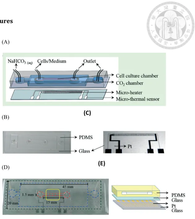
Figure 1. Design and fabrication of the microfluidic chip
(A) Schematic illustration of this device, MD-CaMP. MD-CaMP contains two parts: the micro-temperature control system and the main body of this device, microfluidic chip. Micro-temperature control system includes a micro-heater and a micro thermal sensor. The main body of this device, microfluidic chip, is composed of outer CO2chambers and inner cell culture chambers.(B) CO2 and cell-culture chambers.
(C) Pt micro-heater. (D) Top view of an assembled device. The red dashed line indicates the cell-culture area and between the blue and red lines is the CO2 chamber. The area marked by the yellow square box is the region for observing the cell behavior. (E) Side view of the material of each part. The transparent PDMS facilitates cell observation.
(C)
(E)
(A)
(B)
Figure 2. Migratory abilities of CL1 lung cancer sublines, CL1-0, CL1-1 and CL1-5
(A) Time-lapse images of live CL1 sublines CL1-0, CL1-1 and CL1-5 obtained with a phase-contrast microscope after the indicated intervals after standard wound-scratch procedures. The original width of the area immediately after the scratch was measured as 100%, and for the subsequent 12 and 24 hour, the width of the same locations were measured again to obtain the remaining percentage of cell-free space. (B) Mean wound closure fractions(as percentiles) from three independent experiments. (C) Fold viable cell count of CL1-0, CL1-1 and CL1-5 after 24 hours of culturing. Cell proliferation analysis was performed by counting viable cells using Trypan Blue exclusion assay at the end time point for the wound closure assessment (24h post seeding).
(C)
Figure 3. Level of fibronectin expression on CL1-0, CL1-1 and CL1-5
Western blot analysis of CL1-0, CL1-1 and CL1-5 with fibronectin and α-tubulin (as loading control) antibodies.
(A)
(B)
(C)
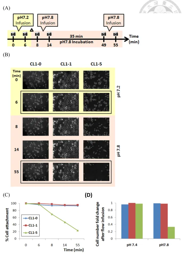
Figure 4.Cell detachment of CL1-0, CL1-1, CL1-5assessed by MD-CaMP
(A) Experimental scheme of cell detachment on MD-CaMP. Cells seeded in MD-CaMP were infused by medium at pH 7.2 for 6 min as control, followed by infusion of medium at pH 7.8 for 6 min. After incubated in the environment at pH 7.8 for 35 min, cells were infused again for 6 min. The velocity of flow we infused
(D)
simulated physiological blood flow (8 mm s−1 in artery = 64 ml h−1 flow rate in our chip). (B) Time-lapse images of the cells in the MD-CaMP upon infusion of buffered media. (C) Quantitative representation of the percentage of cells that remained attached on the chitosan surface at the indicated times. (D) The fold change of cell fractions attached while infusing with pH 7.4 medium as compared to pH7.8 medium.
(A)
(B)
(C)
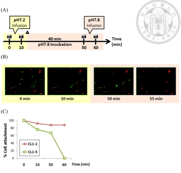
Figure 5. Live cell fractionation in CL1-1,CL1-5 co-cultured system
(A) Experimental scheme of cell detachment in CL1-1 and CL1-5 co-cultured system on MD-CaMP. Cells seeded in MD-CaMP were infused by medium at pH 7.2 for 10 min as control, followed by 40 min of incubation before infusion of medium at pH7.8 for 10 min. (B) Time-lapse images of fluorescently labeled CL1-1 cells (red) and CL1-5 (green) cells co-cultured in the MD-CaMP. Buffered media at the indicated pH were infused in indicated time interval. (C) Quantification of the cells that remained attached to the same region of the chitosan surface was recorded using fluorescence microscopy at the indicated time points.
(A)
Fig. 6 Morphological change of CL1-5 cells on chitosan surface after incubation in different pH media as monitored by DIC microscopy
CL1-5 cells seeded on chitosan surface were incubated in medium at pH 7.4 for 60 min followed by incubation of medium at pH 7.8 for another 60 min.
(A)
(B)
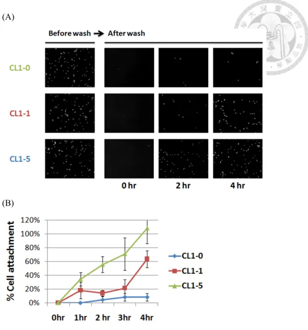
Fig. 7 Cell attachment of CL1-0, CL1-1, CL1-5 on chitosan surface
(A) Images of CL1-0, CL1-1, CL1-5 seeded separately on chitosan surface before wash and wash after 0, 2 and 4 hr of culturing. (B) Quantification of the cells that remained attached on chitosan surface after wash at indicated time point.
(A)
(B)
Fig. 8 Cell fractionation of co-cultured CL1-0, CL1-1, CL1-5 by differential attachment properties on chitosan surface
(A) Images of fluorescently labeled CL1-0 (blue), CL1-1 (red) and CL1-5 (green) cells co-seeded on general TCPS dish (as control) or chitosan surface before and after wash. (B) Quantification of percentages of each sublines attached on chitosan surface after wash at indicated time point.
Appendix
Appendix 1. Schematic representation of the relationship among medium pH, fibronectin adsorption, and cell attachment on chitosan.
研究二 SIK2 藉由磷酸化 CAPS2 調控胰島素分泌之
探討
摘要
動物體的各種器官之協同運作,需仰賴內分泌系統對細胞之調節作用。胰島素即 係一種負責控制血糖濃度之內泌素。當胰島素的生成或分泌發生異常,抑或是體 內細胞出現胰島素失敏之現象,便可能引起糖尿病。佔總糖尿病患數 90 – 95%
之第二型糖尿病,其起因爲體細胞逐漸產生胰島素抗性,進而造成胰島素過量分 泌之代償作用,終而導致 β 細胞劇烈死亡。因此,改善其病徵之方法可為刺激胰 島素分泌及抑制 β 細胞凋亡。Salt inducible kinase 2 (SIK2) 係一屬 AMPK 族 群之酵素,於 β 細胞中調控胰島素之分泌,並藉由抑制 CREB-mediated IRS-2 基 因表現,促使 β 細胞凋亡。本研究室先前研究指出,SIK2 可透過其磷酸酶活性 控制 β 細胞中胰島素小泡之遷移及分泌,而抑制 SIK2 磷酸酶活性可增加胰島素 之分泌。為進一步探討 SIK2 對胰島素分泌調控之機轉,吾人以 Scansite 網站資 料庫預測,發現 calcium-dependent activator protein for secretion 2 (CAPS2) 蛋白具有 SIK2 之磷酸化目標序列,可能為 SIK2 之下游調控因子。欲了解 SIK2 是 否會透過 CAPS2 影響胰島素分泌,本研究首先確認 SIK2 及 CAPS2 均於 β 細胞中
表現,再以免疫沈澱法驗證其交互作用,並以in vitro kinase assay 證明 CAPS2
確為 SIK2 之磷酸化目標。然而,吾人實驗結果亦顯示於 SIK2 及 CAPS2 共同過量 之小鼠 β 細胞中,SIK2 可透過 CAPS2 強化細胞對葡萄糖刺激之反應。綜合上述,
本研究結果指出 SIK2 可藉磷酸化 CAPS2 調控 β 細胞中胰島素之分泌,然其中詳 細之分子機制,尚待未來進一步研究發掘。
Study II: Identification of CAPS2 as a downstream target of SIK2 in regulating insulin secretion
Abstract
Through the endocrine system, communications between cells in various tissues in our body are well coordinated to drive a variety of physiological events properly. Insulin, one of endocrines, is secreted in response to elevated blood concentration of glucose, thereby keeps blood sugar level within certain range. Either failures of production and secretion of insulin, or cellular insulin insensitivity could result in diabetes mellitus.
Type 2 diabetes (T2D) accounting for 90–95% of those with diabetes, is caused by the cellular resistance to insulin stimulation which leads to progressive insulin secretion defect in order to compensate for the less responsive reactions, and finally massive death of β cells. Ways to treat T2D are to stimulate more insulin secretion and to inhibit β cell death. Salt inducible kinase 2 (SIK2), a member of AMPK family, plays important roles in both β cell survival as well as insulin secretion. SIK2 has been reported to suppress β cell survival by repressing CREB-mediated IRS-2 gene expression. Our group found that SIK2 is expressed in insulin-producing β cells and regulates dynamics of insulin secretion via its kinase activity. Inhibition of SIK2 kinase activity resulted in vesicle mobilization to the membrane and increased insulin release.
To further understand the machinery of the regulation of insulin secretion by SIK2, we
applied Scansite database search program and found that CAPS2 (calcium-dependent activator protein for secretion 2), the vesicle related protein containing SIK2 phosphorylation consensus motif, was considered as a putative downstream substrate of SIK2. Herein, we firstly confirmed the physiological protein interaction between CAPS2 and SIK2, and found the interaction was correlated to SIK2 kinase activity. In addition, our in vitro kinase assay results provide clear evidences to show that CAPS2 is the direct substrate of SIK2. However, in SIK2/CAPS2 overexpressing mouse primary β cells, SIK2 might regulate insulin secretion via CAPS2 by sensitizing the responses to glucose in β cells. Taken together, our findings suggest that the function of CAPS2 is involved in insulin secretion and is regulated by protein kinase SIK2. Future studies are needed to reveal the mechanisms of the SIK2/CASP2 pathway in controlling insulin secretion.
Introduction
Definition, classification, cause and prevalence of diabetes mellitus
Diabetes mellitus is a group of metabolic diseases characterized by hyperglycemia resulting from defects in insulin secretion, insulin action, or both [18]. The patients' bodies fail to release enough amounts of the blood glucose-lowering hormone insulin, or/and the target organs is unable to respond to insulin while blood-glucose level increased. The chronic hyperglycemia of diabetes may leads to long-term damage especially to eyes, kidneys, nerves, heart, and blood vessels, causing retinopathy, renal failure, cardiovascular symptoms and so on.
The major cases of diabetes are classified into two categories. Type 1 diabetes, insulin-dependent diabetes (IDDM), also termed Immune-mediated diabetes or juvenile-onset diabetes, accounts for 5–10% of those with diabetes [19]. It is arose from the autoimmune destruction of the pancreatic β cell, leading to absolute insulin deficiency. Patients suffering from type 1 diabetes rely on multiple daily insulin injection or insulin pump therapy with daily blood sugar monitoring or islet transplantation to survive [20]. Type 2 diabetes, noninsulin-dependent diabetes (NIDDM), also referred to as adult-onset diabetes, accounts for 90–95% of those with diabetes. It is caused by the resistant target cells to insulin action, leading to progressive
insulin secretion defect in order to compensate for the resistance, and finally β cell apoptosis [21]. Insulin normally turns down gluconeogenesis to keep blood sugar levels from rising, but in people with insulin resistance, blood sugar levels are elevated because gluconeogenesis continues when it shouldn’t [http://www.science20.com].
Patients with type 2 diabetes depend on medications that stimulate more insulin secretion or inhibit glucose production and release, along with special attentions on diet and physical activities. Drugs such as metformin, the first-line drug for treatment of type 2 diabetes, lower blood glucose by suppressing living releasing glucose and by increasing insulin sensitivity. Other drugs like Sulfonylurea works on increasing insulin production and secretion [22, 23].
The prevalence of diabetes worldwide was reported to be 2.8% in 2000 and will rise to 4.4% in 2030. The total number of people with diabetes was reported to rise from 171 million in 2000 to 366 million in 2030.Similar trend has also happened in Taiwan that there was a more than 70% increase in the total diabetic population, or a 35%
increase in the standardized prevalence rate from 2000 to 2009 [24, 25]. Diabetes has becoming an ever greater public health problem, so it is important to develop therapies to prevent the development of or treat existing diabetes.
Secretory pathway, distinct pools and biphasic secretion of insulin vesicles in
Insulin, the only blood glucose-lowering hormone in the body, plays key roles in glucose homeostasis and maintenances of blood sugar level. It is produced and secreted by pancreatic β cells in islets of Langerhans. Roughly speaking, insulin secretion is a process that involves the packaging of insulin granules, the trafficking of granules to the plasma membrane, and the exocytotic fusion of granules with plasma membrane. After synthesized in the ER and underwent maturation steps, it is packaged in the Golgi into secretory granules allowing further processing to biologically active insulin [26].
Peptide secreting endocrine cells such as chromaffin cells and beta cells pack and store hormones in large dense core vesicles (LDCVs), which contain a big core that is electron-dense under the electron microscope [27].
Insulin secretion is controlled by β-cell electrical activity [28].When the blood glucose level is raised after eating, β cells sense higher ATP production metabolized by glucose. Increased cytosolic ATP/ADP-ratio result in closure of ATP-sensitive potassium-channels (KATP-channels) in the plasma membrane, which leads to depolarization of the cell and activation of the voltage dependent calcium-channels (VDCC). The elevated cytosolic calcium concentration due to influx of calciumion through VDCC then triggers exocytosis and insulin secretion [29].
The secretory vesicles in beta cells can be functionally divided into two distinct pools, readily releasable pool (RRP) and reserve pool (RP), by their release competence
and proximity to the plasma membrane [30]. Each beta cell comprises more than 10,000 insulin-containing LDCVs, but only a small fraction (~ 1%) of these belongs to RRP, which can be released immediately without any further modification upon calcium influx. While the remainder (~99%) which belongs to RP requires a series of ATP-, calcium-, and temperature-dependent mobilization to the plasma membrane to refill the emptied RRP. Recruitment of granules from RP to RRP is a relatively slow process (~ 5 granules/sec., compared with the exocytotic burst of 700 granules/sec.) [31, 32]. The depletion of RRP takes less than a second, whereas its refilling takes up to a minute [33].
Glucose-stimulated insulin exhibits a characteristic biphasic pattern, consisting of a rapid and transient first phase followed by a slowly developing and sustained second phase [34]. The first phase burst secretion occurs within 10 minutes and a second phase can last 2-3 hours. The biphasic insulin secretion reflects the distinct pools of vesicles.
The early rapid component (first phase) involves exocytosis of RRP, while the sustained component (second phase) corresponds to the balance of released RRP by mobilization of new granules from the reserve pool [35] .
Type 2 diabetes features defected insulin action and inability of β-cells to secrete enough insulin to maintain glucose homeostasis. It is characterized by the selective loss of first phase and a reduction of second phase in insulin secretion. Researchers was considering whether the secretory defect seen in type II diabetes results either from
incomplete closure of the KATP channels or interference with the mobilization of granules into the RRP [34]. In addition, restoration of first phase insulin secretion has been shown to improve blood glucose in type II diabetes by suppressing hepatic glucose production and having insulin sensitive tissue readily take up glucose [36]. Clinical treatments of type 2 diabetes focus on inhibiting glucose release from liver, increasing insulin sensitivity as well as stimulating more insulin release, so an understanding of insulin vesicle trafficking and secretion may lead to novel therapeutic strategies [37].
Roles of SIK2 in metabolism and insulin function
Salt Inducible Kinase 2 (SIK2) is among plenty of proteins appealing to us as the key molecule for studying the underlying mechanism in insulin secretion due to its multiple roles in metabolism and insulin function.
SIK2, with SIK1 and SIK3 are three isoforms of SIK that was first cloned from the adrenal glands of rats fed a high salt diet. SIKs are serine/threonine protein kinases which belongs to AMPK (AMP-activated protein kinase) family, essential regulators of cellular metabolism and energy homeostasis [38].
SIK2 is highly expressed in adipose tissue, while SIK1 is abundant in adrenal glands [5–7] and SIK3 has ubiquitous expression pattern [39, 40]. SIK2 has been
reported to play many roles in metabolic functions. Expression of SIK2 in adipocytes might modulate the efficiency of insulin signal transduction via phosphorylation of IRS-1 at Ser794, causing insulin resistance in diabetic animals [39]. CREB (cAMP-response element-binding protein) is required for beta cell survival by driving its direct target, IRS2 gene [41]. CREB also turns on genes for gluconeogenesis in liver during fasting to maintain blood sugar levels. However, SIK2 suppresses beta cell survival and gluconeogenesis by repressing CREB-mediated gene expression via phosphorylation of CREB-coactivator, TORC2, at Ser171, which results in binding of 14-3-3 proteins on TORC2 and thus confines its translocation to nucleus. Yet phosphorylation activity of SIK2 was decreased in response to cAMP by PKA through phosphorylation at SIK2 Ser587 that is important for TORC2 nuclear entry [42].
Overexpression of SIK2-S587A non-phosphorylatable mutant inhibited the PGC-1α and UCP-1 gene expression, indicating the importance of SIK2-TORC2 cascade in
regulation of insulin signaling in brown adipose tissue [43]. Furthermore, SIK2 is critical in the regulation of lipid homeostasis and adipogenesis in vivo, and phosphorylation at SIK2-Ser358 is involved in lipid metabolism [44].
Besides metabolic roles, SIK2 has been reported in diverse biological scenarios, including the regulation of neuronal survival after OGD (oxygen and glucose deprivation) via TORC1-CREB, the repression of melanogenesis, requirement for