國 立 交 通 大 學
生 物 科 技 學 院
生 化 工 程 研 究 所
博士論文
建立循環流動系統壓電式生物感測器用於及時檢測病原菌
大腸桿菌
O157:H7 及登革熱病毒
Establishing a Circulating-flow System of Piezoelectric
Biosensor for In-time Detection of Pathogen, Escherichia coli
O157:H7 and Dengue Virus
研 究 生: 陳思豪
指導教授: 林志生 博士
建立循環流動系統壓電式生物感測器用於及時檢測病原菌大腸桿
菌 O157:H7 及登革熱病毒
Establishing a Circulating-flow System of Piezoelectric Biosensor
for In-time Detection of Pathogen Escherichia coli O157:H7 and
Dengue Virus
研 究 生:陳思豪 Student:Sz-Hau Chen
指導教授:林志生 Advisor:Chih-Sheng Lin Ph.D.
國 立 交 通 大 學
生化工程研究所
博 士 論 文
A ThesisSubmitted to Institute of Biochemical Engineering College of Biological Science and Technology
National Chiao Tung University In partial Fulfillment of the Requirements
For the Degree of Ph.D. In
Biological Science and Technology July 2009
Hsinchu, Taiwan, Republic of China
謝 誌
博士四年的生涯終於結束了,而我也即將邁向了另一個人生的開始,在這過程中不 知有多少人問過我同樣的問題”為什麼妳你想要唸博士?”而我始終如一的回答是”想不 開”。博士的生涯是如此的繁忙與艱辛,整天和研究與書本為伍。在這四年期間實驗室 幾乎成為我第二個家,在每天從實驗室走回宿舍的路上,拖著疲憊的步伐看著皎潔的月 光或剛初昇的太陽,但我知道我正在一步一步達成想要的目標。而這過程必須感謝許多 人給於我的幫助與鼓勵,讓我的博士班生涯多采多姿。 首先我非常感謝指導教授 林志生 老師,指導了我許多做學問的態度、邏輯觀念的 指引與多方角度的思維,並適時的給予鼓勵與肯定,以及生活與人生的方向的關懷與幫 助,讓我在人生的道路上的經歷更加豐富。千言萬語都無法表達我對於您的感謝,能在 您的門下是我這一輩子最大的福氣。另外,也非常感謝美國緬因大學 吳啟華 教授在這 四年來於研究上指導與生活上的關懷,以及擔任口試委員在論文上提供寶貴的意見。亦 非常感謝口試委員兼召集人大同大學 顏聰榮 教授,以及口試委員文化大學 蘇平貴 教 授、中興大學 溫曉薇 教授及交通大學 楊昀良 教授,百忙之中能抽空前來並給予審閱 及斧正,使本論文能更加的完整。 在四年實驗室生活中,往事依舊歷歷在目,感謝博班三人組歌壇唱將建龍學長與冷 面笑將俊旭學長在實驗與生活上的幫助,多少個苦悶的夜晚有你們陪伴一起吃宵夜的日 子,讓生活更添動力;棠青多虧有你的有求必應的個性與幫忙,讓事情作起來格外順利; 筱晶的一流聲音,讓實驗室的每個夜晚都不會寂寞;聖壹與千雅的雙簧二人組更是讓實 驗室充滿歡樂與朝氣;曜禎的體力無限與在實驗上的協助,只能說好在有你阿,接下來 biosensor 組就看你了;証皓的辦事效率及認真態度與停不下來的話匣子,讓大家不感寂 寞,明達的開朗個性與南部人的率直,使實驗室充滿陽光;打字能手的帥氣榕均學妹, 妳做事的細心及在實驗瑣事上的幫助,讓事情趨於簡單;實驗室四朵花-庭妤、郡誼、 瀞韓及子慧,除了在實驗管理上的幫忙,更讓實驗室增色不少;逸柔、家瑋、怡萱、唯 婷、修兆、竣瑋、欣儒、政庭及祥婷在生活上的相互幫忙,感謝你們的支持與幫助,因 為有你們而更覺得豐富。最後僅以這本論文獻給我最親愛的父母及家人,感謝父母的栽培與關懷,你們的期 待與鼓勵是我最大的原動力,沒有你們一路的支持就沒有今日的我,謝謝你們對我的付 出,是我心靈的最佳避風港,對你們獻上無盡的謝意。 陳思豪 謹誌 國立交通大學 生化工程研究所 生醫工程實驗室 中華民國九十八年七月
建立循環流動系統壓電式生物感測器用於及時檢測病原菌大腸桿菌
O157:H7 及登革熱病毒
研究生:陳思豪 指導教授:林志生 博士 國 立 交 通 大 學 生 物 科 技 學 院 生 化 工 程 研 究 所 博 士 班中 文 摘 要
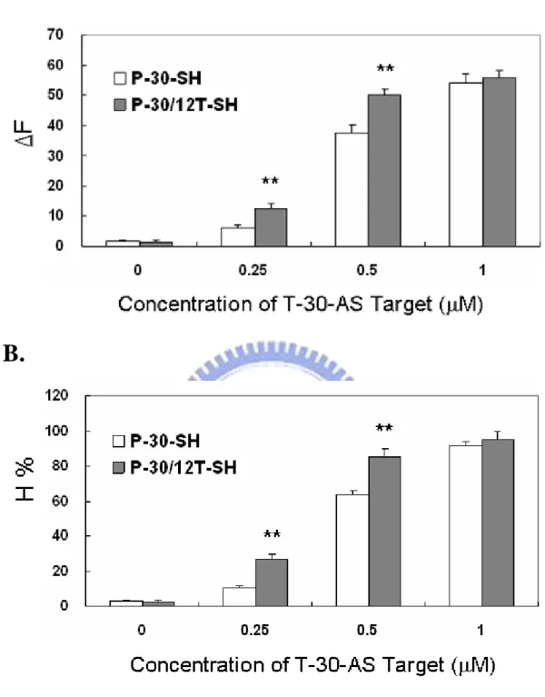
在公共衛生的觀點上,病原菌的感染已經成為嚴重的疾病問題。 然而,在我們的 日常生活中,有許多病原菌類別存在,包括致病性的病毒、細菌、黴菌、寄生蟲、海生 的浮游植物及藍綠藻等。 病原菌的傳播機制不僅只有利用環境的因子如水、空氣或者 是土壤,也包含食品的污染,血液的輸送或接觸的感染等。 由於在每個國家中致死性 的病原菌往往伴隨著經濟的損失及人民的死亡,因此,如何預防及前期偵測病原菌的感 染爆發是一重要的任務。 生物感測器為快速檢測生物分子的一新穎技術,此研究主要為發展在循環流動系統 中一核酸壓電式生物感測方法用以及時偵測病原菌及病毒。 首先設計一具有 30 個去氧 核苷酸及額外具有12 個去氧單磷酸胸腺苷(dT)的專一性核酸探針以解決生物感測系 統上空間的阻礙。 此具有 12 dT 於探針上如同一 spacer,可顯著的增強雜交的效率(P < 0.05)。 結果中指出當探針分別與 30 mer 與 104 mer 的目標物雜交時,spacer 增加雜交 效率分別為1.4 倍及 2 倍。 尤其當探針與較長的核酸目標物雜交時,spacer 減少了固 定化的核酸探針與目標物雜交時的空間阻礙並提供雜交行為的支援。 此核酸壓電式生 物感測系統也被用於從大腸桿菌O157:H7 的 PCR 擴增的 DNA 產物的真實樣本上的檢測 過程中所使用,其雜交效率的結果 PCR 放大的雙股 DNA 可相當於合成的目標物 T-104AS 之單股 DNA。 更進一步地,此循環流動系統之壓電式生物感測器基於奈米金球的放大與驗證方法 被用於一食品中病原菌大腸桿菌O157:H7 的及時檢測。 延續前部分的研究,一含有 12-dT 及修飾硫醇且互補於目標物序列的第二探針被與奈米金球結合,並且作為像一質 量的放大者及序列的檢驗者,用於放大在DNA 壓電式生物感測器上頻率的改變。 在大 腸桿菌O157:H7 的樣品檢測方面,經由後 PCR 的放大後,可藉由此 DNA 壓電式生物感測器所測得1.2 × 10² CFU/ml 的大腸桿菌 O157:H7,並且當大腸桿菌 O157:H7 從 102 至 106 CFU/ml 時其具有線性相關。 在真實的食物樣品的檢測上,此 DNA 壓電式生物感測 器亦可檢測出目標物。 除了病原菌的檢測,病毒的檢測是在壓電式生物感測器上的一個挑戰,考慮到病毒 需要許多時間以傳統的方法進行檢測,此部分基於前面利用及時與可連至電腦的檢測成 果,並結合奈米金球層疊的方法用於快速檢測病毒。 在此研究中,一對在登革熱病毒 外膜蛋白的基因保留區的通用型引子對被設計用來放大其DNA 片段,並且設計兩專一 性探針用以識別在台灣登革熱病毒二型常見亞型。 根據先前的方法,在 DNA 壓電式生 物感測器的表面上,第一探針與目標物進行雜交用以辨識登革熱病毒。 然後,第二探 針結合奈米金球並且與目標物進行雜交,用以增加訊號值與驗證。 為了增加更多的雜 交效率,第一探針被結合至不同大小粒徑的奈米金球並以層疊法引入,且與自由流動的 目標物進行雜交。 接著,第二探針與目標物雜交於另一端,此經由層疊法的結構類似 樹枝狀的結構。 藉由此層疊法,2.1×101 (PFU)/ml 的登革熱病毒二型可藉由此壓電式生 物感測器所測得,其介於頻率變化與病毒濃度的對數從2.1×10⁶ 至 2.1×101 PFU/ml 呈線 性相關。 關鍵字:壓電式生物感測器、奈米金球、病原菌、大腸桿菌O157:H7、登革熱病毒
Establishing a Circulating-flow System of Piezoelectric Biosensor for
In-time Detection of Pathogen Escherichia coli O157:H7 and Dengue Virus
Graduate student: Sz-Hau Chen Advisor: Chih-Sheng Lin Ph.D. Institute of Biochemical Engineering
College of Biological Science and Technology National Chiao Tung University
Abstract
Pathogens infections have been a serious problem on the public health aspect. There are many classes of pathogenic microorganisms, including pathogenic viruses, bacteria, fungi, parasites, marine phytoplankton, and cyanobacteria, etc. in our daily lives. The pathogen transmission mechanism use not only environment factors, ex. water, air or soil, but also the food contamination, blood transfusion or contract infection etc… At every country, the deadly pathogens are usually accompanying economy damage and life loss. Hence, how to prevent and early detect pathogen infection and outbreak is important tasks.
Biosensor is a novel technology for rapid detection of biomolecules. In this study, we develop a DNA piezoelectric biosensing method for In-time detection of pathogens or viruses in a circulating flow system. First, the specific probes of a 30-mer oligonucleotide with additional 12 deoxythymidine 5'-monophosphate (12-dT) is designed to solve steric interference on the biosensor system. The addition of 12-dT to the probes as a spacer, significantly enhanced (P < 0.05) the hybridization efficiency (H%). The results indicate that the spacer enhanced the H% by 1.4- and 2-fold when the probes are hybridized with 30-mer and 104-mer targets, respectively. The spacer reduced steric interference of the support on the hybridization behavior of immobilized oligonucleotides, especially when the probes hybridized with relatively long oligonucleotide targets. The DNA piezoelectric biosensing system is also applied in the detection of PCR-amplified DNA from real samples of Escherichia coli O157:H7. The resultant H% of the PCR-amplified double-strand DNA is comparable to that of the synthetic target T-104AS, a single strand DNA.
Further, a circulating-flow piezoelectric biosensor, based on an Au nanoparticle
amplification and verification method, is used for real-time detection of a foodborne pathogen,
complementary to the target sequence, is conjugated to the Au nanoparticles and used as a “mass enhancer” and “sequence verifier” to amplify the frequency change of the DNA piezoelectric biosensor. The PCR products amplifing from concentrations of 1.2 × 10² CFU/ml of E. coli O157:H7 are detectable by the DNA piezoelectric biosensor. A linear correlation is found when the E. coli O157:H7 detected from 102 to 106 CFU/ml. The piezoelectric biosensor is also able to detect targets from real food samples.
Besides bacteria detection, virus detection is a challenge in the filed of piezoelectric biosensor. It needs a lot of time to detect viruses by traditional methods. This part of study combined the previously developed In-time and on-line work, with Au nanoparticles layer by layer method for rapid detection of virus. In this study, a pair of universal primers of dengue virus envelope gene conserve region was used to amplify cDNA fragment, and two specific probes for the identification of dengue virus type II common subtypes are developed in Taiwan. According to pervious process, first probe hybridizes with the target to identify dengue virus in DNA piezoelectric biosensor surface, then, second probe conjugates with Au nanoparticles and hybridizes with target to enhance signal and verification. In order to increase more hybridization efficiency, the layer by layer method is recommended for the first probe to conjugate to Au nanoparticles in different sizes and to hybridize with free targets. Further, the second probes are hybridized with targets at other terminals. The structure is like dendritic form via layer by layer hybridization. Following layer by layer method, as low as 2.1×101 plaque forming unit (PFU)/ml DENV type 2 can be detected by the DNA
piezoelectric biosensor. Linear correlation between frequency change and logarithmic number of virus concentration is found for DENV from 2.1×10⁶ to 2.1×101 PFU/ml.
Keyword: piezoelectric biosensor, Au nanoparticles, pathogen, Escherichia coli O157:H7,
Content
Acknowledgement ...i
Chinese Abstract...iii
English Abstract...v
Content ...vii
List of Figures ...xi
List of Tables ...xiii
1. Literature review ... 1
2. Pathogen Biosensor ... 2
2.1 Mass sensitivity biosensor... 3
2.2 Electrochemical biosensor ... 6
2.3 Optical biosensor ... 7
2.4. Apply to biosensor related nanotechnology ... 9
2.4.1. Nanoparticles, nanocarbon tube, and nanowires... 9
2.4.2. Quantum dots………...11
2.4.3. Magnetic beads...12
3. Piezoelectric biosensor ... 13
3.1 Piezoelectric effect ... 13
3.2 Piezoelectric quartz crystal ... 13
3.3 Quartz crystal microbalance ... 14
4. Self assembled monolayer for biosensor application ... 17
5. Pathogen diagnosis ... 19
5.1. Escherichia coli O157:H7... 19
5.2. Dengue virus ... 20
7. Materials and methods... 22
7.1 For E. coli O157:H7 detection ... 22
7.1.1 Chemicals... 22
7.1.2 Oligonucleotide primers, probes and targets ... 22
7.1.3 Culture preparation... 23
7.1.4 Food sample studies ... 23
7.1.5. DNA extraction... 24
7.1.6 PCR Conditions... 24
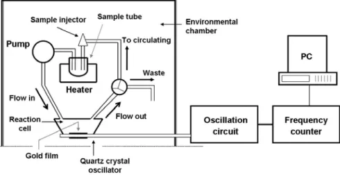
7.1.7 The circulating-flow QCM system ... 25
7.1.8 Gold-QCM device preparations……….…….25
7.1.9 Immobilization of the oligonucleotide probes and hybridization with oligonucleotide targets………..26
7.1.10 Probe oligonucleotide–nanoparticle conjugates... 27
7.1.11. Immobilization of different length probe oligonucleotides and hybridization with target sequences ... 28
7.1.12 Sandwich hybridization by oligonucleotides capped with Au nanoparticles... 28
7.1.13 Data analysis... 29
7.2 For dengue virus detection ... 29
7.2.1 Chemicals and reagents... 29
7.2.2 Oligonucleotide primers, probes and targets design ... 29
7.2.3 Culture preparation of dengue virus... 30
7.2.4 Plaque forming unit assay of DENV ... 30
7.2.5 Dengue virus RNA extraction ... 31
7.2.6 Dengue virus cDNA preparation ... 31
7.2.7 Asymmetric PCR amplification ... 31
7.2.8 The circulating-flow QCM system ... 32
7.2.9 AuNPs preparation and oligonucleotide-modification ... 33
7.2.10 Immobilization of probes, hybridization, and signal amplification by AuNPs-probes ... 33
8. Part I:
In-time detection of E. coli O157:H7 sequences using a
circulating- flow system of quartz crystal microbalance ... 35
8.1 Abstract ... 36
8.2 Introduction ... 37
8.3 Results and discussion... 39
8.3.1 The QCM system and detection... 39
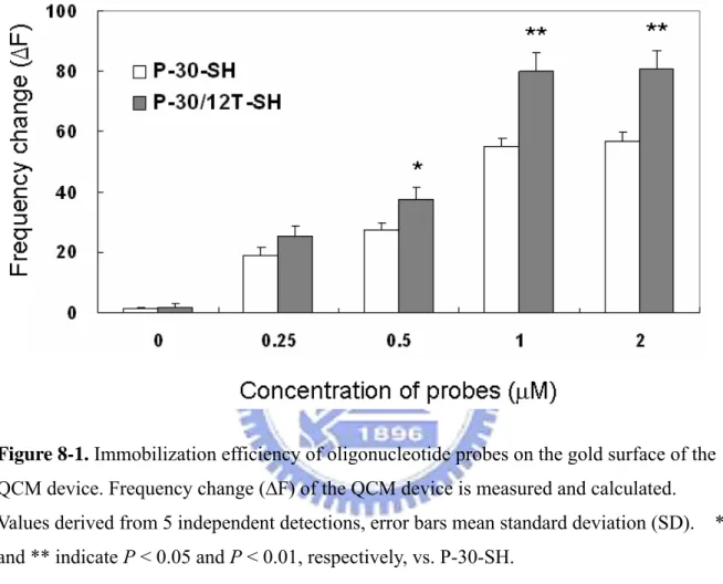
8.3.2 Immobilization of synthesized oligonucleotide probes ... 39
8.3.3 Detection of the short (30 mer) synthesized target oligonucleotides ... 40
8.3.4 Specificity of the QCM detection... 40
8.3.5 Detection of the long (104 mer) synthesized target oligonucleotides... 41
8.3.6 Detection of PCR-amplified DNA of E. coli O157:H7 gene eaeA ... 42
8.3.7 Conclusions... 44
9. Part II:
Using oligonucleotide-functionalized Au nanoparticles to rapidly
detect foodborne pathogens on a piezoelectric biosensor ... 45
9.1 Abstract ... 46
9.2 Introduction ... 47
9.3 Results ... 49
9.3.1 The QCM system and detection... 49
9.3.2 Immobilization of synthesized probe oligonucleotides ... 49
9.3.3 Hybridization of synthesized target oligonucleotides to probes immobilized QCM sensor ... 50
9.3.4 Detection of PCR-amplified DNA of E. coli O157:H7 gene eaeA ... 50
9.3.5 Detection of PCR-amplified DNA of E. coli O157:H7 using oligonucleotide -functionalized Au nanoparticles ... 51
9.3.6 Specificity of the QCM system in detecting E. coli O157:H7... 52
9.3.7 Quantitation of the QCM detection of PCR-amplified DNAs ... 52
9.4 Discussion... 54
9.5 Conclusions ... 57
10. Part III:A method of layer-by-layer gold nanoparticles hybridization in
a quartz crystal microbalance DNA sensing system used to detect dengue
virus... 58
10.1 Abstract ... 59
10.2 Introduction ... 60
10.3 Results and discussion... 62
10.3.1 QCM system and detection ... 62
10.3.2 Identification of AuNPs size ... 62
10.3.3 Effect of AuNPs-probe size on ΔF enhancement... 63
10.3.4 Observation of AuNPs-probes hybridization on QCM chip ... 64
10.3.5 Detection of RT-PCR-amplified DNA from DENV2... 65
10.3.6 Quantitative detection of DENV2 detection in real blood sample... 66
10.4 Conclusions ... 68
11. General conclusions... 69
11.1 Part 1: Real-time detection of Escherichia coli O157:H7 sequences using a circulating- flow system of quartz crystal microbalance ... 69
11.2 Part 2: Using oligonucleotide-functionalized Au nanoparticles to rapidly detect foodborne pathogens on a piezoelectric biosensor ... 69
11.3 Part 3: Using layer by layer gold nanoparticles hybridization method to improve detection limit of dengue virus by a circulating-flow quartz crystal microbalance DNA sensing system... 70
List of Figures
Figure 3-1. Simple molecular model for explaining the piezoelectric effect……... 82 Figure 3-2. Slicing the different angle to the optical z-axis………. 83 Figure 3-3. Reverse piezoelectric effect on quartz crystal……… 84 Figure 3-4. Transducer of Quartz Crystal Analyzer………. 85 Figure 4-1. Schematic representation of a tightly packed alkanethiol monolayer by
SAM……….. 86 Figure 7-1. The real-time and circulating-flow QCM system………. 87 Figure 8-1. Immobilization efficiency of oligonucleotide probes on the gold surface
of the QCM device………. 88 Figure 8-2 Detection of the short target oligonucleotides, T-30-AS, hybridized with the thiolated probes (1.0 μM) immobilized onto the gold surface of the
QCM device……….. 89 Figure 8-3. Detection specificity of the circulating flow QCM system……… 90
Figure 8-4. Detection of the long oligonucleotide targets, T-104AS and T-104S, hybridized with the probes (1.0 μM) immobilized onto the gold surface of the QCM device……….. 91 Figure 8-5. Detection of PCR-amplified DNA………... 92 Figure 8-6. Schematic representation of steric hindrance of the probe and target
DNA hybridization on the QCM device………. 93 Figure 9-1. Time-dependent frequency changes of the circulating-flow QCM
sensor………. 94 Figure 9-2. Immobilization and hybridization efficiencies in the QCM system… 95 Figure 9-3. Sandwich hybridization using the oligonucleotide-functionalized Au
nanoparticles……… 96 Figure 9-4. Gel electrophoresis and QCM detections of PCR-amplified DNA from
E. coli O157:H7 eaeA gene……… 97
Figure 9-5. The responses of the QCM sensor to the PCR-amplified DNAs isolated from different concentrations of E. coli O157:H7………….. 98 Figure 9-6. Detection of E. coli O157:H7 in food samples using the circulating-flow QCM sensor with Au nanoparticles for signal
amplification………. 99 Figure 10-1. Schematic illustration of the steps involved in probe immobilization followed by target DNA hybridization and layer by layer AuNPs signal
amplification……….. 100 Figure 10-2. UV-Vis absorption spectra of the AuNPs with different diameters…. 101
Figure 10-3. The enhancement of detection signal by different sizes of
AuNPs-probes……….. 102 Figure 10-4. SEM images of the chip surface of QCM sensor………... 103 Figure 10-5. Specificity of the layer-by-layer AuNPs-probes (13 nm) for the DENV
sequence detection……… 104 Figure 10-6. Sensitivity of the circulating-flow DNA-QCM sensor combined with the
List of Tables
Table 1-1. Detection technology of foodbrone pathogen……… 107 Table 2-1. Applications of advanced nanomaterials for environmental
monitoring……… 108 Table 7-1. Sequences of the oligonucleotide probes, targets, and primers used in
this study……….. 109 Table 7-2. Sequences of the oligonucleotide probes, targets, and primers used in
experiment……… 110 Table 7-3. Sequences of the probe, target, and primer oligonucleotides for the
dengue virus serotype-2 (DENV2) detection used in this study…….. 111
Table 10-1. Comparisons of the present study with the related detection technologies for dengue virus………. 112
1. Literature review
Pathogens infections have been a serious problem from the public health aspect. There are many classes of pathogenic microorganisms classifications, including pathogenic viruses, bacteria, fungi, parasites, marine phytoplankton, and cyanobacteria, etc. in our daily life. The pathogen transmission mechanism not only uses environment factor, ex. water, air or soil, but also the food contamination, blood transfusion or contract infection etc… At every country, the deadly pathogens are usually accompanying economy damage and life loss. However, how to prevent and early detect pathogen infection outbreak are important tasks.
The foodborne pathogen of bacteria is a common cause of diseases due to food product contamination, accounting for 91% of the total outbreaks of foodborne illness in the USA [Beran et al., 1991; Potter et al., 1997]. An estimated 76 million cases of foodborne disease occur each year in the United States. The great majority of these cases are mild and cause symptoms for only a day or two. Some cases are more serious, and CDC estimates that there are 325,000 hospitalizations and 5,000 deaths related to foodborne diseases in the United States each year [CDC, 2005]. The most severe cases tend to occur in the very old, the very young, those who have an illness already that reduces their immune system function, and in healthy people exposed to a very high dose of an organism. Besides, foodborne diseases are extremely costly. The U.S. Department of Agriculture (USDA) Economic Research Service (ERS) estimates that the medical costs and productivity losses associated with five major pathogens Escherichia coli O157:H7, non-O157 STEC (Shiga Toxin- Producing Escherichia
coli), Salmonella (non-typhoidal serotypes only), Listeria monocytogenes and Campylobacter,
is at least $6.9 billion annually [USDA/ERS, 2002].
Conventional microbiological methods have been standard operating procedures for the detection and identification of pathogens in food for nearly one century and continue to be a reliable standard for ensuring food safety. Detecting foodborne pathogen with conventional procedures can take several days by use of specific agar media to isolate and enumerate viable bacterial cells in samples [Meng et al., 2001]. However, conventional methods are time consuming and labor intensive, and are therefore not suitable for modern food quality
assurance to make a timely response to possible risks. Based on this reason, over the past 25 years, numerous rapid methods have been developed to reduce the assay time.
Recent detection technology to identify foodborne pathogen are present in Table 1-1, including reform method of plating, real-time PCR, ELISA combining with immunomagnetic or nanoparticles, and biosensors platform. These detection methods are usually using
molecular biology technology to rapid detection, include antibody-based methods (immunofluorescence, immunoimmobilization, enzyme-linked immunosorbent assay,
immunomagnetic separation, etc.), nucleic acid-based methods (hybridization and polymerase chain reaction [PCR]), biochemical and enzymatic methods (miniaturized microbiological methods and commercial miniaturized diagnostic kits), and membrane filtration methods (hydrophobic grid membrane filter) [Wu et al., 2004]. In recent years, modern biotechniques such as real time PCR [Yoshitomi et al., 2003; Fu et al., 2005], nanoparticles [Zhao et al., 2004; Mao et al., 2006] and biosensing systems (biosensors) [Campbell and Mutharasan, 2005; Simpson et al., 2005; Mao et al., 2006] have been developed for detection of pathogenic microorganisms. Biosensors are devices that detect biological or chemical complexes in the form of antigen-antibody, nucleic acids, enzyme-substrate, or receptor-ligand compounds. Interest in using biosensors to detect foodborne pathogens is on the rise [Hall, 2002; Patel, 2006; Rasooly and Herold, 2006].
2. Pathogen Biosensor
In recent year, there are many analytic methods that have been developing for the
pathogen rapid detection. One approach for rapid pathogen detection is the use of biosensors, because the biosensor provided fast and simple detection method, causing its application to expand. Until today, biosensors have gradually become rapid biomolecular recognition tools in analytical biological material. The biosensors are also applied to pathogen contamination detection on food safety.
A biosensor is defined as a device that combines a biological component with a
physicochemical detector component for the detection of an analyte. [http://www.biosensors- congress.elsevier.com/about.htm]
It consists 3 parts:
• The sensitive biological element [biological material (eg. tissue, microorganisms, organelles, cell receptors, enzymes, antibodies, nucleic acids, etc), a biologically derived material or biomimic] that can be created by biological engineering.
• The transducer or the detector element (works in a physicochemical way; optical, piezoelectric, electrochemical, etc.) that transforms the signal resulting from the interaction of the analytic with the biological element into another signal (i.e., transducers) that can be more easily measured and quantified;
• Associated electronics or signal processors that is primarily responsible for the display of the results in a user-friendly way.[ Cavalcanti A et al. 2008]
Although the biosensors can rapid detection or have higher sensitivity, the limitation still
exists. The limitation is from unknown pathogen interference and few cells contamination in the food. Hence, the diagnosis with the biosensor has differentiated between pathogens need pre-enrichment or without enrichment, Based on enrichment step, the biosensors can be differentiated between immunoassay or DNA recognition. The immunosensor usually needs pre-enrichment of pathogen cells before detection. Contrary, the DNA recognition biosensor does not need pre-enrichment, because the polymerase chain reaction (PCR) can substitute for the cell culture. The immunoassay or DNA diagnosis methods have different functions at detection. The immunobiosensor are based on exploiting the specific interaction of antibody with antigen. Typically, immunoassays (such as the widely used enzyme-linked
immunosorbent assay technology) employ a label (e.g., enzyme, fluorescent marker) to detect the immunological reaction. On the other hand, the DNA biosensors, commonly, relys on the immobilization of a DNA probe onto the transducer surface, the subsequent hybridization with the DNA target triggering a signal either directly or indirectly.
Based on these detection methods, the biosensors have several classification and numerous biosensors have been developed for detection and enumeration of pathogen and are promising candidates for rapid screening of foods. The biosensors type according to classify on
foodborne pathogen detection have electrochemical, photometric, and mass sensitive etc.
2.1 Mass sensitivity biosensor
The mass sensitivity biosensor meant that it used difference inmass to effect biosensor physical characteristic and then accomplishing objective detection, for example: piezoelectric biosensor. The piezoelectric biosensor can be classified into two main groups: (1) surface acoustic wave-based biosensor (SAW) and (2) quartz crystal microbalance (QCM).
SAW biosensors are based on the detection of mechanical acoustic waves and
incorporate a biological component. These are mass sensitive detectors, which are operated on the basis of an oscillating crystal that resonates at a fundamental frequency. After the crystal has been coated with a biological reagent (such as an antibody) and exposed to the particular antigen a quantifiable change occurs in the resonant frequency of the crystal, which correlates to mass changes at the crystal surface. The vast majority of acoustic wave
are ideal for use in this application due to their ability to generate and transmit acoustic waves in a frequency-dependent manner. The physical dimensions and properties of the
piezoelectric material influence the optimal resonant frequency for the transmission of the acoustic wave. The most commonly used piezoelectric materials include quartz (SiO2) and
lithium niobate (LiTaO3). In order to acquire an active surface for use in a piezoelectric
biosensor the surface must be stable chemically, contain a high number of the actively immobilized biological elements and the coating surface should also be as thin as possible.
SAW biosensors offer label-free, on-line analysis for antigen–antibody interactions, and also provide the option of several immunoassay formats, which allow increased detection sensitivity and specificity. Other advantages include cost effectiveness combined with ease of use. Disadvantages associated with these sensors include relatively long incubation times for the bacteria and biosensor surface, problems with crystal surface regeneration and the number of wishing and drying steps required.
The QCM is another the most popular mass biosensor in the world. The core of the QCM is a specifically manufactured quartz plate and the cut angel with respect to the crystal lattice would result from different type of resonators. Generally, the AT-cut form, which at a 35.25° angle from the Z-axis exhibit tremendous frequency stability of Δf/ f≒10-8 [Janshoff et al., 2000], and when the temperature changes from 0 °C to 60 °C, the frequency change is close to zero [O’Sullivan and Guilbault, 1999].
The crystal is excited to resonance and the effect of molecular absorption monitored [Sauerbrey, 1959]. The QCM is comprised of thin film electrodes, usually gold (Au), deposited on each face of a crystal. Voltage is applied across these electrodes to deform the crystal plate, producing relative motion between the two parallel crystal surfaces. The crystal is induced to oscillate at a specific resonant frequency. Changes in the mass of the material on the surface will alter the resonance frequency of the crystal [Marx, 2003]. A linear relationship exists between deposited mass and frequency response for quartz crystals. The resonance frequency decreases linearly with the increase of mass on the QCM electrode at the nanogram level or less. This characteristic of QCM can be exploited to develop bioanalytical tools on a 10-10 to 10-12 g scale [Zhou et al., 2000]. QCM-based immunoassay has been designed and applied in several different areas [Su et al., 2000; Kurosawa et al., 2003; Su and Li, 2004].
to detect two different microorganisms, Legionella and Escherichia coli, simultaneously. A series of experiments are conducted to optimize the use of the SAW for bacterial detection using a novel protocol of coating bacteria on the sensor surface prior to addition of the antibody. Results are compared with an experiment in which a conventional protocol is utilized, where antibody are coated on the sensor surface prior to exposure to bacteria. The concentration of bacteria that attached to the surface of the SAW device are related to the antibody that specifically bound to it and therefore to frequency in a dose dependent fashion. Unlike conventional microbiological techniques quantitative results can be obtained for
Legionella and E. coli down to 106 cells per ml within 3 h. In addition E. coli are detected down to 105 cells per ml in a modified protocol using sheep IgG as a blocking agent.
Later, Deisingh and Thompson [2000] is report on the amplification by polymerase chain reaction (PCR) of a 509 base sequence unique to E. coli O157:H7. Immobilization of a probe for the bacterium on an acoustic wave sensor by the biotin–neutravidin interaction is employed to detect the on-line hybridization of the sequence with the sample obtained from PCR. The limit of detection is to be 10–8 M.
In the recent, Olsen et al.[2006] are used biosensor for the rapid detection of Salmonella
typhimurium in solution, based on affinity-selected filamentous phage prepared as probes
physically adsorbed to piezoelectric transducers. Specific-bacterial binding resulted in
resonance frequency changes of prepared sensors, which are evaluated using linear regression analysis. Sensors possessed a rapid response time of <180 s, have a low-detection limit of 102 cells/ml and are linear over a range of 101–107 cells/ml with a sensitivity of 10.9 Hz per order of magnitude of S. typhimurium concentration.
Besides SAW biosensor, the QCM biosensor is developed too in recently years. Many
studies is usually used QCM biosensor in air or liquid phase combined antibody-antigen method to detect several foodbrone pathogen, include E. coli O157:H7 [Liu et al., 2007], L.
monocytogenes [Minunni et al., 1996; Vaughana et al., 2001], Salmonella spp. [ Park et al.,
2000; Wong et al., 2002; Su and Li, 2005], S. aureus [Boujday et al., 2008], B. cereus [Vaughan et al., 2003] and P. aertrginosa [Kim et al., 2004] etc..
Recently, the QCM immunosensor is based on nanotechnology rapid developing and application; the detection limit is more improved. Liu et al. [2007] has shown that using antibody-antigen recognition method to develop and to evaluate for detection of E. coli O157:H7 on quartz crystal microbalance (QCM) immunosensor. The immunosensor are fabricated by self-assembling of protein A and affinity-purified anti-E. coli O157:H7
antibodies on the gold electrode of an AT-cut piezoelectric quartz crystal. To enhance the sensitivity of the QCM immunosensor, nanoparticle-antibody conjugates, which are prepared using streptavidin-conjugated nanoparticles (145 nm diameter) and biotinylated anti-E. coli antibodies, are used for signal amplification. Compared to the direct detection of E. coli O157:H7, the binding of the nanoparticle conjugates further resulted in a decrease in resonant frequency and an increase in resonant resistance, and the detection sensitivity are improved by five orders of magnitude by lowering the detection limit from 107 to 102 CFU/mL.
Mao et al. [2006] using quartz crystal microbalance (QCM) DNA sensor, based on the nanoparticle amplification method, is developed for detection of Escherichia coli O157:H7
eaeA gene. The hybridization is induced by exposing the ssDNA probe to the
complementary target DNA, and results in the mass change and therefore frequency change of the QCM. Streptavidin conjugated Fe3O4 nanoparticles are used as “mass enhancers” to
amplify the frequency change. The detection limit is low as 10−12 M synthesized
oligonucleotides and 2.67 × 10² colony forming unit (CFU)/mL E. coli O157:H7 cells can be
detected. Linear correlation between frequency change and logarithmic number of bacterial cell concentration is found for E. coli O157:H7 from 2.67 × 10² to 2.67 × 10⁶ CFU/mL.
2.2 Electrochemical biosensor
Electrochemical biosensors are based on monitoring electro-active species that are either produced or consumed by the action of the biological components (e.g., enzymes and cells). Transduction can be performed using one of several methods under two broad headings: potentiometry and amperometry.
Potentiometric biosensors are based on monitoring the potential of a system at a working electrode, with respect to an accurate reference electrode, under conditions of essentially zero current flow. In operation, potentiometric measurements are related to the analyte activity (of a target species). The use of ion selective and gas sensitive membranes coupled to enzyme systems, linked to the potentiometric sensor, allows the fabrication of a biosensor device specific to the enzyme substrate or product. By measuring either the ions or the gases that are generated or consumed as a result of the enzyme activity, an effective method for measuring the concentration of the target analytic can be realized. Potentiometric biosensors can operate over a wide range (usually several orders of magnitude) of concentrations. The use of potentiometric biosensors for food analysis has not been as widely reported as for
amperometric sensors. Generally, the use of amperometry as the method of transduction has proved to be the most widely reported using an electrochemical approach. Both “one-shot” (disposable) sensors and on-line (multimeasurement) devices have been described, monitoring a wide range of target analytes. In contrast to potentiometric devices, the principle operation of amperometric biosensors is defined by a constant potential applied between a working and a reference electrode. The imposing potential encourages redox reactions to take place, causing a net current to flow. The magnitude of this current is proportional to the concentration of electroactive species present in solution. Both cathodic (reducing) and anodic (oxidizing) reactions can be monitored amperometrically.
Many amperometric biosensors described to date have been based on the use of enzymes. Typically, oxidase enzymes have been the most frequently exploited catalysts used for these biosensor formats. In operation, amperometric biosensors tend to monitor either the oxygen consumed or the hydrogen peroxide generated. Both are electrochemically active; oxygen can be electrochemically reduced, and hydrogen peroxide can be oxidized. The current generated is proportional to the concentration of the enzyme substrate (i.e., the target analytic) present. Biosensor technologists have also adopted other approaches, including the use of mediators ex. ferrocenedicarboxylic acid, N-methylphenazinium cation and
tetracyanoquinodimethane radical anion etc.. These compounds are able to replace oxygen as an electron acceptor and to operate at a much lower operating potential, reducing the effects of other electrochemically active species found in many food matrices.
2.3 Optical biosensor
Optical biosensors are a powerful detection and analysis tool that has vast applications in biomedical research, healthcare, pharmaceuticals, environmental monitoring, food safety, and the battlefield. They are immune to electromagnetic interference, capable of performing remote sensing, and can provide multiplexed detection within a single device. Generally, there are two detection protocols that can be implemented in optical biosensing:
fluorescence-based detection and label-free detection. In fluorescence-based detection, either target molecules or biorecognition molecules are labeled with fluorescent tags, such as dyes; the intensity of the fluorescence indicates the presence of the target molecules and the interaction strength between target and biorecognition molecules. While fluorescence-based detection is extremely sensitive, with the detection limit down to a single molecule, it suffers from laborious labeling processes that may also interfere with the function of a biomolecule.
Quantitative analysis is challenging due to the fluorescence signal bias, as the number of fluorophores on each molecule cannot be precisely controlled. In contrast, in label-free detection, target molecules are not labeled or altered, and are detected in their natural forms. This type of detection is relatively easy and cheap to perform, and allows for quantitative and kinetic measurement of molecular interaction. Additionally, as discussed later, some
label-free detection mechanisms measure refractive index (RI) change induced by molecular interactions, which is related to the sample concentration or surface density, instead of total sample mass. As a result, the detection signal does not scale down with the sample volume. This characteristic is particularly attractive when ultra-small (femtoliter to nanoliter) detection volume is involved and is advantageous over fluorescence-based detection whose signal usually depends on the total number of analytic in the detection volume or on the detection surface. Despite all these differences between fluorescence-based and label-free detection, both protocols are being widely used in optical sensors and provide vital and complementary information regarding interactions among biomolecules, which makes optical sensors more versatile than other types of sensing technologies, such as optical fibers, surface plasmon resonance fluorescence and surface plasmon resonance (SPR).
Fluorescence occurs when a valence electron is excited from its ground state to an excited singlet state. The excitation is produced by the absorption of light of sufficient energy. When the electron returns to its original ground state, it emits a photon at lower energy. Another important feature of fluorescence is the little thermal loss and rapid (<10 ns) light emission taking place after absorption. The emitted light is at a longer wavelength than the absorbed light since some of the energy is lost due to vibrations, this energy gap is termed Stoke’s shift, and it should be large enough to avoid cross talk between excitation and
emission signals. Antibodies may be conjugated to fluorescent compounds, the most
common of which is fluorescein isothiocyanate (FITC) [Li et al., 2004]. There are, however, other fluorescent markers. The use of lanthanides as sources of fluorescence in luminescent assays has very recently been reviewed [Selvin, 2002]. Although lanthanides pose several important advantages (good stability, lowbackground luminescence under normal light conditions and large Stoke’s shift) compared to more traditional fluorophores, their use is very restricted due to safety reasons. Fluorescence detection, in contrast to SPR, is also used in combination with established techniques such as PCR and ELISA. Such is the case of a hand-held real-time thermal cycler recently developed [Higgins et al., 2003]. This analyser measures fluorescence at 490 and 525 nm, which enables the simultaneous detection of more
than one microorganism. Although this work claims detection times of 30 min, it should be pointed that overnight culturing is required to achieve best results. Fluorescence resonance energy transfer (FRET) biosensors [Ko and Grant, 2003] are based on the transfer of energy from a donor fluorophore to an acceptor fluorophore.
SPR biosensors [Cooper, 2003] measure changes in refractive index caused by structural alterations in the vicinity of a thin film metal surface. Current instruments operate as
follows. A glass plate covered by a gold thin film is irradiated from the backside by
p-polarised light (from a laser) via a hemispherical prism, and the reflectivity is measured as a function of the angle of incidence, θ. The resulting plot is a curve showing a narrow dip. This peak is known as the SPR minimum. The angle position of this minimum is
determined by the properties of the gold-solution interface. Hence, adsorption phenomena and even antigen–antibody reaction kinetics can be monitored using this sensitive technique (as a matter of fact, SPR is used to determine antigen–antibody affinity constants). The main drawbacks of this powerful technique lay in its complexity (specialized staff is required), high cost of equipment and large size of most currently available instruments (although
portable SPR kits are also available commercially, as is the case of Texas Instruments’
Spreeta system). SPR has successfully been applied to the detection of pathogen bacteria by means of immunoreactions [Lazcka, et al., 2007].
2.4. Apply to biosensor related nanotechnology
2.4.1. Nanoparticles, nanocarbon tube, and nanowires
A range of nanoparticles (NPs) including nanotubes and nanowires, prepared from metals, semiconductor, carbon or polymeric species, have been widely investigated for their ability to enhance the response of biosensors. NPs can be used in a variety of ways, such as modification of electrode surfaces, or to modify biological receptor molecules such as
enzymes, antibodies or oligonucleotides. Their use in biosensors has been reviewed recently. In particular, they have proved extremely useful in the preparation of DNA biosensors.
There are also many reports of the methods of enzymes and proteins based on NPs-modified sensor. For electrochemical biosensor application, the enhanced electrochemistry is due to the ability of the small NPs to reduce the distance between the redox site of a protein and the electrode, since the rate of electron transfer is inversely dependent on the exponential distance between them [Lin et al. 2008].
of nanotechnology due to their electronic, optical, catalytic and thermal properties[Guo et al, 2007; Kariuki et al., 2006]. Among those, Ag NPs are known for their antimicrobial activity and have been used in water treatment [ Shrivastava et al., 2007; Baker et al., 2005; Sondi and Salopek-Sondi 2004; Morones et al., 2005 ]. Their bactericidal activity is shape and size dependent, with particles of sizes less than 100 nm showing optimal antibacterial activity. Another application is as oligonucleotide labeling tags in electrochemical DNA sensors [Cai et al., 2002]. For example, Au NPs have been intensively used for signal amplification, as a component of bio-immobilization matrixes in biosensors and in a variety of colorimetric and fluorescence assays. A miniaturized battery operated nanomaterial surface energy transfer (NSET) probe designed for detection and screening of mercury in soil, water and fish, with detection limits in the ppt range is an example of this approach [Darbha et al., 2005]. In the NSET system, AuNPs (acting as an acceptor) were combined with an organic dye
(Rhodamine) as a donor. The method is based on the quenching properties of AuNPs through energy- and electron-transfer, providing enhanced sensitivity for the detection of metal ions. Other NPs such as zero valent iron and bimetallic iron NPs coupled to metal catalysts such as Pd, Pt or Ni were used in environmental remediation of chlorinated organics, polychlorinated biphenyls (PCBs) [ He and Zhang, 2005], inorganic ions and heavy metal ions such as As(III), Pb(II), Cu(II), Ni(II) and Cr(VI) [ Theron et al., 2008].
The use of carbon nanotubes (CNTs) in electrochemical sensors has been reviewed. CNTs can be single-walled or multi-walled. Single-walled nanotubes are formed by rolling up a single graphite sheet into a cylinder; whereas, for multi-walled CNTs, several
singlewalled nanotubes of increasing size lie inside one another, like the consecutive skins of an onion. The sidewalls of the CNTs resemble the basal plane of highly orientated pyrolytic graphite (HOPG), a common electrode material, whereas the ends of the tube resemble the edge planes of HOPG. The enhanced electrochemical response at CNTs is due primarily to the large number of edge plane sites, although this may not be the explanation for all
experimental observations. CNTs are often purified with oxidizing acids to introduce surface sites with carboxylate groups, suitable for attachment to electrode surfaces, enzymes or other biological receptors. The large number of sites on the CNTs can result in
attachment of large numbers of enzyme molecules. Single-walled CNTs can self-assemble onto an electrode surface. If they align vertically, they can act as a molecular wire and allow direct communication between an enzyme active site and electrode, eliminating the need for mediator. CNTs can be used in variety of electrode formats, and as an additive in addition to
other materials (e.g. carbon, in screen printed electrodes or carbon paste electrodes). Direct oxidation of hydrogen peroxide and NADH has been reported at screen-printed carbon–CNT composite electrodes, which gives an inexpensive method of mass fabrication of sensors that can be used with oxidases and dehydrogenases.
Boron-doped silicon nanowires (SiNWs) are reported by Cui et al. [2001] to create highly sensitive, real-time electrically based sensors for biological and chemical species. The amine and oxide-functionalized SiNWs exhibited pH-dependent conductance that is linear over a large dynamic range and can be understood in terms of the change in surface charge during protonation and deprotonation. Biotin-modified SiNWs are used to detect streptavidin down to at least a picomolar concentration range. In addition,
antigen-functionalized SiNWs shows reversible antibody binding and
concentration-dependent detection in real time. The small size and capability of these semiconductor nanowires for sensitive, label-free and real-time detection of a wide range of chemical and biological species can be exploited in array-based screening and in vivo diagnostics.
The nanoscale size of these new classes of sensors allows for measurements in the smallest of environments such as individual cells. This property provides opportunities for in vivo monitoring of processes within live cells. Cullum et al. [2000] used optical fibers with a distal-end diameter of less than 1 μm, coated with antibodies, to detect the presence of toxic chemicals within single cells. They are able to measure the concentration of
benzopyrene tetrol (BPT) within human mammary carcinoma cells and rat liver epithelial cells. Tuan [2002] fabricated nanoprobes with optical fibers pulled down to tips with the distal ends having sizes of approximately 30-50 nm [Tuan, 2002]. Using these
nanobiosensors it has become possible to probe chemical species at specific spots. Nanocontrolled release systems have been devised for optical biosensing of peroxide concentration [Choi et al., 2001].
2.4.2. Quantum dots
A group of inorganic fluorophores, including quantum dots, nanocrystals and
functionalized nanoparticles have revolutionized fluorescence-based detection methods. The high quantum yields and high resistance to photo degradation of such probes has led to
semi-conductors that exhibit unique light emitting properties that can be customized by changing the size or composition of the dots. They are typically 2–8 nm in size and are covered with a layer of organic material that allows functionalization of the surface for biomolecule attachment. The narrow emission spectra of nanocrystals give rise to sharper colors and high spectral resolution, which can improve assay sensitivity
2.4.3. Magnetic beads
Magnetic nanoparticles are a powerful and versatile diagnostic tool in biology and medicine. They usually can be prepared in the form of either single domain or
superparamagnetic (Fe3O4), greigite (Fe3S4), maghemite (g-Fe2O3), and various types of
ferrites (MeO_Fe2O3, where Me = Ni, Co, Mg, Zn, Mn, etc.). Bound to biorecognitive
molecules, magnetic nanoparticles can be used to separate or enrich the analyte to be detected. Established techniques such as magnetic cell separation use magnetic field gradients to
manipulate and isolate magnetically labeled cells. Magnetic immunoassay techniques also have been developed in which a magnetic field generated by the magnetically labeled targets is detected directly with a magnetometer.
A new technique has been introduced for rapid detection of biological targets by using superparamagnetic nanoparticles and a ‘‘microscope’’ based on a high-transition temperature superconducting quantum interference device (SQUID). In this technique, a mylar film with bound targets is placed on the microscope. A suspension of magnetic nanoparticles carrying antibodies is added to the mixture in a well, and 1-spulses of magnetic field are applied parallel to the SQUID. In the presence of this aligning field, the nanoparticles develop a net magnetization, which relaxes when the field is turned off. Unbound nanoparticles relax rapidly by Brownian rotation and contribute no measurable signal. Nanoparticles bound to the target are captured and undergo Neel relaxation, producing a slowly decaying magnetic flux, which is detected by the SQUID. The ability to distinguish between bound and unbound labels allows anyone to run homogeneous assays, which do not require separation and removal of unbound magnetic particles. Magnetic nanoparticles or microspheres have been reviewed in detail by [Chen et al., 2004].
Besides forward description, Table 2-1 summarizes the most relevant examples of advanced nanomaterials and nanostructures, their characteristics and potential applications reported in literature.
3. Piezoelectric biosensor
3.1 Piezoelectric effect
The word Piezoelectricity comes from Greek and means “electricity by pressure” (Piezo means pressure in Greek). This name is proposed by Hankel in 1881 to name the
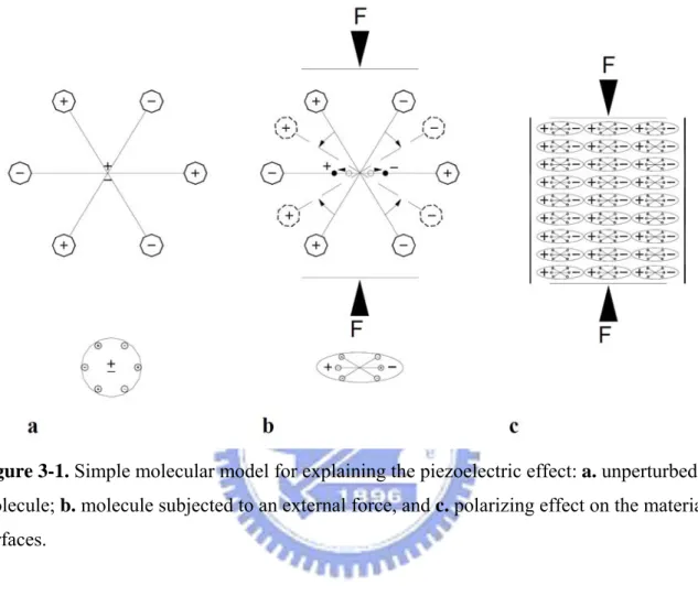
phenomenon discovered a year before by the Pierre and Jacques Curie brothers [Curie and Curie,1880]. They observed that positive and negative charges appeared on several parts of the crystal surfaces when comprising the crystal in different directions, previously analysed according to its symmetry. Figure 3-1A shows a simple molecular model; it explains the generating of an electric charge as the result of a force exerted on the material. Before subjecting the material to some external stress, the gravity centres of the negative and positive charges of each molecule coincide. Therefore, the external effects of the negative and positive charges are reciprocally cancelled. As a result, an electrically neutral molecule appears. When exerting some pressure on the material, its internal reticular structure can be deformed, causing the separation of the positive and negative gravity centres of the molecules and generating little dipoles (Figure 3-1B). The facing poles inside the material are
mutually cancelled and a distribution of a linked charge appears in the material’s surfaces
(Figure 3-1C). That is to say, the material is polarized. This polarization generates an
electric field and can be used to transform the mechanical energy used in the material’s deformation into electrical energy.
The Curie brothers verified, the year after their discovery, the existence of the reverse process, predicted by Lippmann [1881]. That is, if one arbitrarily names direct piezoelectric effect, to the generation of an electric charge, and hence of an electric field, in certain
materials and under certain laws due to a stress, there would also exist a reverse piezoelectric effect by which the application of an electric field, under similar circumstances, would cause deformation in those materials.
3.2 Piezoelectric quartz crystal
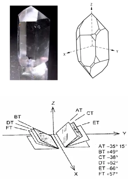
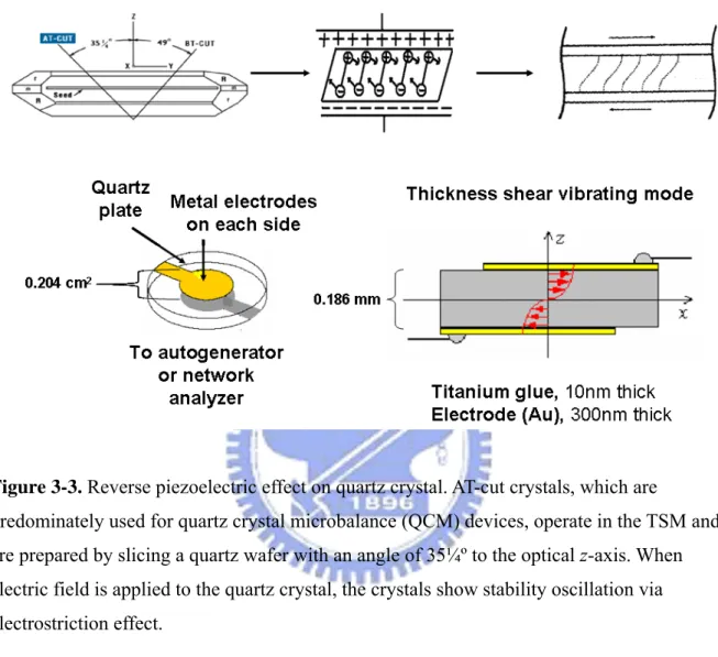
Piezoelectric quartz crystal is a reverse piezoelectric material. A prerequisite for the occurrence of piezoelectricity in crystal is an inversion center. The quartz crystal may provide a large variety of different resonator types depending on the cut angle with respect to the crystal lattice(Figure 3-2). The cut angle determines the mode of induced mechanical
vibration. Resonators operating in the thickness shear mode, face shear mode or flexural mode can be obtained from the mother crystal with eigenfrequencies ranging from 5 × 10² to 3 × 10⁸ Hz. AT-cut crystals, which are predominately used for quartz crystal microbalance (QCM) devices, operate in the TSM and are prepared by slicing a quartz wafer with an angle of 35¼º to the optical z-axis. AT-cut quartz crystals show a tremendous frequency stability of Δf/f ≒10-8 and a temperature coefficient which is close to zero between 0 and 50°C, rendering this particular cut the most suitable for QCM sensors. The technique determines the mass of very thin surface bound layers and simultaneously gives information about their viscoelastic properties [Fredriksson et al., 1998; Fu et al., 2003; Ebarvia et al., 2004; D'Souza et al., 2005]. This offers new opportunities to study conformational changes in layers formed on the sensor surface. Moreover, the technique takes into account water coupled to hydrated layers, in contrast to optical mass measurements, obtained from methods such as surface plasmon resonance and ellipsometry. These unique properties make piezoelectric quartz an invaluable tool for studying macromolecules at surfaces and an important
complement to existing techniques.
Many polymers, ceramics, and molecules such as water are permanently polarized: some parts of the molecule are positively charged, while other parts of the molecule are negatively charged. When an electric field is applied to these materials, these polarized molecules will align themselves with the electric field, resulting in induced dipoles within the molecular or crystal structure of the material. Furthermore, a permanently-polarized material such as quartz or barium titanate (BaTiO3) will produce an electric field when the material changes
dimensions as a result of an imposed mechanical force. These materials are piezoelectric, and this phenomenon is known as the piezoelectric effect. When a stress is applied to a piezoelectric material, an electric field is generated within it [Lee et al., 2004; Li et al., 2004]. Conversely, if an electric field is applied to the material it undergoes a spontaneous strain. Crystals which acquire a charge when compressed, twisted, or distorted are said to be piezoelectric. This provides a convenient transducer effect between electrical and mechanical oscillations. Conversely, an applied electric field can cause a piezoelectric material to change dimensions. This phenomenon is known as electrostriction, or the reverse piezoelectric effect. Quartz demonstrates this property and is extremely stable (Figure 3-3).
Piezoelectric biosensor, known as quartz crystal microbalance (QCM), combines high sensitivity to mass on the surface of the quartz crystal with the high specificity of a
bioreaction. It has been extensively applied as a transducer in hybridization based on DNA biosensors for the detection of gene mutation [Tombelli et al., 2000; Su et al., 2004],
genetically modified organisms [Mannelli et al., 2003], and foodborne pathogens [Ryu et al., 2001; Mo et al., 2002; Wu et al. 2007]. In this study, we utilized QCM as sensing device [Yun et al., 1998; Zhou et al., 1999]. QCM sensors (Figure 3-4) have been widely studied
and developed as detecting tools for humid, biomedical, and environmental monitoring in recent years. They are popular because of its high sensitivity , fast response, small size, low power consumption, low cost, and simplicity of use.
A piezoelectric quartz crystal resonator [Wieliczka et al., 1996; Wegener et al., 1999; Willner et al., 1999] is a precisely cut slab from a natural or synthetic crystal of quartz. A QCM consists of a thin quartz disk with electrodes plated on it (Figure 3-4B).
Pierre and Marie Curie showed in 1880 that crystals of Rochelle salt could produce electricity when pressure is applied in certain crystallographic directions. Later they also showed the converse effect i.e. production of strain by application of electricity. These findings are the discovery of the piezoelectric effect. Piezoelectricity did not receive lot of interest in the beginning and a more detailed study of piezoelectricity is not started until 1917 when it is showed that quartz crystals could be used as transducers and receivers of ultrasound in water. In 1919 several devices of everyday interest based on the piezoelectricity of
Rochelle salt is described i.e. loudspeakers, microphones and sound pick-ups. In 1921 the first quartz crystal controlled oscillator is described. These first quartz crystal controlled oscillators are based on XT-cut crystals, which have the drawback of being very temperature sensitive. Therefore, the XT-cut crystals are nowadays used in applications where the large temperature coefficient is of little importance, such as transducers in space sonars. The dominance of the quartz crystal for all kind of frequency control applications started in 1934 when the AT-cut quartz crystal is introduced [Lasky et al., 1990; Lassalle et al., 2001; Lee et al., 2002]. The advantage with the AT-cut quartz crystal is that it has nearly zero frequency drift with temperature around room temperature. From the very beginning of using quartz crystal resonators as frequency control elements it is common to increase the frequency of the resonator by drawing pencil marks on the electrodes, or decreasing the frequency by rubbing of some electrode material with an eraser. The understanding of this mass induced
a paper that showed that the frequency shift of a quartz crystal resonator is directly proportional to the added mass. Sauerbreys [Barraud et al., 1993; Ben-Dov et al., 1997; Bandyopadhyay et al., 1998; Bizet et al., 1998; Abdelmaksoud et al., 2004] work is generally taken as the breakthrough and the first step towards a new quantitative tool to measure very small masses i.e. the QCM. Hence, one can describe the QCM to be an ultra-sensitive mass sensor. The heart of the QCM is the piezoelectric AT-cut quartz crystal sandwiched between a pair of electrodes. When the electrodes are connected to an oscillator and an AC voltage is applied over the electrodes the quartz crystal starts to oscillate at its resonance frequency due to the piezoelectric effect. This oscillation is generally very stable due to the high quality of the oscillation. If a rigid layer is evenly deposited on one or both of the electrodes the
resonant frequency will decrease proportionally to the mass of the adsorbed layer according to the Sauerbrey equation [Anzai et al., 1998; Bidan et al., 2000; Bizet et al., 2000; Bernhard et al., 2002; Aizawa et al., 2003]:
ΔF = 1/2 2 0 ) ( 2 q q A m f μ ρ Δ −
Where: measured frequency shift = ΔF, resonant frequency of the fundamental mode of the
crystal = f0, mass change per unit area (g/cm2) = Δm, piezo-electrically active area = A,
density of quartz, 2.648 g/cm3 = ρq, shear modulus of quartz, 2.947 × 10¹¹ g/cms2 = μq
There are situations where the Sauerbrey equation does not hold, for example, when the added mass is a) not rigidly deposited on the electrode surface, b) slips on the surface or c) not deposited evenly on the electrode. Therefore, the Sauerbrey equation is only strictly
applicable to uniform, rigid, thin-film deposits. Due to this the QCM is for many years just regarded as a gas-phase mass detector [Ebato et al., 1994; Darder et al., 1999; Gomes et al., 1999]. Not until the beginning of 1980’s scientists realized that a quartz crystal can be excited to a stable oscillation when it is completely immersed in a liquid. Much of the pioneering work in liquid phase QCM measurements have been done by Kanazawa and coworkers [Kanazawa and Gordon., 1985], who showed that the change in resonant frequency of a QCM taken from air into a liquid is proportional to the square root of the liquid’s
density-viscosity product: ΔF= q q L L u f μ πρ η ρ 3 / 2 −
density of liquid in contact with the crystal = ρL, viscosity of liquid in contact with the crystal
= ηL, density of quartz, 2.648 g/cm3 = ρq, shear modulus of quartz, 2.947× 10¹¹ g/cms2 = μq
After it is found out that an excessive viscous loading would not prohibit use of the QCM in liquids and that the response of the QCM is still extremely sensitive to mass changes at the solid-liquid QCMs have been used in direct contact with liquids and/or visco-elastic films to assess changes in mass and visco-elastic properties. Even in air or vacuum, where the damping of layers has been considered to be negligible or small the QCM has been used to probe dissipative processes on the quartz crystal [Okahata et al., 1995; Okahata et al., 1998; Okahata et al., 1999]. This is especially true for soft condensed matters such as thick
polymer layers deposited on the quartz surface.
In the early days of electronic communication as a result of the limited number of quartz resonators available-frequency adjustment is accomplished by a pencil mark depositing a foreign mass layer on the crystal. In 1959 Sauerbrey showed that the shift in resonance frequency of thickness-shear-mode resonators is proportional to the deposited mass. This is the starting point for the development of a new generation of piezoelectric mass-sensitive devices.
The development of new measurement techniques represents one of the major driving forces in biotechnology that positively impacts related research areas such as polymer characterization and biochemistry and is critical to the evolution of the pharmaceutical, biotechnology, and biomaterials industries.
Since QCM is a piezoelectric [Sato et al., 1995; Sato et al., 1998], an oscillating electric field applied across the device induces an acoustic wave that propagates through the crystal and meets minimum impedance when the thickness of the device is a multiple of a half wavelength of the acoustic wave. A QCM is a shear mode device in which the acoustic wave propagates in a direction perpendicular to the crystal surface. To make this happen, the quartz crystal plate must be cut to a specific orientation with respect to the crystal axes. These cuts belonging to the rotated Y-cut family, the AT- and BT-cuts are representative.
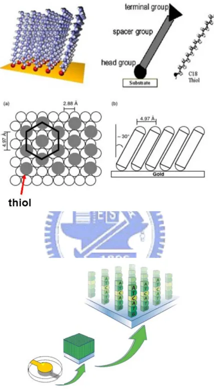
4. Self assembled monolayer for biosensor application
Considerable attention has been drawn during the last two decades to functionalize noble metal surfaces by forming ordered organic films of few nm to several hundred-nm thickness.
Self-assembled monolayer (SAM) provides one simple route to functionalize electrode surfaces by organic molecules (both aliphatic and aromatic) containing free anchor groups such as thiols, disulphides, amines, silanes, or acids. The monolayer produced by
self-assembly allows tremendous flexibility with respect to several applications depending upon their terminal functionality (hydrophilic or hydrophobic control) or by varying the chain length (distance control) (Figure 4-1A). For example, SAM of long chain alkane thiol produces a highly packed and ordered surface, which can provide a membrane like
microenvironment, useful for immobilizing biological molecules. The high selectivity of biological molecules integrated with an electrochemical, optical, or piezoelectric transduction mode of analyte recognition offers great promise to exploit them as efficient and accurate biosensors. It is demonstrated with suitable examples that monolayer design plays a key role in controlling the performance of these SAM based biosensors, irrespective of the
immobilization strategy and sensing mechanism.
The present level of research on new biosensors as well as the development of currently available biosensors e.g. glucose sensors) has increased dramatically over the past decade. The main driving force for this enhanced research activity is the booming demand for miniaturised biosensors, particularly for diagnostic applications. These however, impose rather strict requirements on the size of the device, selective response of the analyte, fast response time and compatibility with the peripheral electronic circuitry. Although the active interest in developing these small sensing devices for biomedical use is growing rapidly, some of the presently available biosensors are inadequate in these respects and several new and improved materials are desired to overcome some of these limitations. Self-assembled monolayers offer several attractive features for these kinds of applications due to various reasons. First, since they use only the bare minimum resources (e.g. a monolayer comprising of 1013 molecules/cm2 or only 10−7 moles/cm2), miniaturisation is easy. Secondly, the high degree of ordered and dense nature of the long chain alkane thiols of SAMs mimics the cellular microenvironment of lipid bilayer structures providing novel substrates for immobilized biomolecules (antibodies, enzymes, nucleic acids) or biological systems (receptors, whole cells). More significantly the easy procedure for SAM formation and compatibility with metal substrates (Au, Ag etc.) for electrochemical measurements
enable special benefits for biosensor applications involving current or potential measurements. Lastly, the chemical stability of the monolayer even after its coupling with the immobilizing molecules (with its associated specificity) for biological sensing integrated with an